. 2021 Jan 15;22(2):820.
doi: 10.3390/ijms22020820.

A Step-by-Step Approach to Improve Clinical Translation of Liposome-Based Nanomaterials, a Focus on Innate Immune and Inflammatory Responses

Affiliations

A Step-by-Step Approach to Improve Clinical Translation of Liposome-Based Nanomaterials, a Focus on Innate Immune and Inflammatory Responses

Giacomo Della Camera et al. Int J Mol Sci. .

Abstract

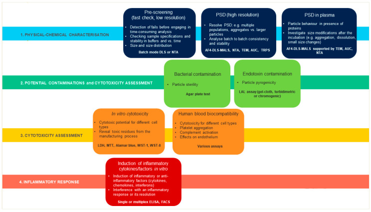
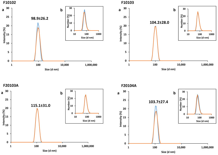
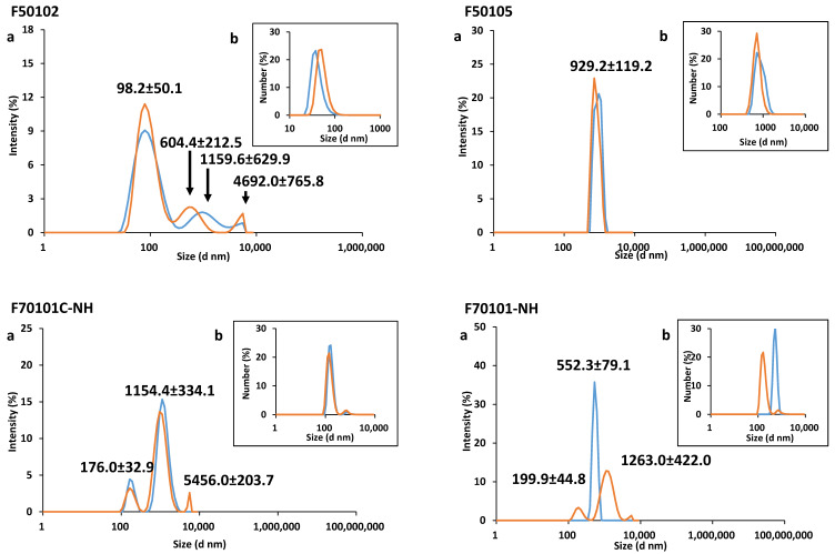
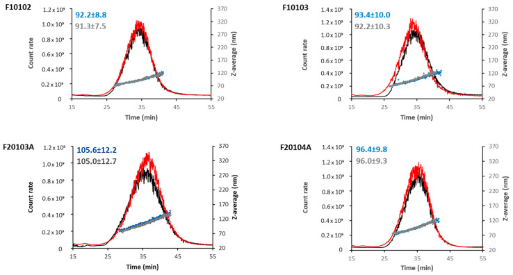
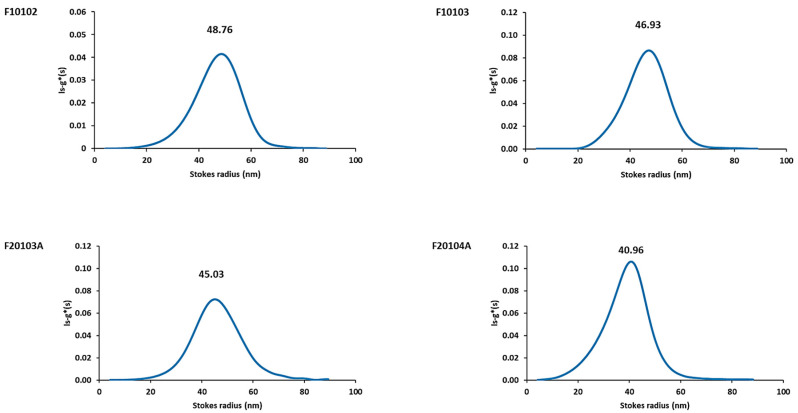
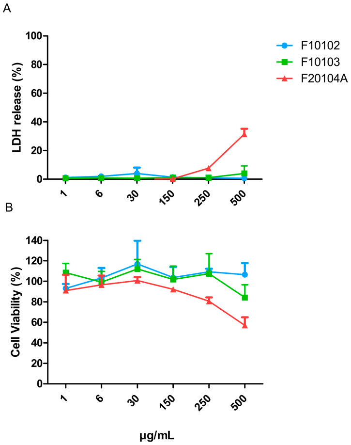
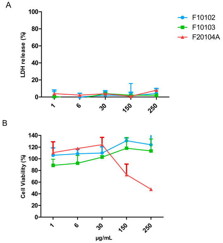
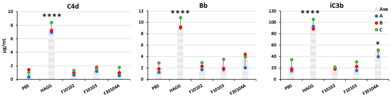
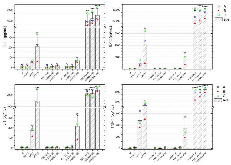
This study aims to provide guidelines to design and perform a robust and reliable physical-chemical characterization of liposome-based nanomaterials, and to support method development with a specific focus on their inflammation-inducing potential. Out of eight differently functionalized liposomes selected as "case-studies", three passed the physical-chemical characterization ( in terms of size-distribution, homogeneity and stability) and the screening for bacterial contamination (sterility and apyrogenicity). Although all three were non-cytotoxic when tested in vitro, they showed a different capacity to activate human blood cells. HSPC/CHOL-coated liposomes elicited the production of several inflammation-related cytokines, while DPPC/CHOL- or DSPC/CHOL-functionalized liposomes did not. This work underlines the need for accurate characterization at multiple levels and the use of reliable in vitro methods, in order to obtain a realistic assessment of liposome-induced human inflammatory response, as a fundamental requirement of nanosafety regulations.

Keywords: Limulus Amoebocyte Lisate (LAL); cytokines; endotoxin; inflammation; interleukin; liposome; nanomaterial; nanomedicine; particle size distribution; physicochemical characterization; safety assessment.


Conflict of interest statement

The authors declare no conflict of interest.

Figures

Figure 1
Figure 1
Each liposome was characterized for its physical-chemical properties and for potential contamination by bacteria and bacterial toxins, and eventually examined for toxicity on human cells and capacity to induce an inflammatory response. PSD, particle size distribution.
Figure 2
Figure 2
Batch-mode DLS data reported (a) as intensity-based size distribution, and (b) as number-based size distribution. Scheme 25 and 50 µg/mL, (red and blue lines, respectively) were prepared in PBS. Average of three measurements are shown. The PDI of all liposomes is reported in Table 2.
Figure 2
Figure 2
Batch-mode DLS data reported (a) as intensity-based size distribution, and (b) as number-based size distribution. Scheme 25 and 50 µg/mL, (red and blue lines, respectively) were prepared in PBS. Average of three measurements are shown. The PDI of all liposomes is reported in Table 2.
Figure 3
Figure 3
AF4-DLS measurements of selected liposomes in PBS (black lines and blue dots) or PBS supplemented with 10% human serum (red lines and grey dots). The elugram of flow mode DLS (scattered intensity and size vs. elution time) are reported for one representative measurement for each selected liposome and condition. Scattering intensity peaks (lines) and hydrodynamic diameters (dots) by DLS are shown. Hydrodynamic diameter values, measured in PBS or in serum, are reported (blue and grey respectively).
Figure 4
Figure 4
AUC measurement results of liposome size distributions. Stock liposomes were diluted in PBS to a final concentration of 50 μg/mL before proceeding with AUC measurements. PBS was used as control in the reference cell. ls-g*(s) sedimentation coefficient distributions of selected liposomes were calculated from AUC measurements using interference optics and transformed to size distributions. A representative measurement for each of the selected liposomes is reported, with the Stokes radius value expressed in nm.
Figure 5
Figure 5
Cell death (% LDH release; A) and cell viability (% MTT metabolic transformation; B) of Hep G2 cells exposed to liposomes F10102, F10103 and F20104A for 24 h. The error bars represent the SD of three independent experiments. Negative controls (incubation in medium) and positive controls (cell lysis with Triton) were used as benchmarks.
Figure 6
Figure 6
Cell death (% LDH release; A) and cell viability (% MTT metabolic transformation; B) of PBMC exposed to liposomes F10102, F10103, and F20104A for 24 h. The error bars represent the SD of three independent experiments. Negative controls (cells incubated with medium) and positive controls (cell lysed with Triton) were used as benchmarks.
Figure 7
Figure 7
Levels of C4d (left), Bb (center), and iC3b (right) in the plasma of healthy volunteers upon incubation with liposomes. Fresh human plasma from three healthy donors was incubated with liposomes F10102, F10103, or F20104A at 150 µg/mL for 1 h at 37 °C. PBS and heat-aggregated gamma globulins (HAGG, 0.5 mg/mL) were used as negative and positive controls, respectively. Data are shown as individual values (dots of different colors for the three donors) and also as mean (shaded columns) ± SD. * p <0.05, **** p < 0.0001.
Figure 8
Figure 8
Whole blood from three healthy donors (represented in blue, red, green) was incubated for 24 h with selected liposomes at three concentrations (6, 30, 150 μg/mL). The production of IL-1α, IL-1β, IL-6, and TNF-α data is here reported. M represents the negative control (whole blood exposed to medium only), whereas LPS was used as a positive control at the concentrations of 0.1, 1 and 10 ng/mL. Data are also shown as mean (shaded bars) ± SD. * p < 0.05, ** p < 0.01, *** p < 0.001, **** p < 0.0001.

References

    1. Sercombe L., Veerati T., Moheimani F., Wu S.Y., Sood A.K., Hua S. Advances and challenges of liposome assisted drug delivery. Front. Pharmacol. 2015;6:286. doi: 10.3389/fphar.2015.00286. - DOI - PMC - PubMed
    1. FDA News Release FDA Approves First Treatment for Certain Types of Poor-Prognosis Acute Myeloid Leukemia. [(accessed on 9 December 2020)];2017 Available online: https://www.fda.gov/news-events/press-announcements/fda-approves-first-t....
    1. Anselmo A.C., Mitragotri S. Nanoparticles in the clinic: An update. Bioeng. Transl. Med. 2019;4:e10143. doi: 10.1002/btm2.10143. - DOI - PMC - PubMed
    1. Krauss A.C., Gao X., Li L., Manning M.L., Patel P., Fu W., Janoria K.G., Gieser G., Bateman D.A., Przepiorka D., et al. FDA Approval Summary: (Daunorubicin and Cytarabine) Liposome for Injection for the Treatment of Adults with High-Risk Acute Myeloid Leukemia. Clin. Cancer Res. 2019;25:2685–2690. doi: 10.1158/1078-0432.CCR-18-2990. - DOI - PubMed
    1. Inglut C.T., Sorrin A.J., Kuruppu T., Vig S., Cicalo J., Ahmad H., Huang H. Immunological and toxicological considerations for the design of liposomes. Nanomaterials. 2020;10:190. doi: 10.3390/nano10020190. - DOI - PMC - PubMed

MeSH terms

LinkOut - more resources