

Systematic Review: Anesthetic Protocols and Management as Confounders in Rodent Blood Oxygen Level Dependent Functional Magnetic Resonance Imaging (BOLD fMRI)-Part B: Effects of Anesthetic Agents, Doses and Timing
- PMID: 33467584
- PMCID: PMC7830239
- DOI: 10.3390/ani11010199
Systematic Review: Anesthetic Protocols and Management as Confounders in Rodent Blood Oxygen Level Dependent Functional Magnetic Resonance Imaging (BOLD fMRI)-Part B: Effects of Anesthetic Agents, Doses and Timing
Abstract
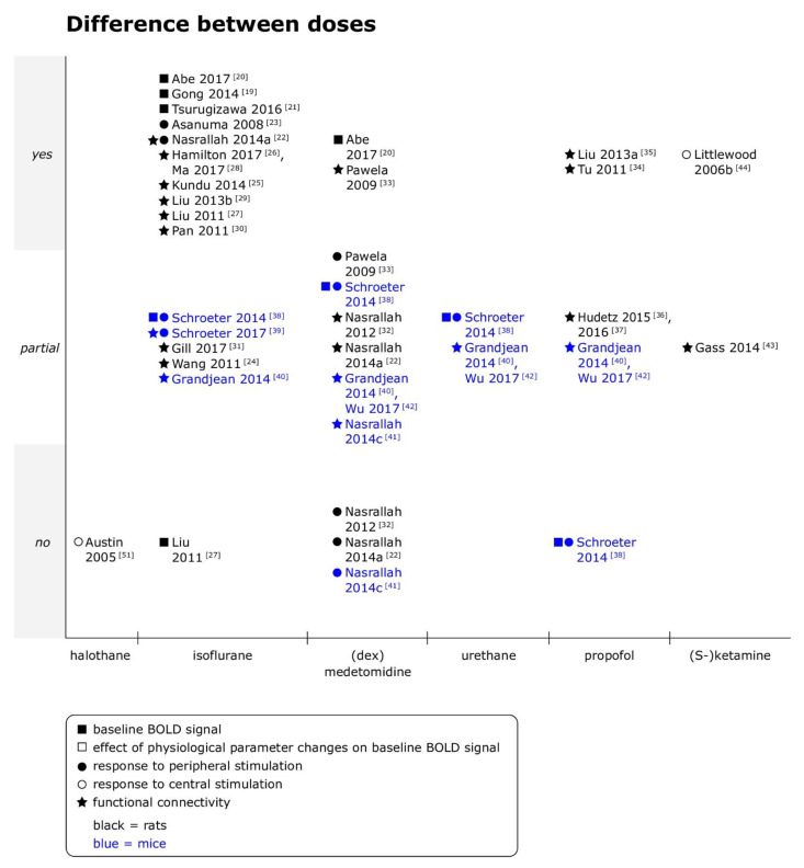
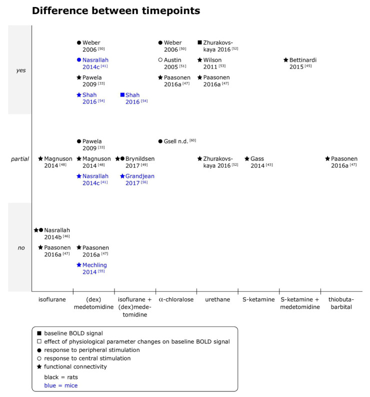
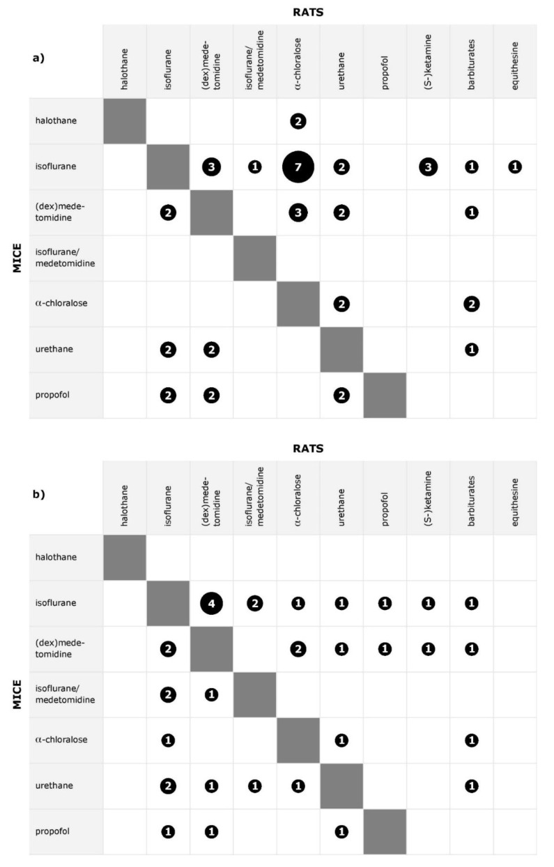
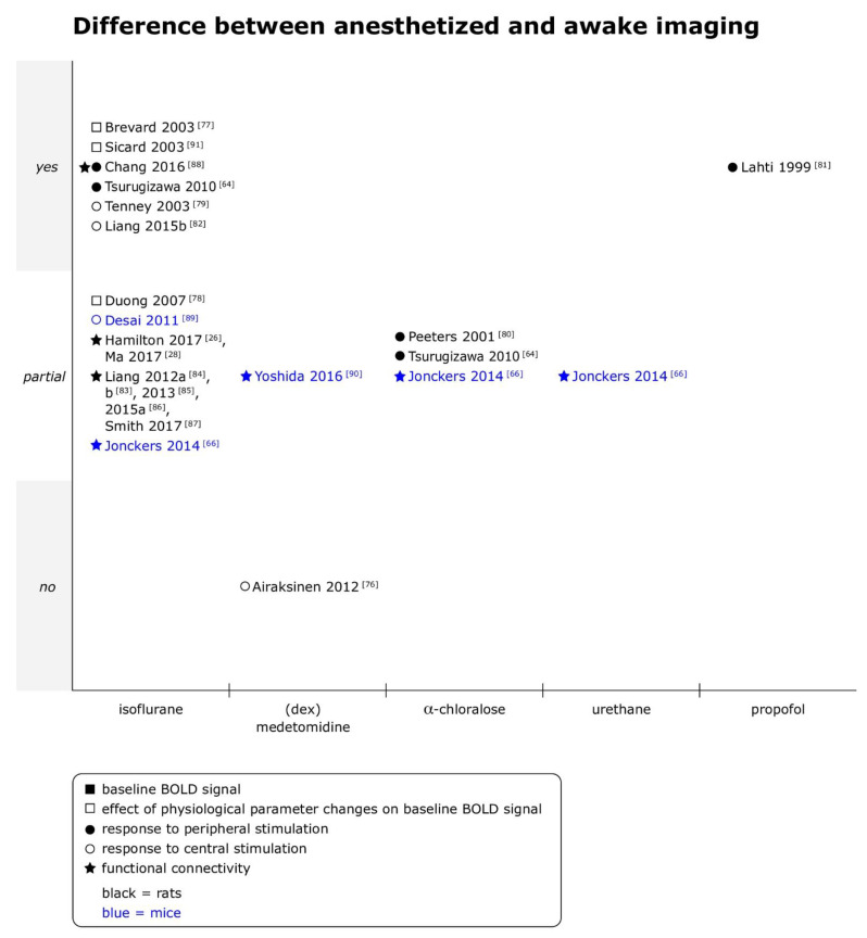
In rodent models the use of functional magnetic resonance imaging (fMRI) under anesthesia is common. The anesthetic protocol might influence fMRI readouts either directly or via changes in physiological parameters. As long as those factors cannot be objectively quantified, the scientific validity of fMRI in rodents is impaired. In the present systematic review, literature analyzing in rats and mice the influence of anesthesia regimes and concurrent physiological functions on blood oxygen level dependent (BOLD) fMRI results was investigated. Studies from four databases that were searched were selected following pre-defined criteria. Two separate articles publish the results; the herewith presented article includes the analyses of 83 studies. Most studies found differences in BOLD fMRI readouts with different anesthesia drugs and dose rates, time points of imaging or when awake status was compared to anesthetized animals. To obtain scientifically valid, reproducible results from rodent fMRI studies, stable levels of anesthesia with agents suitable for the model under investigation as well as known and objectively quantifiable effects on readouts are, thus, mandatory. Further studies should establish dose ranges for standardized anesthetic protocols and determine time windows for imaging during which influence of anesthesia on readout is objectively quantifiable.
Keywords: BOLD fMRI; anesthesia regime; anesthetic protocol; isoflurane; medetomidine; mouse; rat; validity; α-chloralose.
Conflict of interest statement
The authors declare no conflict of interest. The funders had no role in the design of the study; in the collection, analyses, or interpretation of data; in the writing of the manuscript, or in the decision to publish the results.
Figures
Similar articles
-
Where do we stand on fMRI in awake mice?Cereb Cortex. 2024 Jan 14;34(1):bhad478. doi: 10.1093/cercor/bhad478. Cereb Cortex. 2024. PMID: 38100331 Free PMC article.
-
Systematic Review: Anaesthetic Protocols and Management as Confounders in Rodent Blood Oxygen Level Dependent Functional Magnetic Resonance Imaging (BOLD fMRI)-Part A: Effects of Changes in Physiological Parameters.Front Neurosci. 2020 Oct 23;14:577119. doi: 10.3389/fnins.2020.577119. eCollection 2020. Front Neurosci. 2020. PMID: 33192261 Free PMC article.
-
A fully noninvasive and robust experimental protocol for longitudinal fMRI studies in the rat.Neuroimage. 2006 Feb 15;29(4):1303-10. doi: 10.1016/j.neuroimage.2005.08.028. Epub 2005 Oct 11. Neuroimage. 2006. PMID: 16223588
-
Specificity of stimulus-evoked fMRI responses in the mouse: the influence of systemic physiological changes associated with innocuous stimulation under four different anesthetics.Neuroimage. 2014 Jul 1;94:372-384. doi: 10.1016/j.neuroimage.2014.01.046. Epub 2014 Feb 2. Neuroimage. 2014. PMID: 24495809
-
Confounding effects of anesthesia on functional activation in rodent brain: a study of halothane and alpha-chloralose anesthesia.Neuroimage. 2005 Jan 1;24(1):92-100. doi: 10.1016/j.neuroimage.2004.08.011. Neuroimage. 2005. PMID: 15588600
Cited by
-
Promising Cerebral Blood Flow Enhancers in Acute Ischemic Stroke.Transl Stroke Res. 2023 Dec;14(6):863-889. doi: 10.1007/s12975-022-01100-w. Epub 2022 Nov 17. Transl Stroke Res. 2023. PMID: 36394792 Free PMC article. Review.
-
Where do we stand on fMRI in awake mice?Cereb Cortex. 2024 Jan 14;34(1):bhad478. doi: 10.1093/cercor/bhad478. Cereb Cortex. 2024. PMID: 38100331 Free PMC article.
-
Magnetic Resonance Imaging in Tauopathy Animal Models.Front Aging Neurosci. 2022 Jan 25;13:791679. doi: 10.3389/fnagi.2021.791679. eCollection 2021. Front Aging Neurosci. 2022. PMID: 35145392 Free PMC article. Review.
-
The Role of fMRI in Drug Development: An Update.Adv Neurobiol. 2023;30:299-333. doi: 10.1007/978-3-031-21054-9_13. Adv Neurobiol. 2023. PMID: 36928856
-
Low-cost physiology and behavioral monitor for intravital imaging in small mammals.Neurophotonics. 2025 Jan;12(1):015004. doi: 10.1117/1.NPh.12.1.015004. Epub 2025 Jan 25. Neurophotonics. 2025. PMID: 39867131 Free PMC article.
References
Publication types
Grants and funding
LinkOut - more resources
Full Text Sources
Other Literature Sources
Research Materials
