Review
. 2021 Jan 15;13(2):309.
doi: 10.3390/cancers13020309.

IFN-γ and CD38 in Hyperprogressive Cancer Development

Affiliations
Review

IFN-γ and CD38 in Hyperprogressive Cancer Development

Stefania Angelicola et al. Cancers (Basel). .

Abstract

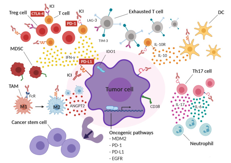
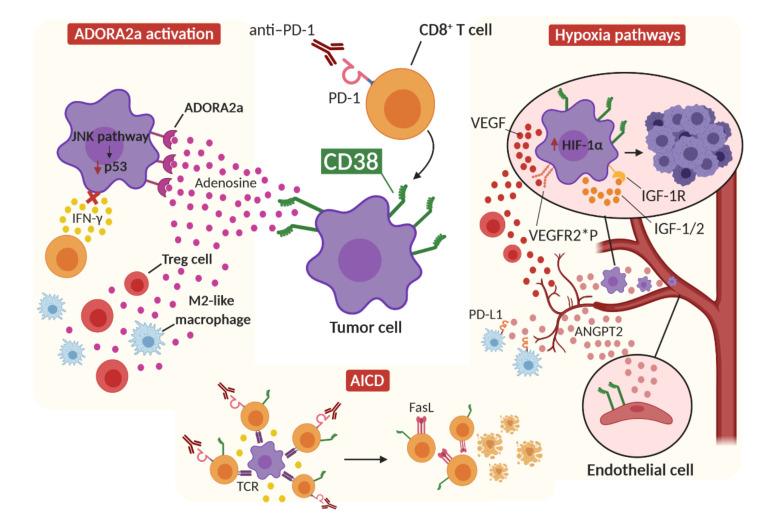
Immune checkpoint inhibitors (ICIs) improve the survival of patients with multiple types of cancer. However, low response rates and atypical responses limit their success in clinical applications. The paradoxical acceleration of tumor growth after treatment, defined as hyperprogressive disease (HPD), is the most difficult problem facing clinicians and patients alike. The mechanisms that underlie hyperprogression (HP) are still unclear and controversial, although different factors are associated with the phenomenon. In this review, we propose two factors that have not yet been demonstrated to be directly associated with HP, but upon which it is important to focus attention. IFN-γ is a key cytokine in antitumor response and its levels increase during ICI therapy, whereas CD38 is an alternative immune checkpoint that is involved in immunosuppressive responses. As both factors are associated with resistance to ICI therapy, we have discussed their possible involvement in HPD with the conclusion that IFN-γ may contribute to HP onset through the activation of the inflammasome pathway, immunosuppressive enzyme IDO1 and activation-induced cell death (AICD) in effector T cells, while the role of CD38 in HP may be associated with the activation of adenosine receptors, hypoxia pathways and AICD-dependent T-cell depletion.

Keywords: CD38; IFN-γ; cancer; hyperprogression; hyperprogressive disease; immune checkpoint inhibitors; immunotherapy; macrophage; tumor microenvironment.


Conflict of interest statement

The authors declare no conflict of interest.

Figures

Figure 1
Figure 1
Possible mechanisms of hyperprogressive disease (HPD) in cancer after immune checkpoint blockade therapy. ICI therapy may functionally activate infiltrating regulatory T cells (Tregs), leading to an immunosuppressive tumor microenvironment. At the same time, compensatory upregulation of alternative immune checkpoints, such as lymphocyte-activation gene 3 (LAG-3), T-cell immunoglobulin and mucin domain-3 (TIM-3) and CTLA-4, on effector T cells after ICI therapy may induce T-cell exhaustion. IC inhibition might also induce the expression of cluster of differentiation 38 (CD38) on tumor cells and IFN-γ-dependent recruitment of CD38-expressing myeloid-derived suppressor cells (MDSCs), resulting in immune suppression. Moreover, ICI therapy may induce the upregulation of the immunosuppressive enzyme indoleamine 2,3-dioxygenase (IDO1) and the increased secretion of immunosuppressive cytokines and soluble molecules, such as interleukin 10 (IL-10), angiopoietin-2 (ANGPT2) and interferon-gamma (IFN-γ) into the tumor microenvironment. IC blockade might also functionally boost T helper 1 (not shown) and T helper 17 (Th17) lymphocytes, resulting in neutrophil recruitment and in an inflammatory immunosuppressive tumor microenvironment enriched in interleukin 6 (IL-6) and interleukin 17 (IL-17). In addition, ICIs may bind to Fc receptors (FcR) on tumor-associated macrophages (TAMs), resulting in a shift from M1 to an immunosuppressive M2 phenotype. IC blockade may also induce an increase in cancer stem cells (CSC) and may activate oncogenic pathways, such as MDM2, PD-1, PD-L1 and EGFR, thus promoting tumor proliferation. DC: dendritic cell.
Figure 2
Figure 2
Proposed IFN-γ-dependent mechanisms of hyperprogression. The release of IFN-γ from CD8+ T cells after ICI therapy can activate the inflammasome pathway by upregulating PD-L1 expression on tumor cells and activating NLRP3 signaling, resulting in immunosuppressive MDSC recruitment in the tumor microenvironment. At the same time, IFN-γ can induce IDO1 activity in tumor cells, which activates the JNK pathway, leading to p53 downregulation and tumor growth. Finally, the concomitant stimulation of tumor-specific CD8+ T cells by ICI therapy and T-cell-receptor (TCR) activation results in a hyperactivated immune environment in which IFN-γ triggers the activation-induced cell death (AICD) mechanism and T-cell Fas-mediated apoptosis.
Figure 3
Figure 3
Proposed CD38-dependent mechanisms of hyperprogression. The upregulation and activation of CD38 after IC blockade therapy, together with the action of enzymes that catabolize ATP and ADP (not shown), lead to the release of adenosine into the tumor microenvironment. The activation of ADORA2a receptors on tumor cells by adenosine leads to the blockade of the IFN-γ pathway, making the tumor cell resistant to cytotoxic IFN-γ action, as well as the activation of oncogenic pathways, such as the JNK signaling pathway, which leads to the downregulation of p53. The action of adenosine also increases Treg cells and the polarization of M2 macrophages in the tumor microenvironment. CD38 also promotes the activation of AICD after the hyperactivation of tumor-specific CD8+ T cells by ICI therapy, and induces the expression of FasL on T cells. CD38 also induces the release of Angiopoietin-2 from endothelial cells, leading to increased angiogenesis, PD-L1 expression on M2-like macrophages and enhanced tumor invasion. Finally, CD38 induces the expression of HIF-1α in tumor cells, which can, in turn, induce the production of VEGF and IGF factors. VEGF can establish paracrine signaling, promoting the recruitment of Treg cells, and autocrine signaling on tumor cells by binding VEGFR2, promoting tumorigenesis and invasion. The polymorphisms rs1870377 A/T and A/A may make this binding stronger. HIF-1α can also induce the expression of IGF-pathway components, leading to autocrine signaling that results in tumor growth and survival. ANGPT2: Angiopoietin 2; IGF: insulin-like growth factor; IGF-1R: IGF-1 receptor; VEGFR2*P: VEGFR2 receptor with rs1870377 A/T or A/A polymorphisms.

References

    1. Darvin P., Toor S.M., Sasidharan Nair V., Elkord E. Immune checkpoint inhibitors: Recent progress and potential biomarkers. Exp. Mol. Med. 2018;50:1–11. doi: 10.1038/s12276-018-0191-1. - DOI - PMC - PubMed
    1. Pardoll D.M. The blockade of immune checkpoints in cancer immunotherapy. Nat. Rev. Cancer. 2012;12:252–264. doi: 10.1038/nrc3239. - DOI - PMC - PubMed
    1. Topalian S.L., Sznol M., McDermott D.F., Kluger H.M., Carvajal R.D., Sharfman W.H., Brahmer J.R., Lawrence D.P., Atkins M.B., Powderly J.D., et al. Survival, durable tumor remission, and long-term safety in patients with advanced melanoma receiving nivolumab. J. Clin. Oncol. 2014;32:1020–1030. doi: 10.1200/JCO.2013.53.0105. - DOI - PMC - PubMed
    1. Ahamadi M., Freshwater T., Prohn M., Li C., de Alwis D., de Greef R., Elassaiss-Schaap J., Kondic A., Stone J. Model-Based Characterization of the Pharmacokinetics of Pembrolizumab: A Humanized Anti-PD-1 Monoclonal Antibody in Advanced Solid Tumors. CPT Pharmacomet. Syst. Pharmacol. 2017;6:49–57. doi: 10.1002/psp4.12139. - DOI - PMC - PubMed
    1. Vaddepally R.K., Kharel P., Pandey R., Garje R., Chandra A.B. Review of indications of FDA-approved immune checkpoint inhibitors per NCCN guidelines with the level of evidence. Cancers. 2020;12:738. doi: 10.3390/cancers12030738. - DOI - PMC - PubMed

LinkOut - more resources