Review
. 2021 Jan 15;11(1):112.
doi: 10.3390/biom11010112.

Evolution and Adaptation of Legionella pneumophila to Manipulate the Ubiquitination Machinery of Its Amoebae and Mammalian Hosts

Affiliations
Review

Evolution and Adaptation of Legionella pneumophila to Manipulate the Ubiquitination Machinery of Its Amoebae and Mammalian Hosts

Christopher T D Price et al. Biomolecules. .

Abstract

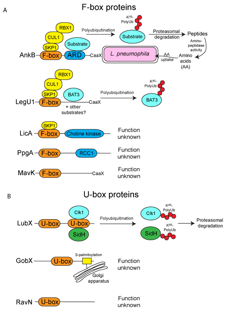
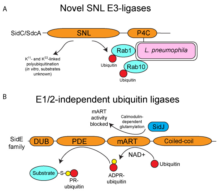
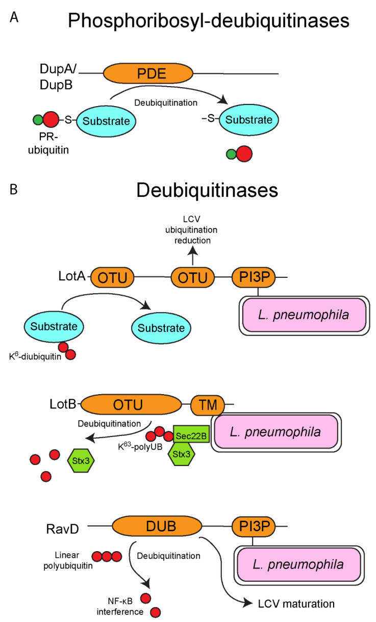
The ubiquitin pathway is highly conserved across the eukaryotic domain of life and plays an essential role in a plethora of cellular processes. It is not surprising that many intracellular bacterial pathogens often target the essential host ubiquitin pathway. The intracellular bacterial pathogen Legionella pneumophila injects into the host cell cytosol multiple classes of classical and novel ubiquitin-modifying enzymes that modulate diverse ubiquitin-related processes in the host cell. Most of these pathogen-injected proteins, designated as effectors, mimic known E3-ubiquitin ligases through harboring F-box or U-box domains. The classical F-box effector, AnkB targets host proteins for K48-linked polyubiquitination, which leads to excessive proteasomal degradation that is required to generate adequate supplies of amino acids for metabolism of the pathogen. In contrast, the SidC and SdcA effectors share no structural similarity to known eukaryotic ligases despite having E3-ubiquitin ligase activity, suggesting that the number of E3-ligases in eukaryotes is under-represented. L. pneumophila also injects into the host many novel ubiquitin-modifying enzymes, which are the SidE family of effectors that catalyze phosphoribosyl-ubiquitination of serine residue of target proteins, independently of the canonical E1-2-3 enzymatic cascade. Interestingly, the environmental bacterium, L. pneumophila, has evolved within a diverse range of amoebal species, which serve as the natural hosts, while accidental transmission through contaminated aerosols can cause pneumonia in humans. Therefore, it is likely that the novel ubiquitin-modifying enzymes of L. pneumophila were acquired by the pathogen through interkingdom gene transfer from the diverse natural amoebal hosts. Furthermore, conservation of the ubiquitin pathway across eukaryotes has enabled these novel ubiquitin-modifying enzymes to function similarly in mammalian cells. Studies on the biological functions of these effectors are likely to reveal further novel ubiquitin biology and shed further lights on the evolution of ubiquitin.

Keywords: Dot/Icm; E1/2-independent ubiquitin ligase; E3-ubiquitin ligase; F-box; SNL E3-ligase; U-box; amoebae; deubiquitinase; effectors; evolution; legionella pneumophila; ubiquitin.


Conflict of interest statement

No potential conflicts of interest were reported by the authors.

Figures

Figure 1
Figure 1
The “classical” E3-ubiquitin ligases of L. pneumophila. L. pneumophila has multiple effectors that show homology to the classical F-box and U-box families that are widespread in eukaryotes. (A) F-box effectors of L. pneumophila. There are five known L. pneumophila F-box effectors. Firstly, AnkB, which harbors an F-box, an ankyrin repeat domain (ARD) and a C-terminal CaaX motif that is targeted for farnesylation, enabling localization to the Legionella-containing vacuole, (LCV) membrane. The F-box domain of AnkB interacts with the host Skp1-Cul1 (SCF) complex to promote the decoration of the LCV with K48-linked polyubiquitinated proteins. These proteins are ultimately degraded by the host proteasomes to generate copious quantities of amino acids required for metabolism of L. pneumophila. LegU1 also interacts with the SCF complex and directs the polyubiquitination of the host protein, BAT3. It is possible LegU1 also targets other as yet unidentified host proteins. Similar to AnkB, LegU1 also has a C-terminal CaaX motif, targeting this effector to host membranes. LicA interacts with the Skp1 component of the SCF complex but not Cul1, suggesting a LicA forms a non-canonical complex, and the functional role of this effector is unknown. The functional roles of MavK, which also has a C-terminal CaaX motif and PpgA are unknown. (B) U-box effectors of L. pneumophila. L. pneumophila injects three effectors with the U-box domain. LubX ubiquitinates the host factor Clk1. Additionally, LubX functions as a metaeffector and directs ubiquitination of the injected effector, SidH, which is then targeted for proteasomal-mediation degradation. GobX is targeted to the Golgi apparatus where it is embedded into the membrane through S-palmitoylation modification, however the functional role of GobX is unknown. RavN also harbors the U-box domain, but the role of this effector is unknown.
Figure 2
Figure 2
Novel ubiquitin ligases of L. pneumophila. L. pneumophila possesses multiple effectors that exhibit ubiquitin ligase activity but do not show any homology to any known eukaryotic ligases. (A) Novel SNL E3-ligases of L. pneumophila. L. pneumophila injects two paralogous effectors (SidC and SdcA) that possess the SidC N-terminal ubiquitin ligase (SNL) domain, which catalyzes K11- and K33-linked polyubiquitin linkages (in vitro), and direct monoubiquitination of host Rab1 and Rab10. Both effectors associate with the outer leaflet of the LCV membrane via their C-terminal PI4P binding domain. SidC/SdcA contribute to ER-derived vesicle recruitment to the LCV. The SNL domain has not been found in eukaryotic ubiquitin ligases. (B) E1/2-independent ubiquitin ligases of L. pneumophila. The E1/2-independent ubiquitin ligases of the SidE family are the first known ubiquitin ligases that function completely independently of the canonical activity of the E1 and E2 enzymes. The mART domain of the SidE family catalyzes the formation of ADP-ribosylated ubiquitin using NAD+ as a substrate. The PDE domain of the SidE family can then use ADP-ribosylated ubiquitin to add phosphoribosylated-ubiquitin to serine residues to substrate proteins, such as host Rab proteins and Rtn4. Activity of the SidE family is regulated by the metaeffector, SidJ, which catalyzes calmodulin-dependent glutamylation of the mART domain, and this blocks activity of this domain.
Figure 3
Figure 3
Deubiquitinases of L. pneumophila. Besides possessing several ubiquitin ligase, L. pneumophila also injects into the cytosol of host cells several deubiquitinases. (A) Phosphoribosyl-deubiquitinases of L. pneumophila. The SidE family-dependent phosphoribosyl-ubiquitination of proteins can be reversed by the deubiquitinase activity of the two injected effectors, DupA and DupB. Both DupA and DupB possess a PDE domain homologous to those found in the SidE family, but in contrast these domains catalyze the removal of phosphoribosyl-ubiquitin moieties from substrate proteins. (B) Deubiquitinases of L. pneumophila. L. pneumophila also possesses several deubiquitinases that specifically remove ubiquitin moieties from targeted proteins. LotA harbors two OTU domains and is localized to the LCV outer leaflet via its C-terminal PI3P-binding domain. LotA has been shown to reduce the overall ubiquitination of the LCV, and additionally specifically deubiquitinate proteins that are modified with a K6-diubiquitin moiety, though the functional importance of this effector is unknown. Similar to LotA, LotB also harbors an OTU domain and has a C-terminal transmembrane domain to allow localization to the LCV membrane. LotB specifically deubiquitinates K63-linked polyubiquitin chains from the host SNARE, Sec22B, resulting in the release of SNARE, Stx3. Finally, RavD harbors a deubiquitinase domain that specifically deubiquitinates linear polyubiquitin chains, and this may disrupt NF-κB signaling. Similar to LotA, RavD has the C-terminal PI3P-binding domain to allow localization of the effector of the LCV membrane.

Similar articles

Cited by

References

    1. Vere G., Kealy R., Kessler B.M., Pinto-Fernandez A. Ubiquitomics: An Overview and Future. Biomolecules. 2020;10:1453. doi: 10.3390/biom10101453. - DOI - PMC - PubMed
    1. Kliza K., Husnjak K. Resolving the Complexity of Ubiquitin Networks. Front. Mol. Biosci. 2020;7:21. doi: 10.3389/fmolb.2020.00021. - DOI - PMC - PubMed
    1. Song L., Luo Z.-Q. Post-translational regulation of ubiquitin signaling. J. Cell Biol. 2019;218:1776–1786. doi: 10.1083/jcb.201902074. - DOI - PMC - PubMed
    1. Grice G.L., Nathan J.A. The recognition of ubiquitinated proteins by the proteasome. Cell. Mol. Life Sci. 2016;73:3497–3506. doi: 10.1007/s00018-016-2255-5. - DOI - PMC - PubMed
    1. Kudriaeva A.A., Belogurov A.A. Proteasome: A Nanomachinery of Creative Destruction. Biochemistry. 2019;84:159–192. doi: 10.1134/S0006297919140104. - DOI - PubMed

Publication types

MeSH terms

LinkOut - more resources