. 2021 Jan 27;288(1943):20202095.
doi: 10.1098/rspb.2020.2095. Epub 2021 Jan 20.

The extensibility of the plantar fascia influences the windlass mechanism during human running

Affiliations

The extensibility of the plantar fascia influences the windlass mechanism during human running

Lauren Welte et al. Proc Biol Sci. .

Abstract

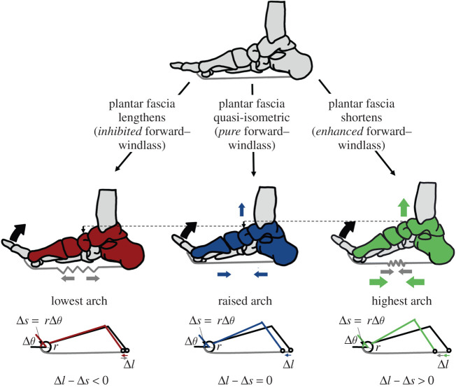
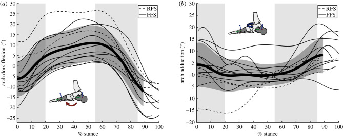
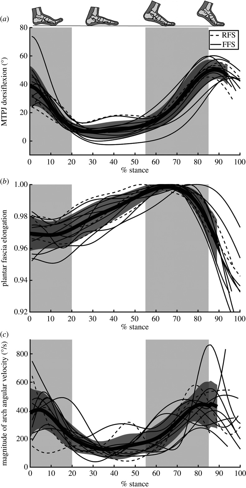
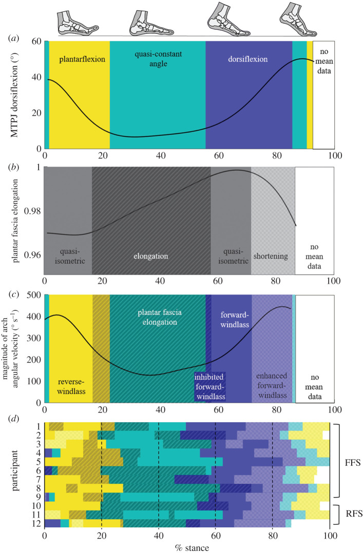
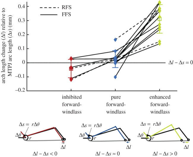
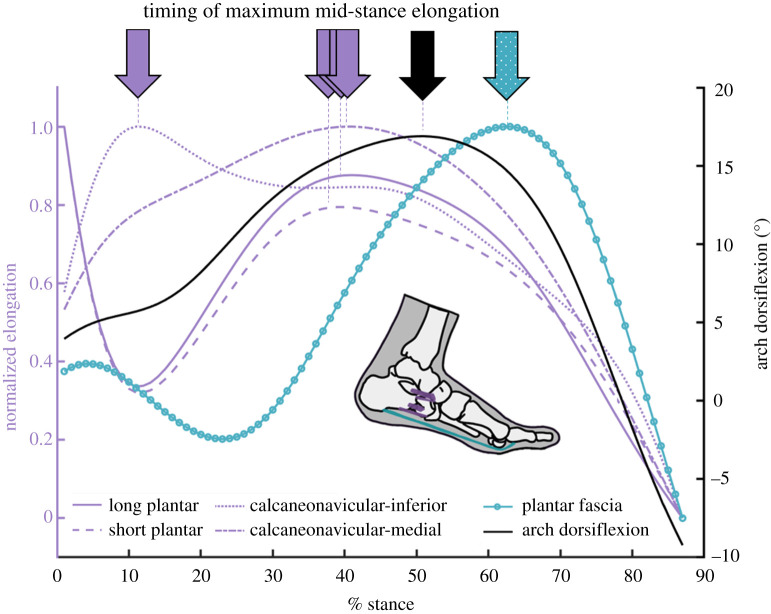
The arch of the human foot is unique among hominins as it is compliant at ground contact but sufficiently stiff to enable push-off. These behaviours are partly facilitated by the ligamentous plantar fascia whose role is central to two mechanisms. The ideal windlass mechanism assumes that the plantar fascia has a nearly constant length to directly couple toe dorsiflexion with a change in arch shape. However, the plantar fascia also stretches and then shortens throughout gait as the arch-spring stores and releases elastic energy. We aimed to understand how the extensible plantar fascia could behave as an ideal windlass when it has been shown to strain throughout gait, potentially compromising the one-to-one coupling between toe arc length and arch length. We measured foot bone motion and plantar fascia elongation using high-speed X-ray during running. We discovered that toe plantarflexion delays plantar fascia stretching at foot strike, which probably modifies the distribution of the load through other arch tissues. Through a pure windlass effect in propulsion, a quasi-isometric plantar fascia's shortening is delayed to later in stance. The plantar fascia then shortens concurrently to the windlass mechanism, likely enhancing arch recoil at push-off.

Keywords: arch-spring; biplanar videoradiography; foot arch biomechanics; plantar fascia; running; windlass mechanism.


Conflict of interest statement

The authors declare no competing interests.

Figures

Figure 1.
Figure 1.
Theoretical framework. The forward-windlass mechanism's ability to shorten and raise the arch can be theoretically influenced if the plantar fascia simultaneously lengthens, shortens or is quasi-isometric. The pure forward-windlass mechanism occurs if there is a 1 : 1 ratio of arch deformation (Δl) to arc length change from MTPJ dorsiflexion (Δs = rΔθ). If the plantar fascia shortens concurrently to the forward-windlass mechanism, it could enhance arch rising. If the plantar fascia lengthens simultaneously to the forward-windlass mechanism, the arch rising effect could be limited. (Online version in colour.)
Figure 2.
Figure 2.
Arch angles. The (a) dorsiflexion and (b) adduction angle between the first metatarsal and the calcaneus during running stance phase. The thickest line and dark grey region represent the mean ± 1 s.d. The subject's strike pattern is indicated with either a dashed (rear-foot strike, RFS) or solid (fore-foot strike, FFS) line. The light grey shaded regions highlight early stance (0–20%), mid-stance (20–55%) and propulsion (55–85%) to facilitate comparison with figure 4. (Online version in colour.)
Figure 3.
Figure 3.
MTPJ angle, plantar fascia elongation and magnitude of arch angular velocity. (a) MTPJ dorsiflexion, (b) plantar fascia elongation and (c) the magnitude of 3D arch angular velocity over the stance phase of running gait. The thickest line and dark grey region represent the mean ± 1 s.d. The subject's strike pattern is indicated with either a dashed (rear-foot strike, RFS) or solid (fore-foot strike, FFS) line. The light grey shaded regions highlight early stance (0–20%), mid-stance (20–55%) and propulsion (55–85%) to facilitate comparison with figure 4.
Figure 4.
Figure 4.
Classification of windlass mechanism phases. (a) The mean of all participants' metatarsophalangeal joint (MTPJ) angle is classified into phases based on whether it is plantarflexing (yellow, light), staying within the 0.5° per 1% stance threshold as quasi-constant (teal, medium), or dorsiflexing (purple, dark). (b) The plantar fascia elongation is classified as elongation (dark diagonal lines), staying within the 0.0005 normalized elongation per 1% of stance threshold as quasi-isometric (no pattern), or shortening (light checkerboard). (c) The magnitude of the three-dimensional arch angular velocity is super-imposed over the classification of windlass mechanism behaviour. The plantar fascia elongation phases in (b) are matched with the windlass engagement (MTPJ dorsiflexion) periods in (a) to highlight the phases of inhibited, pure or enhanced forward-windlass, as well as the reverse-windlass phase. (d) The phases for each participant using the same thresholds as were selected for the mean MTPJ angle and plantar fascia elongation.
Figure 5.
Figure 5.
Windlass mechanism coupling with arch length change. For the pure forward-windlass mechanism, as the MTPJ dorsiflexes, the change in arc length (Δs) should be equal to the change in the length of the arch (Δl), such that the difference between them is 0 (i.e. Δl − Δs = 0). The difference between arch length change and MTPJ arc length change is averaged during the phases as classified in figure 4. There is reduced arch length change during the inhibited forward-windlass, and additional arch length change in the enhanced forward-windlass compared with the same participant's pure forward-windlass mechanism, as indicated by connecting lines (dashed, rear-foot strike, RFS; solid, fore-foot strike, FFS). (Online version in colour.)
Figure 6.
Figure 6.
Arch ligament deformation. The arch dorsiflexion over the stance phase of running (black, right y-axis) shows similar maximum timing to the elongation of the arch-spanning ligaments. Elongation of the arch-spanning ligaments normalized to the range of elongation are measured on the left y-axis (purple). The plantar fascia's normalized elongation is measured on the left y-axis but is shown in teal dots to highlight the later peak elongation. (Online version in colour.)

Similar articles

Cited by

References

    1. D'Août K, Aerts P. 2008. The evolutionary history of the human foot. Maastricht, The Netherlands: Shaker Publishing.
    1. Holowka NB, Lieberman DE. 2018. Rethinking the evolution of the human foot: insights from experimental research. J. Exp. Biol. 221, jeb174425 (10.1242/jeb.174425) - DOI - PubMed
    1. Ker RF, Bennett MB, Bibby SR, Kester RC, Alexander RMcN. 1987. The spring in the arch of the human foot. Nature 325, 147–149. (10.1038/325147a0) - DOI - PubMed
    1. Hicks JH 1954. The mechanics of the foot. II. The plantar aponeurosis and the arch. J. Anat. 88, 25–30. - PMC - PubMed
    1. Bojsen-Moller F, Flagstad KE. 1976. Plantar aponeurosis and internal architecture of the ball of the foot. J. Anat. 121, 599–611. - PMC - PubMed

Publication types

LinkOut - more resources