. 2021 Jan 19;21(1):28.
doi: 10.1186/s12883-021-02057-3.

Abnormal eye movements in spinocerebellar ataxia type 3

Affiliations

Abnormal eye movements in spinocerebellar ataxia type 3

Junyu Lin et al. BMC Neurol. .

Abstract

Background: Abnormal eye movements are common in spinocerebellar ataxias Type 3 (SCA3). We conducted the research to explore the frequency of abnormal eye movements in Chinese patients with SCA3, to compare the demographic and clinical characteristics between SCA3 patients with and without each type of abnormal eye movement, and to explore the correlation between abnormal eye movements and the severity of ataxia.

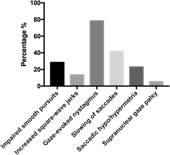
Methods: Seventy-four patients with SCA3 were enrolled in this cross-sectional study. Six types of abnormal eye movements including impaired smooth pursuit, increased square-wave jerks (SWJ), gaze-evoked nystagmus (GEN), slowing of saccades, saccadic hypo/hypermetria and supranuclear gaze palsy were evaluated by experienced neurologists. The severity of ataxia was evaluated by Scale for the Assessment and Rating of Ataxia (SARA).

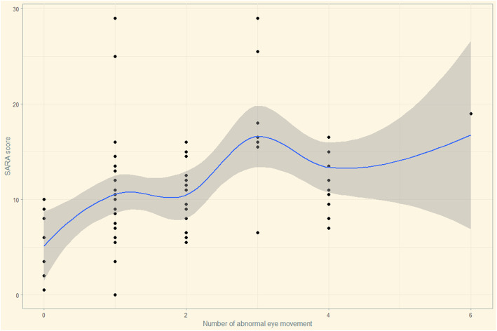
Results: The prevalence of impaired smooth pursuit, increased SWJ, GEN, slowing of saccades, saccadic hypo/hypermetria and supranuclear gaze palsy in Chinese SCA3 patients was 28.4, 13.5, 78.4, 41.9, 23.0, and 5.4%, respectively. SCA3 patients with GEN had higher scores of International Cooperative Ataxia Rating Scale (ICARS-IV) and total ICARS, and longer length of CAG repeat than patients without GEN. SCA3 patients with slowing of saccades had a longer disease duration, higher scores of ICARS-I, ICARS-II, total ICARS and SARA than patients without slowing of saccades. SCA3 patients with saccadic hypo/hypermetria had higher scores of ICARS-III, ICARS-IV, and SARA than patients without saccadic hypo/hypermetria. The demographic and clinical characteristics did not differ significantly between SCA3 patients with and without impaired smooth pursuit, increased SWJ, or supranuclear gaze palsy. Multivariate linear regression showed that the number of abnormal eye movements (0-6), disease duration, Hamilton Depression Rating Scale-24 (HDRS-24) score, and CAG repeat length were positively correlated with SARA score, whereas Montreal Cognitive Assessment (MoCA) score was negatively correlated with SARA score in SCA3.

Conclusions: An increased number of abnormal eye movement types correlated with the severity of ataxia in SCA3.

Keywords: Eye movements; Severity; Spinocerebellar Ataxia type 3.


Conflict of interest statement

The authors declare that they have no competing interests.

Figures

Fig. 1
Fig. 1
Frequency of each type of abnormal eye movement in SCA3. X-axis demonstrates the type of each abnormal eye movement, and Y-axia shows the frequency of each abnormal eye movement. The frequency of impaired smooth pursuit, increased square-wave jer ks, gaze-evoked nystagmus, slowing of saccades, saccadic hypo/hypermetria and supranuclear gaze palsy was 28.4, 13.5, 78.4, 41.9, 23.0, and 5.4% respectively
Fig. 2
Fig. 2
Hypothesis testing for the multivariate linear regression model diagnosis. 1) Residuals vs Fitted figure (upper left) was used to explore the linearity between fitted values and residuals. Data points were uniformly distributed on the both side of y = 0 and the line presented a stationary curve without obvious shape feature, which suggested the linearity was good. 2) Normal Q-Q figure (upper right) was used to examine the normality of standardized residuals. Data points were arranged in diagonal lines, tending to a straight line, and passing diagonally through it, which indicated the residuals were normally distributed. 3) Square root of standardized residuals and fitted figure (lower left) was used to examine the homoscedasticity of standardized residuals. Data points were uniformly distributed on the both side of y = 0 and the line presented a stationary curve without obvious shape feature, which suggested that the residual values of all predicted dependent variables are approximately equal. 4) Standardized residuals vs leverage figure (lower right) was used to check the outlier. There were no significant outliers that can affect the regression results in this model
Fig. 3
Fig. 3
Scatter plots between SARA scores and the numbers of abnormal eye movement. X-axis represents the numbers of abnormal eye movement, and Y-axia represents the SARA scores

Similar articles

Cited by

References

    1. Klockgether T, Mariotti C, Paulson HL. Spinocerebellar ataxia. Nat Rev Dis Primers. 2019;5(1):24. doi: 10.1038/s41572-019-0074-3. - DOI - PubMed
    1. Stephen CD, Schmahmann JD. Eye movement abnormalities are ubiquitous in the Spinocerebellar ataxias. Cerebellum. 2019;18(6):1130–1136. doi: 10.1007/s12311-019-01044-2. - DOI - PubMed
    1. Jacobi H, Hauser TK, Giunti P, Globas C, Bauer P, Schmitz-Hubsch T, et al. Spinocerebellar ataxia types 1, 2, 3 and 6: the clinical spectrum of ataxia and morphometric brainstem and cerebellar findings. Cerebellum. 2012;11(1):155–166. doi: 10.1007/s12311-011-0292-z. - DOI - PubMed
    1. Rosini F, Pretegiani E, Battisti C, Dotti MT, Federico A, Rufa A. Eye movement changes in autosomal dominant spinocerebellar ataxias. Neurol Sci. 2020;41(7):1719–1734. doi: 10.1007/s10072-020-04318-4. - DOI - PubMed
    1. Rivaud-Pechoux S, Durr A, Gaymard B, Cancel G, Ploner CJ, Agid Y, et al. Eye movement abnormalities correlate with genotype in autosomal dominant cerebellar ataxia type I. Ann Neurol. 1998;43(3):297–302. doi: 10.1002/ana.410430306. - DOI - PubMed