. 2021 Jan 20;20(1):19.
doi: 10.1186/s12943-021-01314-w.

Circular RNA circSDHC serves as a sponge for miR-127-3p to promote the proliferation and metastasis of renal cell carcinoma via the CDKN3/E2F1 axis

Affiliations

Circular RNA circSDHC serves as a sponge for miR-127-3p to promote the proliferation and metastasis of renal cell carcinoma via the CDKN3/E2F1 axis

Junjie Cen et al. Mol Cancer. .

Abstract

Background: There is increasing evidence that circular RNAs (circRNAs) have significant regulatory roles in cancer development and progression; however, the expression patterns and biological functions of circRNAs in renal cell carcinoma (RCC) remain largely elusive.

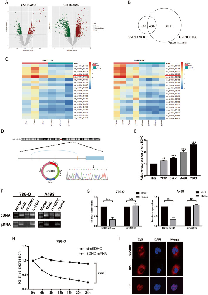
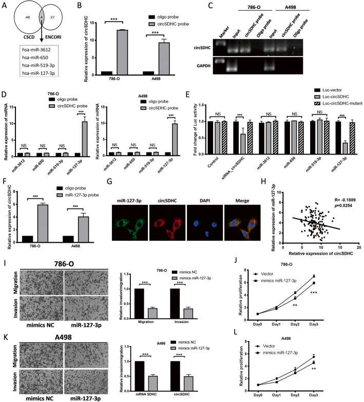
Method: Bioinformatics methods were applied to screen for circRNAs differentially expressed in RCC. Analysis of online circRNAs microarray datasets and our own patient cohort indicated that circSDHC (hsa_circ_0015004) had a potential oncogenic role in RCC. Subsequently, circSDHC expression was measured in RCC tissues and cell lines by qPCR assay, and the prognostic value of circSDHC evaluated. Further, a series of functional in vitro and in vivo experiments were conducted to assess the effects of circSDHC on RCC proliferation and metastasis. RNA pull-down assay, luciferase reporter and fluorescent in situ hybridization assays were used to confirm the interactions between circSDHC, miR-127-3p and its target genes.

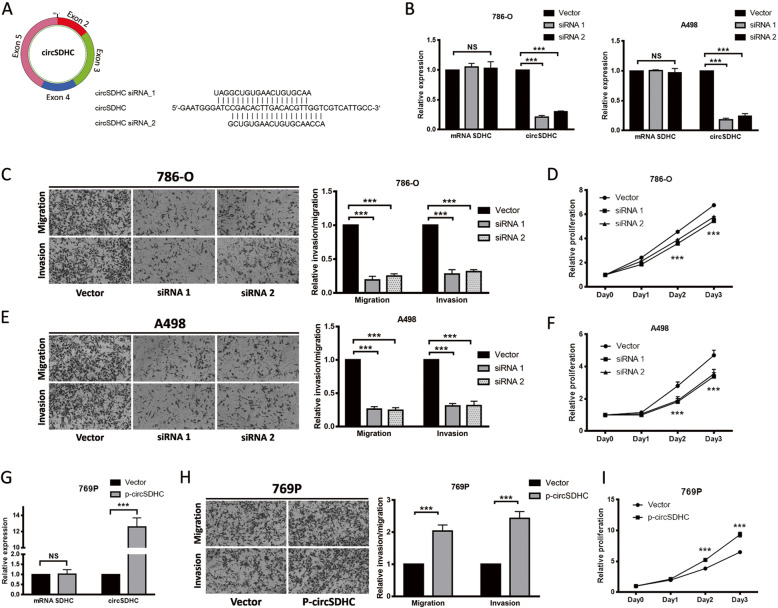
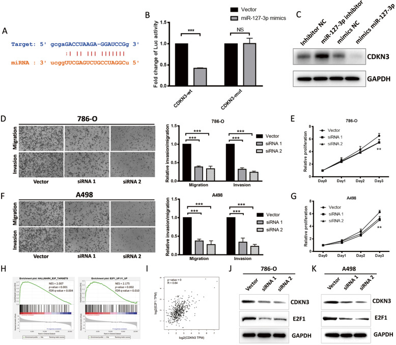
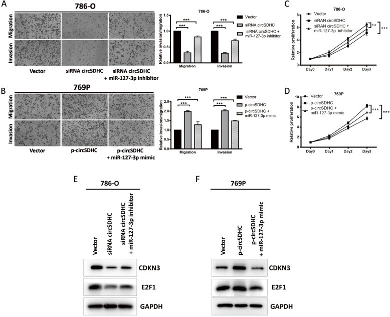
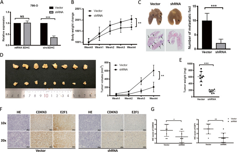
Results: Clinically, high circSDHC expression was correlated with advanced TNM stage and poor survival in patients with RCC. Further, circSDHC promoted tumor cell proliferation and invasion, both in vivo and in vitro. Analysis of the mechanism underlying the effects of circSDHC in RCC demonstrated that it binds competitively to miR-127-3p and prevents its suppression of a downstream gene, CDKN3, and the E2F1 pathway, thereby leading to RCC malignant progression. Furthermore, knockdown of circSDHC caused decreased CDKN3 expression and E2F1 pathway inhibition, which could be rescued by treatment with an miR-127-3p inhibitor.

Conclusion: Our data indicates, for the first time, an essential role for the circSDHC/miR-127-3p/CDKN3/E2F1 axis in RCC progression. Thus, circSDHC has potential to be a new therapeutic target in patients with RCC.

Keywords: E2F1 pathway; Renal cell carcinoma; circSDHC; miR-127-3p.


Conflict of interest statement

The authors declare that they have no competing interests.

Figures

Fig. 1
Fig. 1
Oncogenic circRNAs discovery and characterization of circSDHC in RCC. a. Volcano plot of GSE137836 and GSE100186. Compared to the primary tumors (in GSE137836) and adjacent normal tissue (in GSE100186), red dots represent significantly up-regulated circRNAs, while green dots represent significantly down-regulated circRNAs. Grey dots represent circRNAs that are not significant. b. Venn plot of the two datasets. Common circRNAs with p < 0.05, |log2 (fold change)| > 1 are chosen. c. Heatmaps of the two datasets. The red color represents high expression whereas the blue color represents low expression. d. Schematic illustration of the circSDHC formation from SDHC gene in the chromosome 1. The back splicing junction was verified by Sanger sequencing. Black arrow indicates the specific junction. e. The expression level of circSDHC in normal kidney cell line HK2 and different RCC cell lines, measured by qRT-PCR. f. The existence of circSDHC was confirmed by RT-PCR and gel electrophoresis using convergent and divergent primers. circSDHC can only be amplified in cDNA. GAPDH serves as control. g. Stability of circSDHC and linear SDHC was assessed by RNase treatment followed by qRT-PCR. h. Stability of circSDHC and linear SDHC was assessed by Actinomycin D treatment followed by qRT-PCR at different time points. i. Cellular localization of circSDHC was detected by FISH. Nuclear was label with DAPI dye. The majority of circSDHC is within the cytoplasm. Data are mean ± SD, n = 3
Fig. 2
Fig. 2
CircSDHC promotes the proliferation and aggressiveness of RCC. a. Target sites of siRNAs used in the knockdown experiment. Both siRNAs target back-splice junction of circPRMT5. b. The knockdown efficiency was measured by qRT-PCR in 786-O and A498 cell lines. c and e. Cell migration and invasion abilities of 786-O and A498 transfected with circSDHC siRNAs or control vector. Number of cells migrated or invaded was determined by counting the cells on five random microscopic field and calculating the mean. Level of migration and invasion were normalized to the control vector group. d and f. Cell proliferation ability of 786-O and A498 transfected with circSDHC siRNAs or control vector. The relative proliferative rate at different time points were normalized to day 0. g. The overexpression plasmid of circSDHC or control vector were transfected into 769P cell line, and expression level of circSDHC was measured by qRT-PCR. h. Cell migration and invasion abilities of 769P transfected with overexpression plasmid or control vector. i. Cell proliferation abilities of 769P transfected with overexpression plasmid or control vector. Data are mean ± SD, n = 3
Fig. 3
Fig. 3
Tumor suppressor miR-127-3p is a target of circSDHC in RCC. a. Four miRNAs were predicted as the potential target of circSDHC in CSCD and ENCORI databases. b and c. Relative expression detected by qRT-PCR and gel electrophoresis of circSDHC in 786-O and A498 lysates after RNA pull down with circSDHC specific probe or oligo probe. Expression levels were normalized to oligo probe. GAPDH was used as negative control. d. Relative levels of candidate miRNAs were detected by qRT-PCR after being pull down by circSDHC probe or oligo probe. e. Luciferase reporter assay of 786-O with Luc-vector, Luc-circSDHC or Luc-circSDHC-mutant co-transfected with different candidate miRNAs. f. Relative level of circSDHC was detected by qRT-PCR after being pull down by miR-127-3p probe or oligo probe. g. Cellular localization of miR-127-3p (FAM) and circSDHC (Cy3) detected by FISH. Nuclear was label with DAPI dye. h. circSDHC expression was negatively correlative with miR-127-3p in our own patient cohort (n = 140). i and k. Cell migration and invasion abilities of 786-O and A498 transfected with mimics miR-127-3p or mimics NC. j and l. Cell proliferation ability of 786-O and A498 transfected with mimics miR-127-3p or mimics NC
Fig. 4
Fig. 4
miR-127-3p inhibits RCC progression through downregulation of CDKN3/E2F1 axis. a. The binding site between miR-127-3p (orange) and CDKN3 (blue) predicted by ENCORI. b. Luciferase reporter assay of 786-O with Luc-CDKN3-wt or Luc-CDKN3-mut co-transfected with mimics miR-127-3p or mimics NC. c. Western blot of CDKN3 levels after 786-O cells were treated with miR-127-3p inhibitor, mimics miR-127-3p or negative control. d and f. Cell migration and invasion abilities of 786-O and A498 transfected with CDKN3 siRNAs or control vector. e and g. Cell proliferation ability of 786-O and A498 transfected with CDKN3 siRNAs or control vector. h. GSEA analysis of E2F1 pathway in TCGA patients with high and low CDKN3 expression (NES, normal enrichment score; FDR, false discovery rate). i. Correlation analysis between CDKN3 and E2F1 from GEPIA website (TCGA dataset, n = 523). j and k. Western blot of CDKN3 and E2F1 levels after 786-O and A498 cells were transfected with CDKN3 siRNAs or control vector
Fig. 5
Fig. 5
circSDHC regulates CDKN3/E2F1 and promotes RCC progression by the sponge effect towards miR-127-3p. a. Cell migration and invasion abilities of 786-O transfected with control vector, circSDHC siRNA alone or circSDHC siRNA plus miR-127-3p inhibitor. b. Cell migration and invasion abilities of 769P transfected with control vector, circSDHC overexpression plasmid alone or circSDHC overexpression plasmid plus miR-127-3p mimics. c. Cell proliferation ability of 786-O transfected with control vector, circSDHC siRNA alone or circSDHC siRNA plus miR-127-3p inhibitor. d. Cell proliferation ability of 769P transfected with control vector, circSDHC overexpression plasmid alone or circSDHC overexpression plasmid plus miR-127-3p mimics. e. Western blot of CDKN3 and E2F1 levels after 786-O transfected with control vector, circSDHC siRNA alone or circSDHC siRNA plus miR-127-3p inhibitor. f. Western blot of CDKN3 and E2F1 levels after 786-O transfected with control vector, circSDHC overexpression plasmid alone or circSDHC overexpression plasmid plus miR-127-3p mimics
Fig. 6
Fig. 6
Knockdown of circSDHC inhibits the progression of RCC tumor in vivo. a. Stable circSDHC knockdown 786-O cell line establishment. Relative expression levels of linear SDHC and circSDHC were measured by qRT-PCR. Expression level were normalized to control vector group. b. Weekly mice body weight change in the metastasis experiment. 1 × 106 stable circSDHC knockdown or control 786-O cells were injected via tail vein (n = 8 in each group). c. Representative images of gross and microscopic HE stain of the tumor-infiltrated lung (left). The metastatic foci in each mouse from two groups were counted under microscope and summarized (right). d. The picture of the gross tumors in dissected from subcutaneous xenograft model (left). 5 × 106 stable circSDHC knockdown or control 786-O cells were inoculated subcutaneously into the left side of the body. (n = 8 in each group). Weekly tumor volume change was recorded and showed (right). e. The final tumor weights in the subcutaneous xenograft model. f. HE and IHC staining of the tumors from subcutaneous xenograft model. g. Summary and statistical analysis of CDKN3 and E2F1 IHC scores

Similar articles

Cited by

References

    1. Scelo G, Larose TL. Epidemiology and Risk Factors for Kidney Cancer. J Clin Oncol. 2018:JCO2018791905. - PMC - PubMed
    1. Ljungberg B, et al. Corrigendum to "the epidemiology of renal cell carcinoma" [Eur Urol 2011;60:615-21] Eur Urol. 2011;60(6):1317. doi: 10.1016/j.eururo.2011.09.001. - DOI - PubMed
    1. De Meerleer G, et al. Radiotherapy for renal-cell carcinoma. Lancet Oncol. 2014;15(4):e170–e177. doi: 10.1016/S1470-2045(13)70569-2. - DOI - PubMed
    1. Blanco AI, Teh BS, Amato RJ. Role of radiation therapy in the management of renal cell cancer. Cancers (Basel) 2011;3(4):4010–4023. doi: 10.3390/cancers3044010. - DOI - PMC - PubMed
    1. Moch H, et al. Biomarkers in renal cancer. Virchows Arch. 2014;464(3):359–365. doi: 10.1007/s00428-014-1546-1. - DOI - PubMed

Publication types

MeSH terms