

Human mesenchymal stem cell therapy promotes retinal ganglion cell survival and target reconnection after optic nerve crush in adult rats
- PMID: 33468246
- PMCID: PMC7814601
- DOI: 10.1186/s13287-020-02130-7
Human mesenchymal stem cell therapy promotes retinal ganglion cell survival and target reconnection after optic nerve crush in adult rats
Abstract
Background: Optic-nerve injury results in impaired transmission of visual signals to central targets and leads to the death of retinal ganglion cells (RGCs) and irreversible vision loss. Therapies with mesenchymal stem cells (MSCs) from different sources have been used experimentally to increase survival and regeneration of RGCs.
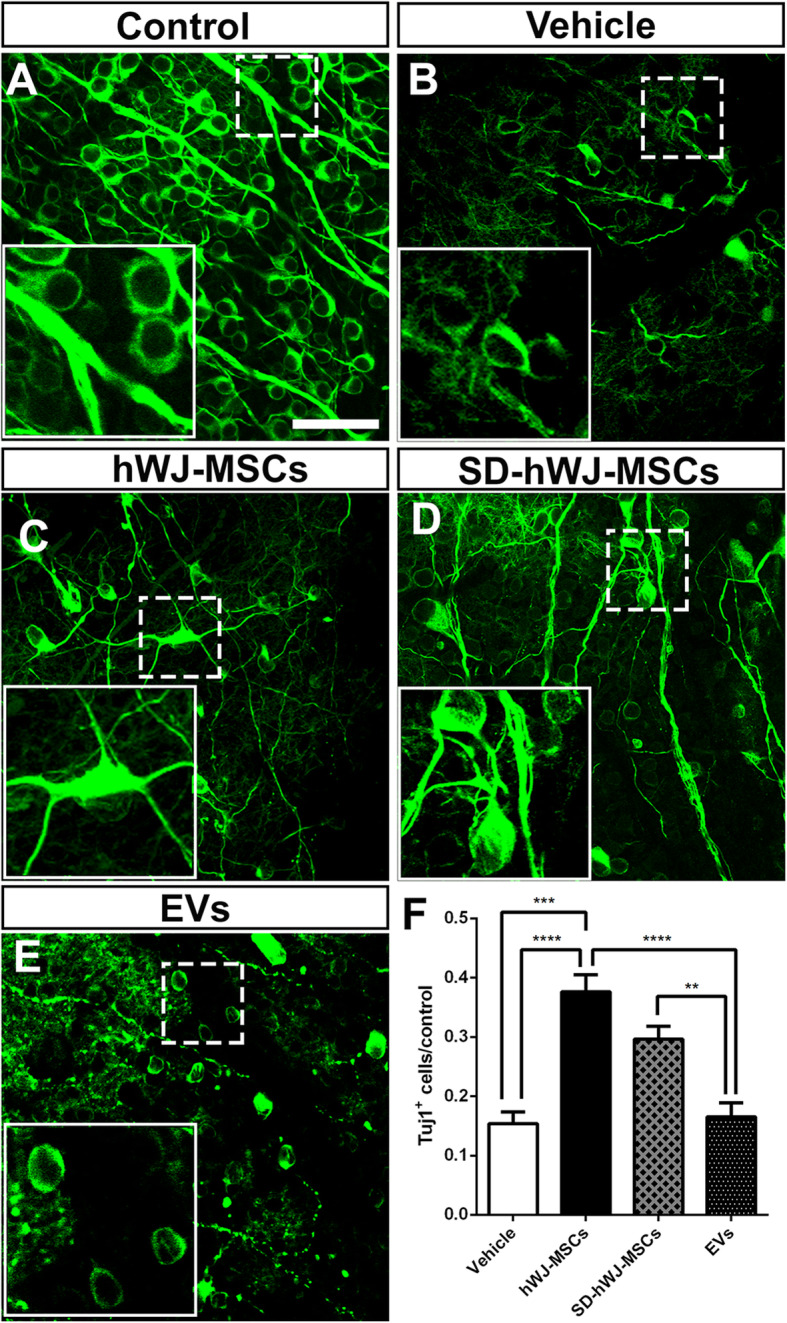
Methods: We investigated the efficacy of human umbilical Wharton's jelly-derived MSCs (hWJ-MSCs) and their extracellular vesicles (EVs) in a rat model of optic nerve crush.
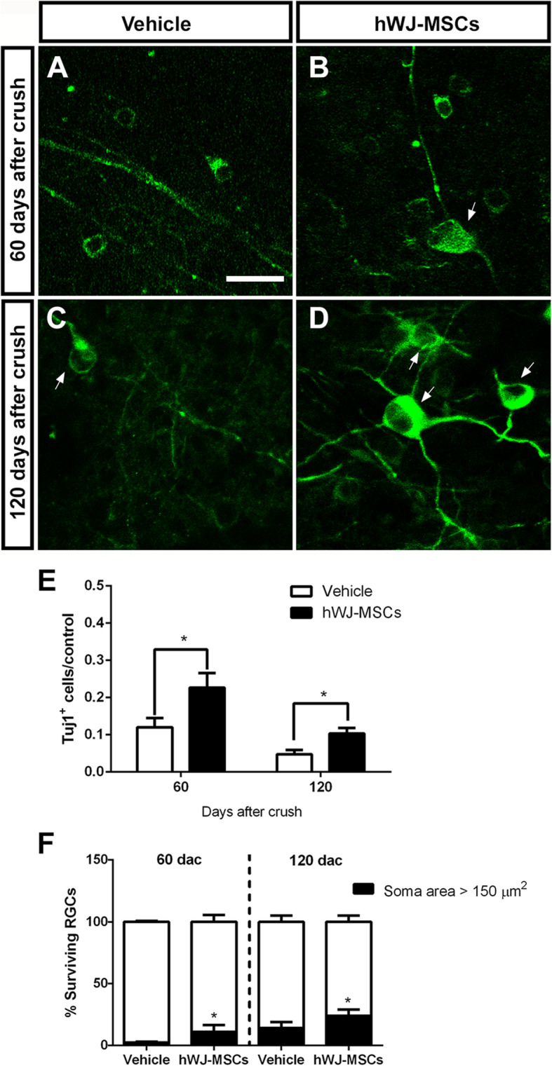
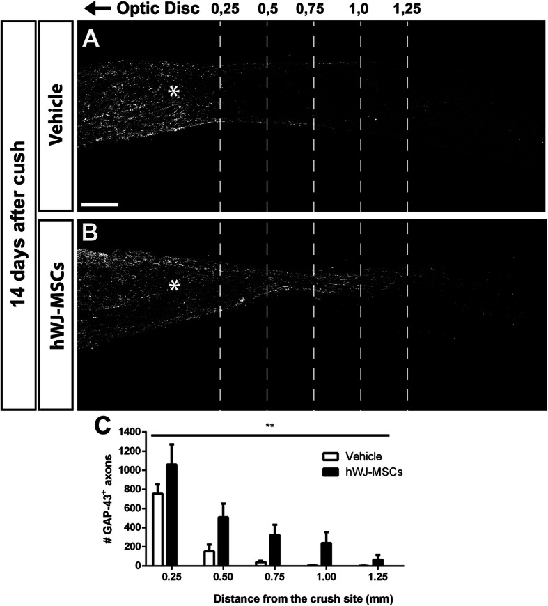
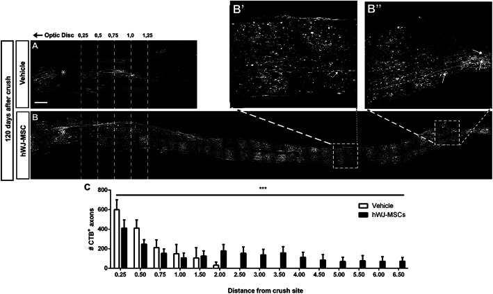
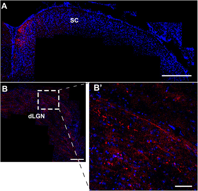
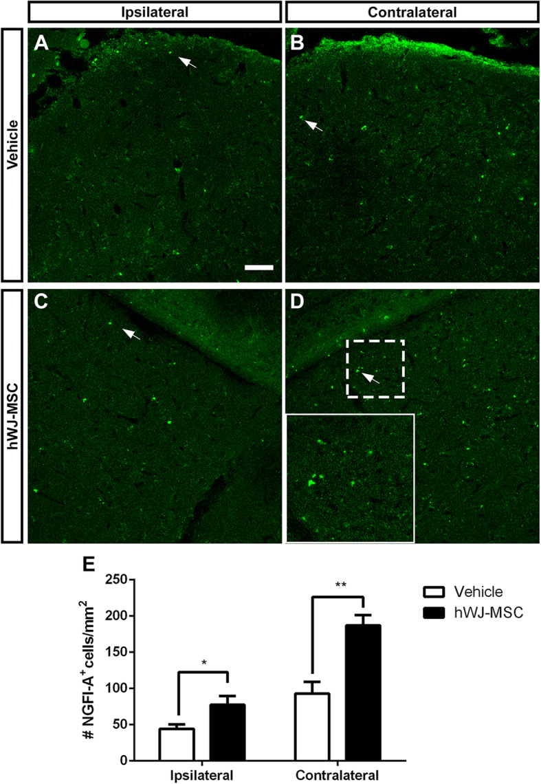
Results: hWJ-MSCs had a sustained neuroprotective effect on RGCs for 14, 60, and 120 days after optic nerve crush. The same effect was obtained using serum-deprived hWJ-MSCs, whereas transplantation of EVs obtained from those cells was ineffective. Treatment with hWJ-MSCs also promoted axonal regeneration along the optic nerve and reinnervation of visual targets 120 days after crush.
Conclusions: The observations showed that this treatment with human-derived MSCs promoted sustained neuroprotection and regeneration of RGCs after optic nerve injury. These findings highlight the possibility to use cell therapy to preserve neurons and to promote axon regeneration, using a reliable source of human MSCs.
Keywords: Cell therapy; Central nervous system; Mesenchymal stem cells; Nerve regeneration; Neuroprotection; Optic nerve injury.
Conflict of interest statement
The authors declare that they have no competing interests.
Figures
Similar articles
-
Long-term neuronal survival, regeneration, and transient target reconnection after optic nerve crush and mesenchymal stem cell transplantation.Stem Cell Res Ther. 2019 Apr 17;10(1):121. doi: 10.1186/s13287-019-1226-9. Stem Cell Res Ther. 2019. PMID: 30995945 Free PMC article.
-
Traditional two-dimensional mesenchymal stem cells (MSCs) are better than spheroid MSCs on promoting retinal ganglion cells survival and axon regeneration.Exp Eye Res. 2019 Aug;185:107699. doi: 10.1016/j.exer.2019.107699. Epub 2019 Jun 13. Exp Eye Res. 2019. PMID: 31202832
-
Extracellular vesicles derived from human ES-MSCs protect retinal ganglion cells and preserve retinal function in a rodent model of optic nerve injury.Stem Cell Res Ther. 2020 May 27;11(1):203. doi: 10.1186/s13287-020-01702-x. Stem Cell Res Ther. 2020. PMID: 32460894 Free PMC article.
-
Exploring Optic Nerve Axon Regeneration.Curr Neuropharmacol. 2017;15(6):861-873. doi: 10.2174/1570159X14666161227150250. Curr Neuropharmacol. 2017. PMID: 28029073 Free PMC article. Review.
-
Optic nerve regeneration in mammals: Regenerated or spared axons?Exp Neurol. 2017 Oct;296:83-88. doi: 10.1016/j.expneurol.2017.07.008. Epub 2017 Jul 14. Exp Neurol. 2017. PMID: 28716559 Free PMC article. Review.
Cited by
-
Mesenchymal stem cells for repairing glaucomatous optic nerve.Int J Ophthalmol. 2024 Apr 18;17(4):748-760. doi: 10.18240/ijo.2024.04.20. eCollection 2024. Int J Ophthalmol. 2024. PMID: 38638254 Free PMC article. Review.
-
Cell-Based Neuroprotection of Retinal Ganglion Cells in Animal Models of Optic Neuropathies.Biology (Basel). 2021 Nov 15;10(11):1181. doi: 10.3390/biology10111181. Biology (Basel). 2021. PMID: 34827174 Free PMC article. Review.
-
Neuroprotection and Axonal Regeneration Induced by Bone Marrow Mesenchymal Stromal Cells Depend on the Type of Transplant.Front Cell Dev Biol. 2021 Nov 4;9:772223. doi: 10.3389/fcell.2021.772223. eCollection 2021. Front Cell Dev Biol. 2021. PMID: 34805178 Free PMC article.
-
Evaluating the impact of mesenchymal stem cell therapy on visual acuity and retinal nerve fiber layer thickness in optic neuropathy patients: a comprehensive systematic review and meta-analysis.BMC Ophthalmol. 2024 Jul 29;24(1):316. doi: 10.1186/s12886-024-03588-2. BMC Ophthalmol. 2024. PMID: 39075477 Free PMC article.
-
Diversified Treatment Options of Adult Stem Cells for Optic Neuropathies.Cell Transplant. 2022 Jan-Dec;31:9636897221123512. doi: 10.1177/09636897221123512. Cell Transplant. 2022. PMID: 36165292 Free PMC article. Review.
References
-
- Levkovitch-Verbin H, Harris-Cerruti C, Groner Y, Wheeler LA, Schwartz M, Yoles E. RGC death in mice after optic nerve crush injury: oxidative stress and neuroprotection. Invest Ophthalmol Vis Sci. 2000;41(13):4169–4174. - PubMed
Publication types
MeSH terms
LinkOut - more resources
Full Text Sources
Other Literature Sources
