. 2021 Jan 19;12(1):2.
doi: 10.1186/s13229-020-00412-8.

Major motor and gait deficits with sexual dimorphism in a Shank3 mutant mouse model

Affiliations

Major motor and gait deficits with sexual dimorphism in a Shank3 mutant mouse model

Emmanuel Matas et al. Mol Autism. .

Abstract

Background: Contrasting findings were reported in several animal models with a Shank3 mutation used to induce various autism spectrum disorder (ASD) symptoms. Here, we aimed at investigating behavioral, cellular, and molecular consequences of a C-terminal (frameshift in exon 21) deletion in Shank3 protein in mice, a mutation that is also found in clinical conditions and which results in loss of major isoforms of Shank3. A special focus was made on cerebellar related parameters.

Methods: All three genotypes were analyzed [wild type (WT), heterozygote (Shank3+/ΔC) and homozygote (Shank3 ΔC/ΔC)] and males and females were separated into two distinct groups. Motor and social behavior, gait, Purkinje cells (PC) and glutamatergic protein levels were determined. Behavioral and cellular procedures used here were previously validated using two environmental animal models of ASD. ANOVA and post-hoc analysis were used for statistical analysis.

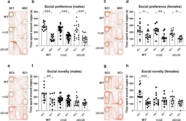
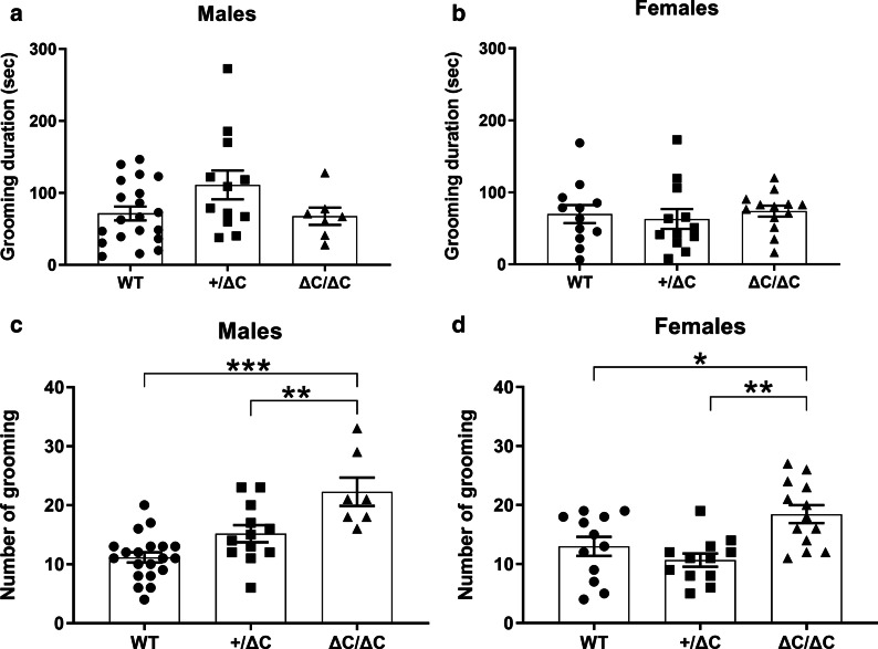
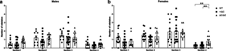
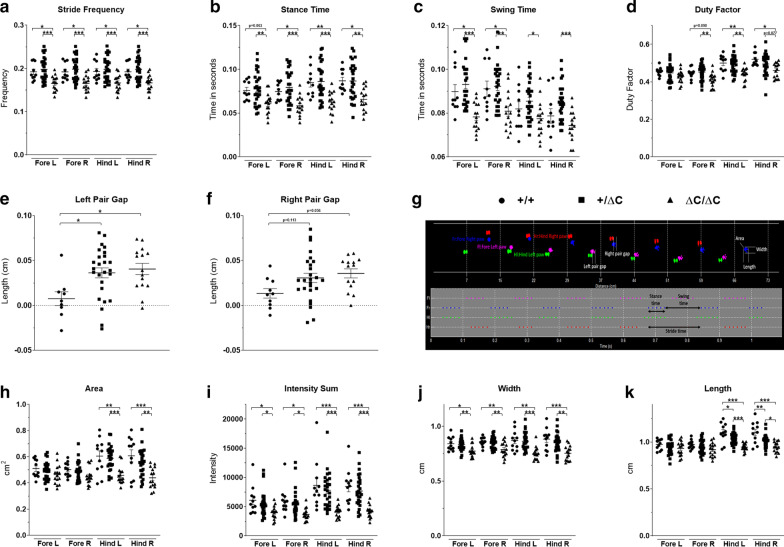
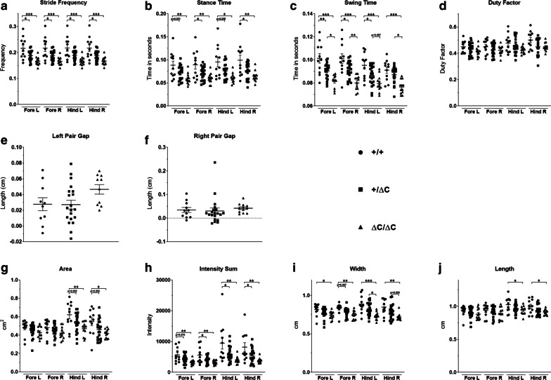
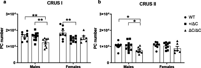
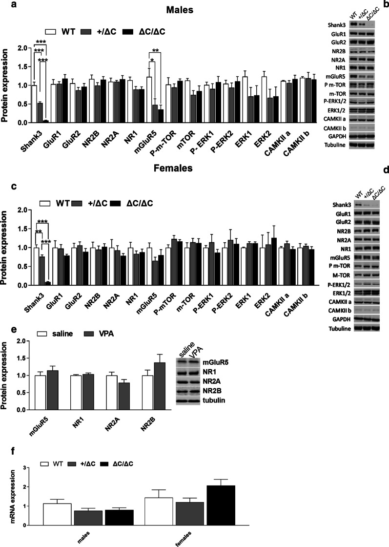
Results: Shank3 ΔC/ΔC mice showed significant impairments in social novelty preference, stereotyped behavior and gait. These were accompanied by a decreased number of PC in restricted cerebellar sub-regions and decreased cerebellar expression of mGluR5. Females Shank3 ΔC/ΔC were less affected by the mutation than males. Shank3+/ΔC mice showed impairments only in social novelty preference, grooming, and decreased mGluR5 expression and that were to a much lesser extent than in Shank3 ΔC/ΔC mice.

Limitations: As Shank3 mutation is a haploinsufficiency, it is of interest to emphasize that Shank3+/ΔC mice showed only mild to no deficiencies compared to Shank3 ΔC/ΔC.

Conclusions: Our findings indicate that several behavioral, cellular, and molecular parameters are affected in this animal model. The reported deficits are more pronounced in males than in females. Additionally, male Shank3 ΔC/ΔC mice show more pronounced alterations than Shank3+/ΔC. Together with our previous findings in two environmental animal models of ASD, our studies indicate that gait dysfunction constitutes a robust set of motor ASD symptoms that may be considered for implementation in clinical settings as an early and quantitative diagnosis criteria.

Keywords: Cerebellum; Crus I; Crus II; Gait; Motor coordination; Purkinje cells; Sociability; mGluR5.


Conflict of interest statement

The authors declare that they have no competing interests.

Figures

Fig. 1
Fig. 1
Social preference in Shank3 mice assessed through the three-chambers test (3-CT). aceg Representative tracks of mice in the 3-CT. bdfh Social behavior analysis. bd. Following habituation, mice were put in the 3-CT with one chamber containing an encaged mouse (social chamber 1: SC1) and one chamber with an empty cage (Non-social chamber: NSC) to evaluate social preference. Note that all mice, whatever the sex and the genotype, spent more time in SC1 than in NSC. fh Then, a novel mouse was introduced in the chamber 2 (social chamber 2: SC2) in addition to the encaged mouse in SC1 to evaluate social novelty recognition. Note that Shank3 ΔC/ΔC and Shank3+/ΔC mice of both sexes spend equal time interacting with the novel mouse in SC2 than with the mouse in SC1. In comparison, wild type mice spent more time interacting with SC2 than with SC1. All data are expressed as means ± SEM; One-way ANOVA followed by Tukey’s multiple analysis was performed (*p < 0.05, **p < 0.01, ***p < 0.001). WT males n = 13; Shank3+/ΔC males n = 20; Shank3 ΔC/ΔC males, n = 18; WT females n = 14; Shank3+/ΔC females n = 12; Shank3 ΔC/ΔC females, n = 16
Fig. 2
Fig. 2
Stereotyped behavior in Shank3 mice. ad Grooming time and frequency in Shank3 mice. Mice were placed in a clean cylinder and videotaped. ac There was no difference in the time spent grooming within all groups of mice. bd However, compared to WT, grooming frequency was increased in Shank3 ΔC/ΔC and Shank3+/ΔC males. Only Shank3 ΔC/ΔC females showed increased grooming compared to WT and Shank3+/ΔC (B). All data are expressed as means ± SEM. One-way ANOVA followed by Tukey’s multiple analysis was performed (*p < 0.05, **p < 0.01, ***p < 0.001) WT males n = 20; Shank3+/ΔC males n = 12; Shank3 ΔC/ΔC males, n = 7; WT females n = 12; Shank3+/ΔC females n = 12; Shank3 ΔC/ΔC females, n = 13
Fig. 3
Fig. 3
Motor coordination deficits in Shank3 mice. Time to traverse the beam was only slightly affected in the last section of the beam, and which is the narrower, in Shank3 ΔC/ΔC females. This is indicative of motor coordination deficits. All data are expressed as means ± SEM. One-way ANOVA followed by Tukey’s multiple analysis was performed (*p < 0.05, **p < 0.01, ***p < 0.001) WT males n = 10; Shank3+/ΔC males n = 20; Shank3 ΔC/ΔC males, n = 14; WT females n = 14; Shank3+/ΔC females n = 17; Shank3 ΔC/ΔC females, n = 12
Fig. 4
Fig. 4
Gait analysis in Shank3 males. Gait was analyzed during spontaneous walk using an automated gait analysis system (Viewpoint, France). Ten parameters were analyzed: a stride frequency, b stance time, c swing time, d duty factor, e, f pair gap, h area, i intensity sum, j width, k length and g representative spatio-temporal gait analysis. All data are expressed as means ± SEM. One way ANOVA followed by Tukey’s multiple analysis was performed. (*p < 0.05, **p < 0.01, ***p < 0.001) WT males n = 11; Shank3+/ΔC males n = 27; Shank3 ΔC/ΔC males, n = 15
Fig. 5
Fig. 5
Gait analysis in Shank3 females. Gait was analyzed during spontaneous walk as with males. The following parameters were analyzed: a stride frequency, b stance time, c swing time, d duty factor, e, f pair gap, g area, h intensity sum, i width, j length. All data are expressed as means ± SEM. One way ANOVA followed by Tukey’s multiple analysis was performed. (*p < 0.05, **p < 0.01, ***p < 0.001) WT females n = 11; Shank3+/ΔC females n = 21; Shank3 ΔC/ΔC females, n = 12
Fig. 6
Fig. 6
Purkinje cell (PC) number in Shank3 mice. Stereological PC counts on coronal sections of the cerebellum in the Crus I (a) and the Crus II (b) sub-regions. Only male Shank3 ΔC/ΔC mice showed a decrease in the number of PC in the crus I and Crus II cerebellar sub-regions. Female + /ΔC mice showed a slight decrease in PC number in the Crus I subregion. All data are expressed as means ± SEM. Two-way ANOVA was performed (*p < 0.05, **p < 0.01, ***p < 0.001) WT males n = 8; Shank3+/ΔC males n = 11; Shank3 ΔC/ΔC males, n = 8; WT females n = 8; Shank3+/ΔC females n = 9; Shank3 ΔC/ΔC females, n = 6
Fig. 7
Fig. 7
Brain levels in PSD proteins and mRNA in ASD mouse models. ac Expression profile of Shank3 and associated postsynaptic scaffolding proteins in males and females Shank3 mice within the cerebellum by immunoblot analysis. bd Representative figures of the western blot. Note that only mGluR5 was significantly decreased in Shank3+/ΔC and ΔC/ΔC males compared to wild type. No decreases were present in Shank3 ΔC/ΔC and Shank3+/ΔC females. Experiments for western blots were repeated at least three times and statiscal analysis were performed with a one-way ANOVA followed by Tukey’smultiple analysis (*p < 0.05, **p < 0.01, ***p < 0.001) WT males n = 4; Shank3+/ΔC males n = 8; Shank3 ΔC/ΔC males, n = 8; WT females n = 5; Shank3+/ΔC females n = 8; Shank3 ΔC/ΔC females, n = 9. e No difference in the protein levels of several glutamate receptors was found in the male cerebellum of VPA ASD mouse model whatever the prenatal treatment (Saline, n = 5; VPA, n = 5, Student’s t test, p > 0.05). f No difference in the mGluR5 mRNA levels was found in the cerebellum whatever the genotype or sex (wild type males (n = 7), Shank3+/ΔC males (n = 8), Shank3 ΔC/ΔC males (n = 8), wild type females (n = 7), Shank3+/ΔC females (n = 8), Shank3 ΔC/ΔC females (n = 8), One-way ANOVA, p > 0.05). All data are expressed as means ± SEM

References

    1. Association AP. Diagnostic and statistical manual of mental disorders (DSM-5®) Philadelphia: American Psychiatric Pub; 2013.
    1. Loomes R, Hull L, Mandy WPL. What is the male-to-female ratio in autism spectrum disorder? A systematic review and meta-Analysis. J Am Acad Child Adolesc Psychiatry. 2017;56(6):466–474. doi: 10.1016/j.jaac.2017.03.013. - DOI - PubMed
    1. Baron-Cohen S, Knickmeyer RC, Belmonte MK. Sex differences in the brain: implications for explaining autism. Science. 2005;310(5749):819–823. doi: 10.1126/science.1115455. - DOI - PubMed
    1. Auyeung B, Baron-Cohen S, Ashwin E, Knickmeyer R, Taylor K, Hackett G. Fetal testosterone and autistic traits. Br J Psychol Lond Engl. 2009;100(Pt 1):1–22. - PubMed
    1. Noor A, Whibley A, Marshall CR, Gianakopoulos PJ, Piton A, Carson AR, et al. Disruption at the PTCHD1 Locus on Xp22.11 in Autism spectrum disorder and intellectual disability. Sci Transl Med. 2010;2(49):49ra68. doi: 10.1126/scitranslmed.3001267. - DOI - PMC - PubMed

Publication types

MeSH terms

LinkOut - more resources