. 2021 Mar 18;65(4):e02095-20.
doi: 10.1128/AAC.02095-20. Print 2021 Mar 18.

Deletion of pknG Abates Reactivation of Latent Mycobacterium tuberculosis in Mice

Affiliations

Deletion of pknG Abates Reactivation of Latent Mycobacterium tuberculosis in Mice

Mehak Zahoor Khan et al. Antimicrob Agents Chemother. .

Abstract

Eradication of tuberculosis (TB), caused by Mycobacterium tuberculosis (Mtb), has been a challenge due to its uncanny ability to survive in a dormant state inside host granulomas for decades. Mtb rewires its metabolic and redox regulatory networks to survive in the hostile hypoxic and nutrient-limiting environment, facilitating the formation of drug-tolerant persisters. Previously, we showed that protein kinase G (PknG), a virulence factor required for lysosomal escape, aids in metabolic adaptation, thereby promoting the survival of nonreplicating mycobacteria. Here, we sought to investigate the therapeutic potential of PknG against latent mycobacterium. We show that inhibition of PknG by AX20017 reduces mycobacterial survival in in vitro latency models such as hypoxia, persisters, and nutrient starvation. Targeting PknG enhances the bactericidal activity of the frontline anti-TB drugs in peritoneal macrophages. Deletion of pknG resulted in 5- to 15-fold-reduced survival of Mtb in chronically infected mice treated with anti-TB drugs. Importantly, in the Cornell mouse model of latent TB, the deletion of pknG drastically attenuated Mtb's ability to resuscitate after antibiotic treatment compared with wild-type and complemented strains. This is the first study to investigate the sterilizing activity of pknG deletion and inhibition for adjunct therapy against latent TB in a preclinical model. Collectively, these results suggest that PknG may be a promising drug target for adjunct therapy to shorten the treatment duration and reduce disease relapse.

Keywords: Mycobacterium tuberculosis; adjunct therapy; bacterial protein kinase; bacterial signal transduction; drug discovery; hypoxia; latency; latent infection; persistence; persisters; phosphorylation.


Figures

FIG 1
FIG 1
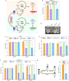
AX inhibits Mtb survival in the in vitro models of latency. (a) Model illustrating the role of PknG during latency. GarA, a central metabolic regulator, inhibits α-ketoglutarate (α-KG) decarboxylase (KGD) and glutamate dehydrogenase (GDH) and activates glutamate synthase (GS). The phosphorylation of GarA by PknG alleviates the inhibition of KGD and GDH and the activation of GS. The PknG-GarA signaling axis is necessary for fine-tuning the tricarboxylic acid (TCA) cycle and glutamate metabolism, thus maintaining cellular redox homeostasis and abetting the survival of Mtb under latency-like conditions. (b to f) In the case of Rv plus AX (Rv+AX), 1 mM AX was added to Rv at the start of the experiment. (b) Rv, RvΔG, Rv+AX, and RvΔG::G were inoculated at an A600 of ∼0.1 in Sauton’s medium, and bacillary survival was enumerated at day 6. (c) Early-log-phase cultures were resuspended in PBS, and CFU were enumerated on day 7. (d) Pictorial representation of the growth of the indicated strains in acidified 7H9-ADC medium at day 7. (e) Single-cell suspensions of Rv, RvΔG, Rv+AX, and RvΔG::G were inoculated at an A600 of ∼0.1 in acidified 7H9-ADC medium, and CFU were enumerated on day 7. (f) Bacterial strains were inoculated at an A600 of ∼0.1 in 7H9-ADC medium containing methylene blue in tightly sealed tubes. CFU were enumerated on days 0, 20, and 40. (g) Schematic representation of the hypoxia experiment. Twenty days after the start of the experiment, 1 mM AX was injected using a fine needle, and tubes were resealed. A parallel group was left untreated (Rv). CFU were enumerated on day 40. Bars depict means ± SD (n = 3), representative of data from two biologically independent experiments. *, P < 0.05; **, P < 0.005; ***, P < 0.0005.
FIG 2
FIG 2
Inhibition of PknG suppresses the emergence of drug-resistant and -tolerant cells. (a and b) The sensitivities of wild-type and pknG mutant Msm (a) and Mtb (b) strains to INH were determined with the help of an alamarBlue assay. ND and NC indicate no-drug and no-cell controls, respectively. (c) A total of 108 cells of Msm, MsmΔG, and MsmΔG::G were plated on 7H11-OADC plates containing 10 μg/ml isoniazid. (d and e) The antibiotic resistance frequency was determined by spotting 108 cells of Msm (d) and Mtb (e) strains on 7H11-OADC plates containing 100 μg/ml (d) and 10 μg/ml (e) INH, respectively. (f) Experiment performed as described above for panel e, with 1 mM AX added to Rv, where indicated. For panels d to f, the mutation rate is calculated as the number of colonies obtained on antibiotic-containing plates/number of colonies obtained on plain plates. The bar diagram represents means ± standard errors of the means (SEM) and is representative of results from a minimum of two independent biological replicates (n = 7). (g and h) Mtb strains were inoculated in 7H9-ADS medium containing 5 μg/ml INH (g) or 1 μg/ml RIF (h). Survival was monitored on days 0 and 7, and the survival obtained at day 0 was normalized to 100%. Percent survival at day 7 was calculated with respect to survival at day 0 for each strain. Data are represented as mean percent survival ± SD from one of three biologically independent experiments, each performed in triplicates (n = 3). *, P < 0.05; **, P < 0.005; ***, P < 0.0005.
FIG 3
FIG 3
Adjunct therapy with AX20017 lowers drug tolerance inside murine macrophages. (a) Schematic representation of the peritoneal macrophage infection experiment. (b and c) Peritoneal macrophages were infected with Rv, RvΔG, and RvΔG::G at a multiplicity of infection (MOI) of 1:10. After 4 h, 0.5 μg/ml INH (b) or RIF (c) was added to the infected macrophages. At 48 h postinfection, macrophage cells were lysed, and bacillary survival was examined. CFU obtained for untreated infected macrophages at 48 h postinfection were normalized to 100%. The mean percent intracellular survival (±SD) (n = 3) of the bacterial strain in the treated macrophages was calculated with respect to (w.r.t) the corresponding untreated cells for that strain. (d to g) The same experiment as in panels b and c except with an additional Rv-infected sample, wherein 25 μM AX was added at 4 h postinfection (Rv+AX). Infected macrophages were either left untreated (d) or treated with 0.5 μg/ml INH (e), 0.5 μg/ml RIF (f), or 1 μg/ml BDQ (g). The mean percent intracellular survival (±SD) (n = 3) of the bacterial strain in the treated macrophages was calculated with respect to the corresponding untreated cells for that strain. Data are representative of results from two biologically independent experiments, each performed in triplicates. *, P < 0.05; **, P < 0.005; ***, P < 0.0005.
FIG 4
FIG 4
PknG abets drug tolerance in a murine model of tuberculosis infection. (a) Schematic representation of the murine infection experiment. Mice (n = 6 per group at each time point) were infected with Rv, RvΔG, and RvΔG::G. After the establishment of infection for 4 weeks, antibiotics (INH or RIF) were added to drinking water for the next 4 weeks. A parallel group was left untreated (control). Bacillary survival was assessed at day 1 and weeks 4 and 8. (b and c) Bacillary loads in lungs (b) and spleen (c) of infected mice at 4 and 8 weeks postinfection (n = 6). Each data point represents the log10 CFU of an infected animal in individual organs, and the error bar depicts the SD for each group. *, P < 0.05; **, P < 0.005; ***, P < 0.0005. (d) Table depicting mean log10 CFU ± SD in the lungs and spleen of infected animals at the indicated time points.
FIG 5
FIG 5
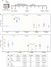
PknG is required for the resuscitation of Mtb in the modified Cornell mouse model of infection. (a) Schematic representation of the modified Cornell mouse model of tuberculosis infection experiment. Mice (n = 6 to 10 per group for each time point/strain) were infected with Rv, RvΔG, and RvΔG::G. After the establishment of infection for 4 weeks, antibiotics (INH and RIF) were added to drinking water for 12 weeks to eliminate actively replicating bacilli. Following 4 weeks of rest, dexamethasone (Dx) was injected for 4 weeks for immune suppression. Bacillary survival was examined at day 1 and weeks 4, 16, 30, and 34. (b and c) Bacillary loads in lungs (b) and spleen (c) of infected mice at the indicated time points postinfection (n = 6 to 10). Each data point represents log10 CFU of an infected animal in individual organs, and the error bar depicts the SD for each group. *, P < 0.05; **, P < 0.005; ***, P < 0.0005. N.D., not determined. (d) Table depicting mean log10 CFU ± SD in the lungs and spleen of infected animals at the indicated time points.
FIG 6
FIG 6
Model depicting the therapeutic potential of AX20017. In response to Mtb infection, the host immune system contains the bacilli inside a granuloma. A granuloma is an organized and compact bunch of immune cells such as macrophages, multinucleated giant cells, dendritic cells, T cells, and B cells. Depending on the severity of the infection, granulomas can be of various kinds. The bacterium exploits this niche as its “safe haven” to survive and disseminate, leading to disease progression. The microaerophilic conditions found inside the granulomas drive cells of Mtb toward its nonreplicating antibiotic-tolerant persister phase. PknG helps Mtb adapt to hypoxic conditions by waning its metabolism. Deletion of pknG or its inhibition reduces Mtb’s ability to survive under in vivo latency-like conditions and attenuates its ability to thrive upon antibiotic stress. Hence, adjunct therapy with a PknG inhibitor could potentially suppress reactivation and lower the disease relapse rate and, hence, the transmission of the disease. The proof of principle for the interventions depicted in the schematic has been established with the help of various in vitro, ex vivo, and in vivo models of latency in this study.

Similar articles

Cited by

References

    1. Mitchison DA. 2005. Shortening the treatment of tuberculosis. Nat Biotechnol 23:187–188. doi:10.1038/nbt0205-187. - DOI - PubMed
    1. Getahun H, Matteelli A, Chaisson RE, Raviglione M. 2015. Latent Mycobacterium tuberculosis infection. N Engl J Med 372:2127–2135. doi:10.1056/NEJMra1405427. - DOI - PubMed
    1. Comstock GW, Livesay VT, Woolpert SF. 1974. The prognosis of a positive tuberculin reaction in childhood and adolescence. Am J Epidemiol 99:131–138. doi:10.1093/oxfordjournals.aje.a121593. - DOI - PubMed
    1. Lin PL, Flynn JL. 2010. Understanding latent tuberculosis: a moving target. J Immunol 185:15–22. doi:10.4049/jimmunol.0903856. - DOI - PMC - PubMed
    1. Getahun H, Matteelli A, Abubakar I, Aziz MA, Baddeley A, Barreira D, Den Boon S, Borroto Gutierrez SM, Bruchfeld J, Burhan E, Cavalcante S, Cedillos R, Chaisson R, Chee CB-E, Chesire L, Corbett E, Dara M, Denholm J, de Vries G, Falzon D, Ford N, Gale-Rowe M, Gilpin C, Girardi E, Go U-Y, Govindasamy D, Grzemska M, Harris R, Horsburgh CR, Jr, Ismayilov A, Jaramillo E, Kik S, Kranzer K, Lienhardt C, LoBue P, Lonnroth K, Marks G, Menzies D, Migliori GB, Mosca D, Mukadi YD, Mwinga A, Nelson L, Nishikiori N, Oordt-Speets A, Rangaka MX, Reis A, Rotz L, Sandgren A, Sane Schepisi M, et al.. 2015. Management of latent Mycobacterium tuberculosis infection: WHO guidelines for low tuberculosis burden countries. Eur Respir J 46:1563–1576. doi:10.1183/13993003.01245-2015. - DOI - PMC - PubMed

Publication types

LinkOut - more resources