Review
. 2021 Apr 21;203(10):e00547-20.
doi: 10.1128/JB.00547-20. Print 2021 Apr 21.

Seeing and Touching the Mycomembrane at the Nanoscale

Affiliations
Review

Seeing and Touching the Mycomembrane at the Nanoscale

Albertus Viljoen et al. J Bacteriol. .

Abstract

Mycobacteria have unique cell envelopes, surface properties, and growth dynamics, which all play a part in the ability of these important pathogens to infect, evade host immunity, disseminate, and resist antibiotic challenges. Recent atomic force microscopy (AFM) studies have brought new insights into the nanometer-scale ultrastructural, adhesive, and mechanical properties of mycobacteria. The molecular forces with which mycobacterial adhesins bind to host factors, like heparin and fibronectin, and the hydrophobic properties of the mycomembrane have been unraveled by AFM force spectroscopy studies. Real-time correlative AFM and fluorescence imaging have delineated a complex interplay between surface ultrastructure, tensile stresses within the cell envelope, and cellular processes leading to division. The unique capabilities of AFM, which include subdiffraction-limit topographic imaging and piconewton force sensitivity, have great potential to resolve important questions that remain unanswered on the molecular interactions, surface properties, and growth dynamics of this important class of pathogens.

Keywords: adhesins; atomic force microscopy; binding force; chemical properties; drugs; growth dynamics; mycobacterial envelope; ultrastructure.


Figures

FIG 1
FIG 1
Two different basic AFM modes to study mycobacterial cells. (A) AFM imaging, in which the AFM probe is raster scanned across the sample, allows study of the nanoscale topography of cell walls along with correlative fluorescence microscopy imaging of cellular processes (e.g., markers of cellular division as illustrated by the fluorescent beads inside of the bacterium). (B) Force spectroscopy measurements with chemically (e.g., small molecules) or biologically sensitive (e.g., the ligand of an adhesin, such as human fibronectin) tips allows characterization of local physicochemical properties and of the strength of adhesin-ligand complexes.
FIG 2
FIG 2
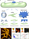
Interactions between mycobacterial surfaces and their environment. (A) Mycobacteria rely on hydrophobic properties of their surfaces to associate with aerosol droplets and to adhere to each other and form cords. Specialized adhesins stimulate mycobacterium-mycobacterium interactions as well as adhesion of mycobacteria to extracellular matrix proteins and cells. (B) Using AFM force spectroscopy to probe mycobacterial morphology, chemical properties, and adhesin interactions. (Left) A 3D projection of a height image showing a microcolony of Mycobacterium abscessus cells. (Center, top left) AFM probes exposing hydrophobic methyl groups have unraveled hydrophobic properties of mycobacterial cells. (Center, top right) Striking hydrophobic (lighter yellow, maximum of 2.5 nN) and hydrophilic (darker brown, minimum of 0 nN) nanodomains were seen on very high-resolution adhesion maps recorded on the surfaces of M. abscessus smooth variant (glycopeptidolipid+) cells. (Center, bottom) Typical force-distance curves obtained as well as the histogram plot of the frequency distributions of hydrophobic adhesion forces. The red star is indicative of when hydrophobic adhesive forces were registered, whereas the green star indicates when no hydrophobic adhesive forces were registered. Adapted with permission from reference . (Right) AFM probe exposing single molecules of the host extracellular matrix protein, fibronectin (Fn), to which mycobacterial antigen 85 (Ag85, yellow oval) binds. The adhesion map in the top right corner shows a homogenous distribution of Ag85 on the surface of an M. abscessus cell. The white pixels represent single adhesins, with the lightest shade representing a maximum adhesion strength of 250 pN and the darkest pixels representing zero adhesion force. Typical force-distance curves of this specific receptor-ligand interaction are shown at the bottom. The sawtooth unbinding peaks highlighted by red arrows relate to sequential unfolding of repeat domains in Fn. Adapted from reference .
FIG 3
FIG 3
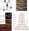
Studying cell growth dynamics with a correlative AFM-optical microscope. (A) Schematic representation of a correlative AFM-optical microscope. The field of view of an inverted fluorescence microscope is aligned with the cantilever of an AFM in order to acquire correlated images. (B) AFM used as a nanomanipulation tool. Schematics and time-lapse AFM of growing mycobacteria. (Top) Sibling cells are kept in their original position after cell division. Poles of mother and daughter cells are in close contact. (Bottom) The AFM cantilever was used to remove one of the sibling cells to avoid physical constraints on the new pole (NP, OP being the old pole). Arrowheads point to the division site. Adapted from reference . (C) AFM stiffness measurement of cell surface at the division site between the emergence of the precleavage furrow (PCF) and cleavage. Lighter colors represent a higher stiffness. (D). Three-dimensional rendered AFM topographic images of Mycobacterium smegmatis before cell cleavage and enlargements of the area around precleavage furrow. The arrow indicates increasing time. The top panel was scanned from bottom to top, the middle panel was scanned from top to bottom and the bottom panel was scanned from bottom to top. (C, D) Adapted from reference with permission. (E) Schematic representation of the consecutive events leading to cell division in Mycobacterium smegmatis studied by correlative AFM-optical microscopy. (1) At cell birth (division of the mother cell), Wag31-green fluorescent protein (GFP) (dark green) is localized only at the poles. The cell surface comprises wavelike morphological features, and the subsequent cell division occurs at the centermost trough near midcell. (2) FtsZ-GFP (light green) localizes at the central wave trough and forms a circumferential ring. (3) Formation of the precleavage furrow starts, which is colocalized with the FtsZ ring. (4) Stress builds up, whereas membrane strength decreases at precleavage furrow. Septum formation and cytokinesis occur. Wag31-GFP localizes to the future division side, whereas the FtsZ ring disassembles. (5) The tensile stress increases. (6) Further increase of turgor pressure cumulates in physical cell separation by rapid mechanical rupture leading to newborn sibling cells. (The space between the sibling cells after division was inserted for visualization purposes. In reality, the sibling cells stay in proximity to each other.) (7) Pre-NETO phase: low growth rate of the NP. Reallocation of Wag31 from the OP to the NP. (8) Post-NETO phase: growth rate change (NETO) is followed by a high growth rate of the NPs. The OPs grow in both phases with a constant, high rate.

References

    1. Jarlier V, Nikaido H. 1994. Mycobacterial cell wall: structure and role in natural resistance to antibiotics. FEMS Microbiol Lett 123:11–18. 10.1111/j.1574-6968.1994.tb07194.x. - DOI - PubMed
    1. Dulberger CL, Rubin EJ, Boutte CC. 2020. The mycobacterial cell envelope—a moving target. Nat Rev Microbiol 18:47–59. 10.1038/s41579-019-0273-7. - DOI - PubMed
    1. Falkinham JO. 2003. Mycobacterial aerosols and respiratory disease. Emerg Infect Dis 9:763–767. 10.3201/eid0907.020415. - DOI - PMC - PubMed
    1. Cambier CJ, Falkow S, Ramakrishnan L. 2014. Host evasion and exploitation schemes of Mycobacterium tuberculosis. Cell 159:1497–1509. 10.1016/j.cell.2014.11.024. - DOI - PubMed
    1. Jankute M, Nataraj V, Lee OY-C, Wu HHT, Ridell M, Garton NJ, Barer MR, Minnikin DE, Bhatt A, Besra GS. 2017. The role of hydrophobicity in tuberculosis evolution and pathogenicity. Sci Rep 7:1315. 10.1038/s41598-017-01501-0. - DOI - PMC - PubMed

Publication types

MeSH terms

LinkOut - more resources