. 2021 Jan 26;118(4):e2022790118.
doi: 10.1073/pnas.2022790118.

Long-range structural defects by pathogenic mutations in most severe glucose-6-phosphate dehydrogenase deficiency

Affiliations

Long-range structural defects by pathogenic mutations in most severe glucose-6-phosphate dehydrogenase deficiency

Naoki Horikoshi et al. Proc Natl Acad Sci U S A. .

Abstract

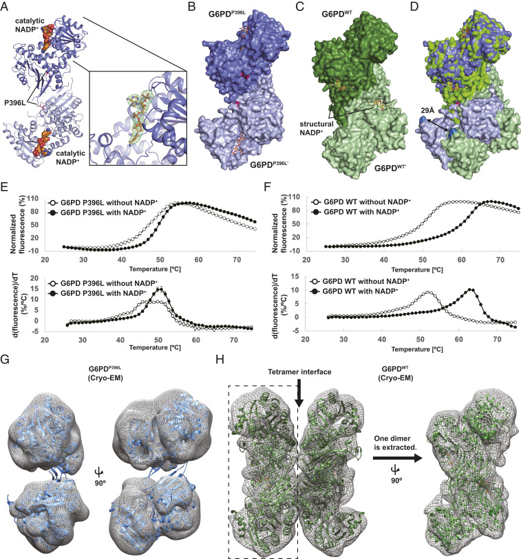
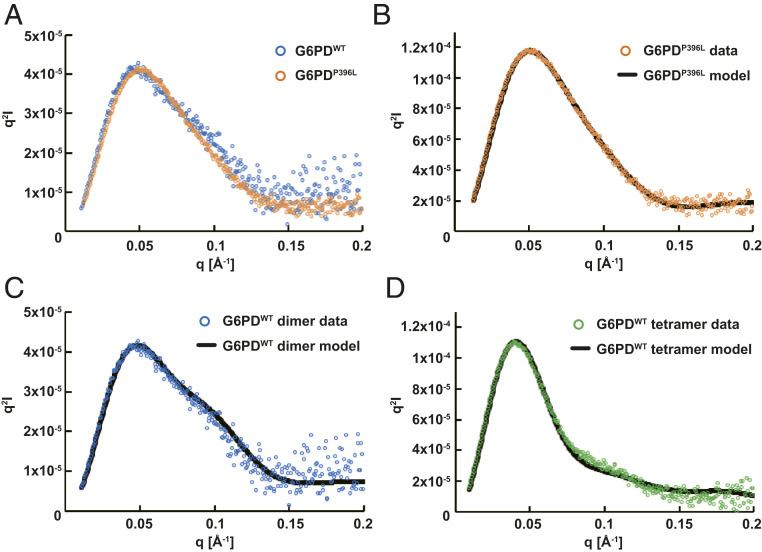
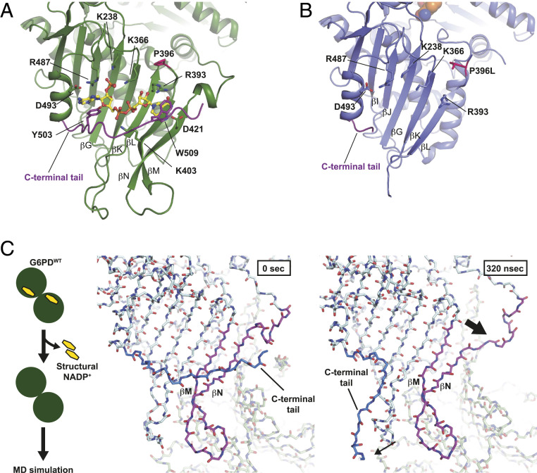
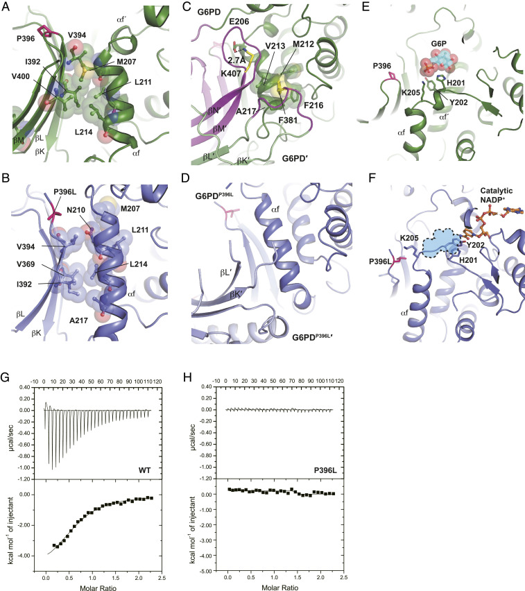
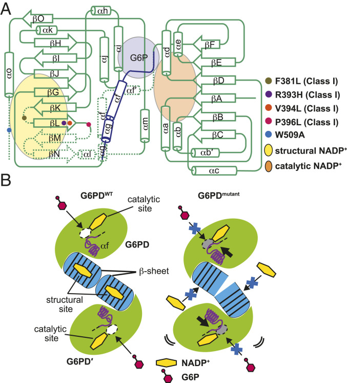
Glucose-6-phosphate dehydrogenase (G6PD) deficiency is the most common blood disorder, presenting multiple symptoms, including hemolytic anemia. It affects 400 million people worldwide, with more than 160 single mutations reported in G6PD. The most severe mutations (about 70) are classified as class I, leading to more than 90% loss of activity of the wild-type G6PD. The crystal structure of G6PD reveals these mutations are located away from the active site, concentrating around the noncatalytic NADP+-binding site and the dimer interface. However, the molecular mechanisms of class I mutant dysfunction have remained elusive, hindering the development of efficient therapies. To resolve this, we performed integral structural characterization of five G6PD mutants, including four class I mutants, associated with the noncatalytic NADP+ and dimerization, using crystallography, small-angle X-ray scattering (SAXS), cryogenic electron microscopy (cryo-EM), and biophysical analyses. Comparisons with the structure and properties of the wild-type enzyme, together with molecular dynamics simulations, bring forward a universal mechanism for this severe G6PD deficiency due to the class I mutations. We highlight the role of the noncatalytic NADP+-binding site that is crucial for stabilization and ordering two β-strands in the dimer interface, which together communicate these distant structural aberrations to the active site through a network of additional interactions. This understanding elucidates potential paths for drug development targeting G6PD deficiency.

Keywords: G6PD deficiency; NADP+; enzymopathy; hemolytic anemia; structural defects.


Conflict of interest statement

The authors declare no competing interest.

Figures

Fig. 1.
Fig. 1.
Overall structure of the class I mutant, G6PDP396L. (A) Overall structure of the dimer G6PDP396L. Two G6PD molecules and the catalytic NADP+ are shown in blue, light blue, and orange, respectively. The catalytic NADP+-binding site is shown in close-up view on the Right. The Fo-DFc map for the catalytic NADP+ is contoured at 3.0σ level. (B) Space filling model of the dimer G6PDP396L. (C) Space filling model of the dimer G6PDWT. (D) Superimposition of the dimer G6PDP396L and G6PDWT. Distance between Cα-carbon atoms of E93 residues in the G6PDP396L and G6PDWT structures are shown. (E and F) The thermal denaturation curves of the G6PDP396L and G6PDWT with or without NADP+ (Top). The error bars indicate the SDs (n = 4). Bottom shows the derivative values of the thermal denaturation curves on the Top. (G) Crystal structure of G6PDP396L dimer is superimposed with the cryo-EM map of G6PDP396L. The G6PDP396L dimer on the Left is rotated by 90° on the Right. (H) Crystal structure of the G6PDWT tetramer (PDB ID: 6E08) is superimposed with the cryo-EM map of G6PDWT. The cryo-EM map of the Left half on the Left (enclosed with a dashed rectangle) is extracted and rotated by 90°. The crystal structure of the G6PDWT dimer (PDB ID: 6E08) is superimposed with the cryo-EM map on the Right.
Fig. 2.
Fig. 2.
Structural analysis of the G6PDP396L in solution. SEC-SAXS experiments were performed to study the G6PDWT dimer (blue), G6PDP396L (orange), and G6PDWT tetramer (green). (A) Kratky plots of G6PDWT dimer and G6PDP396L. The plot of G6PDP396L is scaled to that of G6PDWT dimer. Curvatures in the middle q region are significantly different. (BD) The program CORAL was employed for SAXS modeling. The theoretical Kratky plot of the best CORAL model is fitted with the experimental data.
Fig. 3.
Fig. 3.
Loss of the structural NADP+ caused a partial deformation of the β-sheet. (A) Close-up view of the structural NADP+-binding site in G6PDWT. G6PDWT molecule is shown in green. Residues responsible for the structural NADP+ binding are shown. C-terminal tail is shown in purple. Structural NADP+ is shown in yellow. (B) Close-up view of the structural NADP+-binding site in G6PDP396L. A G6PDP396L monomer is shown in blue. Residues responsible for the structural NADP+ binding in G6PDWT are shown. (C) MD simulation of the structure of G6PDWT (PDB ID: 6E08). Scheme of the MD simulation is shown at Left. Snapshots of the G6PD dimer without the structural NADP+ at 0 (Middle) and 320 ns (Right), respectively.
Fig. 4.
Fig. 4.
Loss of the structural NADP+ causes the structural change in the G6P-binding site. (A) Close-up view of the interface between the β-sheet and αf helix in G6PDWT. (B) Close-up view of the interface between the β-sheet and αf helix in G6PDP396L. (C) Dimer interface in the G6PDWT structure. Two G6PDWT molecules are shown in green and light green, respectively. Disordered regions in G6PDP396L are colored in magenta. F381 and K407 residues are shown in yellow. (D) Dimer interface in the G6PDP396L structure. Two G6PDP396L molecules are shown in blue and light blue, respectively. (E) Close-up view of the G6P-binding site in G6PDWT (PDB ID: 2BHL) (15). A G6P molecule is shown in cyan. Phosphorus and oxygen atoms are shown in orange and red, respectively. (F) Close-up view of the G6P-binding site in G6PDP396L. Catalytic NADP+ is shown in orange. G6P-binding site in G6PDWT is shown in sky blue. (G) The isothermal titration calorimetry analysis of G6PDWT with G6P. (H) The isothermal titration calorimetry analysis of G6PDP396L with G6P.
Fig. 5.
Fig. 5.
Summary of structural changes caused by the class I pathogenic mutations associated with the structural NADP+ binding. (A) Comparison of the arrangements of secondary structure elements of G6PD monomer between the wild type and the mutants. Binding sites of the catalytic NADP+, structural NADP+, and G6P are shown in orange, yellow, and purple, respectively. The shifted αg, αf, and stretched αf´ helices in G6PDmutant are shown in blue. Disordered regions in G6PDmutant are indicated by dotted lines. (B) Schematic representations of the G6PDWT dimer (Left) and the G6PDmutant dimer (Right). White and gray ovals indicate the open and occluded G6P-binding sites, respectively. Yellow hexagon indicates NADP+ and its binding sites. Blue barrels at the center indicate the β-sheets that the structural NADP+ molecules are bound to in G6PDWT but not in G6PDmutant.

Similar articles

Cited by

References

    1. Minucci A., et al. , Glucose-6-phosphate dehydrogenase (G6PD) mutations database: Review of the “old” and update of the new mutations. Blood Cells Mol. Dis. 48, 154–165 (2012). - PubMed
    1. Bhutani V. K., et al. , Neonatal hyperbilirubinemia and rhesus disease of the newborn: Incidence and impairment estimates for 2010 at regional and global levels. Pediatr. Res. 74 (suppl. 1), 86–100 (2013). - PMC - PubMed
    1. Luzzatto L., Nannelli C., Notaro R., Glucose-6-phosphate dehydrogenase deficiency. Hematol. Oncol. Clin. North Am. 30, 373–393 (2016). - PubMed
    1. Cunningham A. D., Hwang S., Mochly-Rosen D., Glucose-6-phosphate dehydrogenase deficiency and the need for a novel treatment to prevent kernicterus. Clin. Perinatol. 43, 341–354 (2016). - PMC - PubMed
    1. Nkhoma E. T., Poole C., Vannappagari V., Hall S. A., Beutler E., The global prevalence of glucose-6-phosphate dehydrogenase deficiency: A systematic review and meta-analysis. Blood Cells Mol. Dis. 42, 267–278 (2009). - PubMed

Publication types

MeSH terms

LinkOut - more resources