. 2021 Jan 19;12(1):e02801-20.
doi: 10.1128/mBio.02801-20.

Trehalose Recycling Promotes Energy-Efficient Biosynthesis of the Mycobacterial Cell Envelope

Affiliations

Trehalose Recycling Promotes Energy-Efficient Biosynthesis of the Mycobacterial Cell Envelope

Amol Arunrao Pohane et al. mBio. .

Abstract

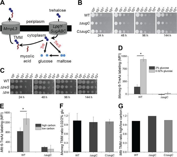
The mycomembrane layer of the mycobacterial cell envelope is a barrier to environmental, immune, and antibiotic insults. There is considerable evidence of mycomembrane plasticity during infection and in response to host-mimicking stresses. Since mycobacteria are resource and energy limited under these conditions, it is likely that remodeling has distinct requirements from those of the well-characterized biosynthetic program that operates during unrestricted growth. Unexpectedly, we found that mycomembrane remodeling in nutrient-starved, nonreplicating mycobacteria includes synthesis in addition to turnover. Mycomembrane synthesis under these conditions occurs along the cell periphery, in contrast to the polar assembly of actively growing cells, and both liberates and relies on the nonmammalian disaccharide trehalose. In the absence of trehalose recycling, de novo trehalose synthesis fuels mycomembrane remodeling. However, mycobacteria experience ATP depletion, enhanced respiration, and redox stress, hallmarks of futile cycling and the collateral dysfunction elicited by some bactericidal antibiotics. Inefficient energy metabolism compromises the survival of trehalose recycling mutants in macrophages. Our data suggest that trehalose recycling alleviates the energetic burden of mycomembrane remodeling under stress. Cell envelope recycling pathways are emerging targets for sensitizing resource-limited bacterial pathogens to host and antibiotic pressure.IMPORTANCE The glucose-based disaccharide trehalose is a stress protectant and carbon source in many nonmammalian cells. Mycobacteria are relatively unique in that they use trehalose for an additional, extracytoplasmic purpose: to build their outer "myco" membrane. In these organisms, trehalose connects mycomembrane biosynthesis and turnover to central carbon metabolism. Key to this connection is the retrograde transporter LpqY-SugABC. Unexpectedly, we found that nongrowing mycobacteria synthesize mycomembrane under carbon limitation but do not require LpqY-SugABC. In the absence of trehalose recycling, compensatory anabolism allows mycomembrane biosynthesis to continue. However, this workaround comes at a cost, namely, ATP consumption, increased respiration, and oxidative stress. Strikingly, these phenotypes resemble those elicited by futile cycles and some bactericidal antibiotics. We demonstrate that inefficient energy metabolism attenuates trehalose recycling mutant Mycobacterium tuberculosis in macrophages. Energy-expensive macromolecule biosynthesis triggered in the absence of recycling may be a new paradigm for boosting host activity against bacterial pathogens.

Keywords: Mycobacterium; mycomembrane; oxidative stress; starvation; trehalose.


Figures

FIG 1
FIG 1
Mycomembrane synthesis and degradation are active under carbon limitation. (A) Mycomembrane synthesis and degradation. TMM, trehalose monomycolate; TDM, trehalose dimycolate; AG, arabinogalactan; AGM, arabinogalactan mycolates; MA, free mycolic acids; TDMH, TDM hydrolase. (B) TDM turnover under nutrient deprivation. M. smegmatis was cultured in 0.02% glucose-supplemented medium in the presence of metabolic probes O-AlkTMM (primarily labels AGM), N-AlkTMM (labels TDM), or HADA (labels cell wall peptidoglycan). After 24 h, the cultures were washed and resuspended in probe-free medium. Aliquots were removed 0, 4, and 8 h into the chase and fixed with 2% formaldehyde. Alkynes were detected by copper-catalyzed azide-alkyne cycloaddition (CuAAC) reaction with carboxyrhodamine-110 azide. Fluorescence was quantitated by flow cytometry, with the median fluorescence intensities (MFIs) were normalized to the initial, 0-h time point for each probe. The experiment was performed three times in triplicate; the results of one representative experiment are shown. (C) Metabolic labeling of M. smegmatis in 0.02% glucose-supplemented medium with O-AlkTMM, N-AlkTMM, and alkDala (labels peptidoglycan). Alkynes were detected by CuAAC reaction with carboxyrhodamine-110 azide. Data were normalized to labeling in 2% glucose-supplemented medium and plotted from four independent experiments. (D) Quantitation of TLC of different mycomembrane components for M. smegmatis in 0.02% glucose-supplemented medium. TDM, trehalose dimycolate; CS-MA, free, culture supernatant mycolic acids; AGM-MA, mycolic acids released from arabinogalactan. TLC results were scanned and processed in ImageJ (99). The data are normalized to TLC results from samples taken from M. smegmatis cultured in 2% glucose-supplemented medium and plotted from three independent experiments. (For representative TLC results, see Fig. S2.) (E) PI staining of M. smegmatis during adaptation to low carbon. M. smegmatis was cultured in 0.02% glucose-supplemented medium. Aliquots were removed at 13, 24, and 48 h and incubated with PI. Fluorescence was quantitated by flow cytometry, and the MFI was plotted. The experiment was performed three times in triplicate; the results of a representative experiment are shown. (F) O-AlkTMM labeling of M. smegmatis AGM in 2 or 0.02% glucose-supplemented medium. Alkynes were detected by CuAAC reaction with carboxyrhodamine-110 azide. (Left) Fluorescence microscopy. Scale bars, 5 μm. (Right) The cellular fluorescence was quantitated for cells lacking visible septa from three independent experiments. The signal was normalized to both cell length and total fluorescence intensity. Cells were oriented such that the brighter pole is on the right-hand side of the graph. A.U., arbitrary units. (G) Quantification of trehalose from supernatants of M. smegmatis wild-type and ΔsugC strains cultured in 2 or 0.02% glycerol-supplemented medium. The experiment was performed at least three times in triplicate; the results of one representative experiment are shown. Error bars, standard deviations. The statistical significance of 0.02% versus 2% glucose or glycerol samples from three independent experiments was assessed by two-tailed Student t test. *, P < 0.05; **, P < 0.005.
FIG 2
FIG 2
Trehalose cycling supports mycomembrane metabolism during carbon limitation. (A) Potential fates of recycled trehalose in catabolism (trehalase [Tre] or TreS) or in trehalose monomycoate (TMM) biosynthesis. (B and C) Survival of wild-type, ΔsugC, complemented ΔsugC (CΔsugC), ΔtreS, and Δtre M. smegmatis strains in 0.02% glucose-supplemented medium. Tenfold serial dilutions were plated at the indicated time points. The experiment was performed two times with similar results; the results of one experiment are shown. (D and E) 6-TreAz labeling of wild-type and ΔsugC M. smegmatis (Msmeg) and M. tuberculosis (Mtb) cultured in low- or high-carbon medium. Azides were detected by strain-promoted azide-alkyne cycloaddition (SPAAC) with DBCO-Cy5 label. The fluorescence was detected by flow cytometry, with MFI values from controls lacking 6-TreAz (but subjected to SPAAC) subtracted from the sample MFI. The experiment was performed at least three times in triplicate; the results of one representative experiment are shown. (F and G) TMM abundance in M. smegmatis and M. tuberculosis cultured in low- or high-carbon medium. TLC results were scanned and processed in ImageJ (99). The data are normalized to the TLC results from mycobacteria cultured in high-carbon medium and plotted from two (M. tuberculosis) or three (M. smegmatis) independent experiments. (For representative TLC results, see Fig. S3B and C.) Error bars, standard deviations. The statistical significance of low- versus high-carbon samples was assessed by two-tailed Student t test. *, P < 0.05.
FIG 3
FIG 3
Trehalose recycling promotes redox and energy homeostasis under carbon limitation. (A and B) Sensitivity of carbon-deprived wild-type, ΔsugC, and complemented ΔsugC (CΔsugC) M. smegmatis (A) or M. tuberculosis (B) strains to hydrogen peroxide. Tenfold serial dilutions were plated. White triangles highlight the most sensitive strain or condition. The sensitivity of each strain or condition was assessed at least three independent times; representative data are shown. (C) Staining of M. smegmatis cultured in 0.02% glucose-supplemented medium by superoxide indicator dye dihydroethidium (DHE). Fluorescence was detected by flow cytometry, and the MFI was plotted. The experiment was performed three times in triplicate; the results of one representative experiment are shown. (D) Oxygen consumption of M. smegmatis cultured in 0.02% glucose-supplemented medium. Strains were incubated with or without methylene blue, and the absorbance at 665 nm was measured. The absorbance from untreated samples was subtracted and then values were normalized to those of the wild-type. The data are plotted for three independent experiments performed in triplicate. (E) ATP levels of M. smegmatis cultured in 0.02% glucose-supplemented medium. Protein concentration-normalized cell lysates were incubated with BacTiter-Glo reagent, and the luminescence was measured in relative light-forming units (RLU). The experiment was performed at least three times in triplicate; the results of one representative experiment are shown. (F) Cartoon summary of Fig. 3 and Fig. S5. Error bars, standard deviation. For panels C to E, the statistical significance of ΔsugC or complement strains versus the wild type from at least three independent experiments was assessed by a two-tailed Student t test. *, P < 0.05.
FIG 4
FIG 4
Trehalose anabolism disrupts redox balance under carbon limitation. (A) Anabolic and catabolic pathways for trehalose. Light blue, phosphorylated glucose intermediates; purple, α-glucan polymer. (B) Expression of trehalose biosynthesis genes by qRT-PCR. Wild-type and ΔsugC M. smegmatis strains were cultured in 0.02% glucose-supplemented medium. Expression data were first normalized to the housekeeping gene sigA and then plotted as a ratio of the ΔsugC mutant to the wild type. The data are combined from three independent experiments performed in triplicate. (C) Glucose-6-phosphate (G6P) levels of M. smegmatis cultured in 0.02% glucose-supplemented medium. Protein concentration-normalized cell lysates were incubated with G6P working solution, and the G6P level was measured in a 96-well plate by monitoring the absorbance ratio at 575 nm/605 nm. The data are plotted for three independent experiments performed in duplicate. G6P levels normalized to those of the wild type. (D) Sensitivity of carbon-deprived M. smegmatis to hydrogen peroxide upon trehalase overexpression. Tenfold serial dilutions were plated at the indicated time points. White triangles highlight the difference in sensitivity with or without otsA. –Tre, plasmid backbone only; +Tre, plasmid with gene encoding trehalase under acetamide-inducible promoter; Acet, acetamide. The sensitivity of each strain or condition was assessed at least three independent times; representative data shown. Error bars, standard deviations. The statistical significance of expression in the ΔsugC mutant relative to the wild-type (B) or of other strains versus the wild type (C) was assessed by two-tailed Student t test. *, P < 0.05; **, P < 0.005.
FIG 5
FIG 5
Trehalose recycling promotes M. tuberculosis survival in macrophages. (A) Survival of wild-type, ΔsugC, and complemented ΔsugC (CΔsugC) M. tuberculosis strains in immortalized C57BL/6 bone marrow-derived macrophages (iBMDM) with or without IFN-γ treatment at 3 days postinfection. The experiment was performed at least three times in duplicate or triplicate; the results of one representative experiment are shown. (B) Wild-type and ΔsugC M. tuberculosis strain survival in IFN-γ-stimulated iBMDM at 0, 2, and 5 days postinfection. Log10-transformed data are combined from three to seven independent experiments performed in duplicate or triplicate. (C, left) Survival of wild-type, ΔsugC, and ΔlpqY M. tuberculosis strains in IFN-γ-activated iBMDM with or without bedaquiline (BDQ) or rifampin (RIF) at 2 days postinfection. The CFU from each condition were normalized to the untreated wild type. (Raw data are shown in Fig. S8B.) (Right) Bliss independence scores for mutant-drug interactions were obtained by subtracting the expected values for inhibition from the observed values. The expected values were calculated as described in Materials and Methods. Combined data from five (RIF) or six (BDQ) independent experiments are shown. Error bars, standard deviations. Statistical significance was assessed by a two-tailed Student t test on log10-transformed data at each time point (B) or by comparing expected and observed values for mutant-drug interactions (C, right). *, P < 0.05.
FIG 6
FIG 6
Model for the role of trehalose recycling in mycomembrane remodeling under nutrient or host stress. (Bottom left) Mycobacteria growing under carbon-replete conditions synthesize peptidoglycan (PG; green) and arabinogalactan mycolates (AGM; red) primarily at the poles of the cell. (Bottom right) Mycobacteria respond to growth-limiting carbon deprivation by turning over trehalose dimycolate (TDM) and synthesizing AGM along the entire cell periphery. Peptidoglycan metabolism, in contrast, is relatively inactive. (Top left) In carbon-deprived wild-type cells, the TMM building blocks are obtained at least in part from trehalose recycled by LpqY-SugABC. Trehalose may also be funneled to central carbon metabolism via TreS- or trehalase (Tre)-mediated catabolism. (Top right) In carbon-deprived mutants unable to recycle trehalose, TMM is supplied by de novo trehalose synthesis (dark arrow), which in turn depletes ATP, drives respiration, and confers ROS sensitivity.

Similar articles

Cited by

References

    1. Puffal J, Garcia-Heredia A, Rahlwes KC, Siegrist MS, Morita YS. 2018. Spatial control of cell envelope biosynthesis in mycobacteria. Pathog Dis 76. doi:10.1093/femspd/fty027. - DOI - PubMed
    1. Lee WB, Kang JS, Choi WY, Zhang Q, Kim CH, Choi UY, Kim-Ha J, Kim YJ. 2016. Mincle-mediated translational regulation is required for strong nitric oxide production and inflammation resolution. Nat Commun 7:11322. doi:10.1038/ncomms11322. - DOI - PMC - PubMed
    1. Yang Y, Kulka K, Montelaro RC, Reinhart TA, Sissons J, Aderem A, Ojha AK. 2014. A hydrolase of trehalose dimycolate induces nutrient influx and stress sensitivity to balance intracellular growth of Mycobacterium tuberculosis. Cell Host Microbe 15:153–163. doi:10.1016/j.chom.2014.01.008. - DOI - PMC - PubMed
    1. Gebhardt H, Meniche X, Tropis M, Kramer R, Daffe M, Morbach S. 2007. The key role of the mycolic acid content in the functionality of the cell wall permeability barrier in Corynebacterineae. Microbiology (Reading) 153:1424–1434. doi:10.1099/mic.0.2006/003541-0. - DOI - PubMed
    1. Galagan JE, Minch K, Peterson M, Lyubetskaya A, Azizi E, Sweet L, Gomes A, Rustad T, Dolganov G, Glotova I, Abeel T, Mahwinney C, Kennedy AD, Allard R, Brabant W, Krueger A, Jaini S, Honda B, Yu WH, Hickey MJ, Zucker J, Garay C, Weiner B, Sisk P, Stolte C, Winkler JK, Van de Peer Y, Iazzetti P, Camacho D, Dreyfuss J, Liu Y, Dorhoi A, Mollenkopf HJ, Drogaris P, Lamontagne J, Zhou Y, Piquenot J, Park ST, Raman S, Kaufmann SH, Mohney RP, Chelsky D, Moody DB, Sherman DR, Schoolnik GK. 2013. The Mycobacterium tuberculosis regulatory network and hypoxia. Nature 499:178–183. doi:10.1038/nature12337. - DOI - PMC - PubMed

Publication types

MeSH terms

LinkOut - more resources