. 2021 Jan 19;12(1):e03226-20.
doi: 10.1128/mBio.03226-20.

Exploring the Impact of Ketodeoxynonulosonic Acid in Host-Pathogen Interactions Using Uptake and Surface Display by Nontypeable Haemophilus influenzae

Affiliations

Exploring the Impact of Ketodeoxynonulosonic Acid in Host-Pathogen Interactions Using Uptake and Surface Display by Nontypeable Haemophilus influenzae

Sudeshna Saha et al. mBio. .

Abstract

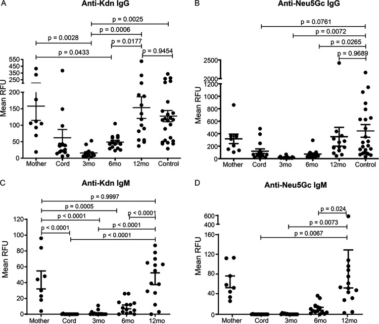
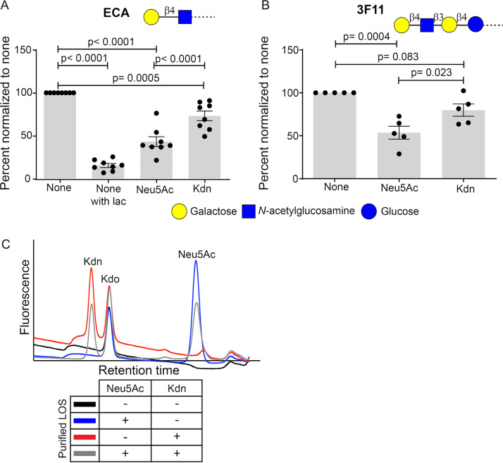
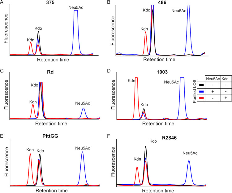
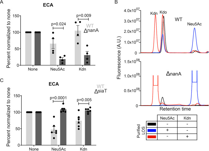
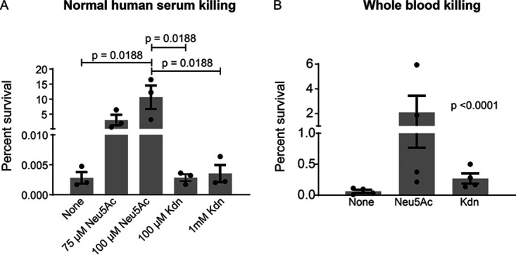
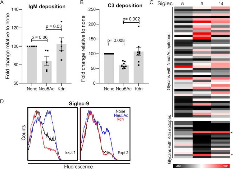
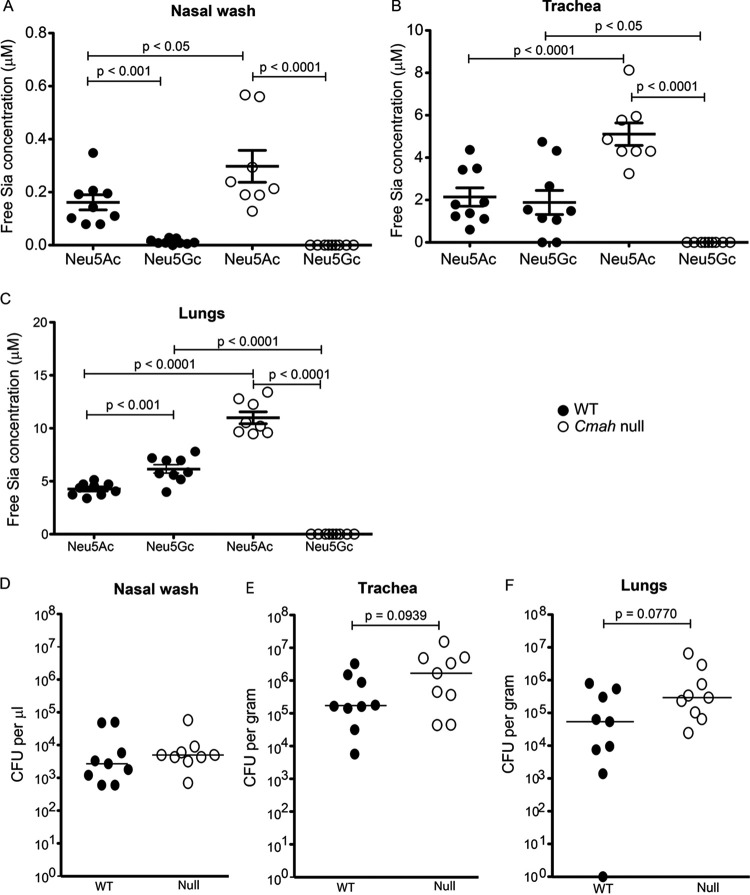
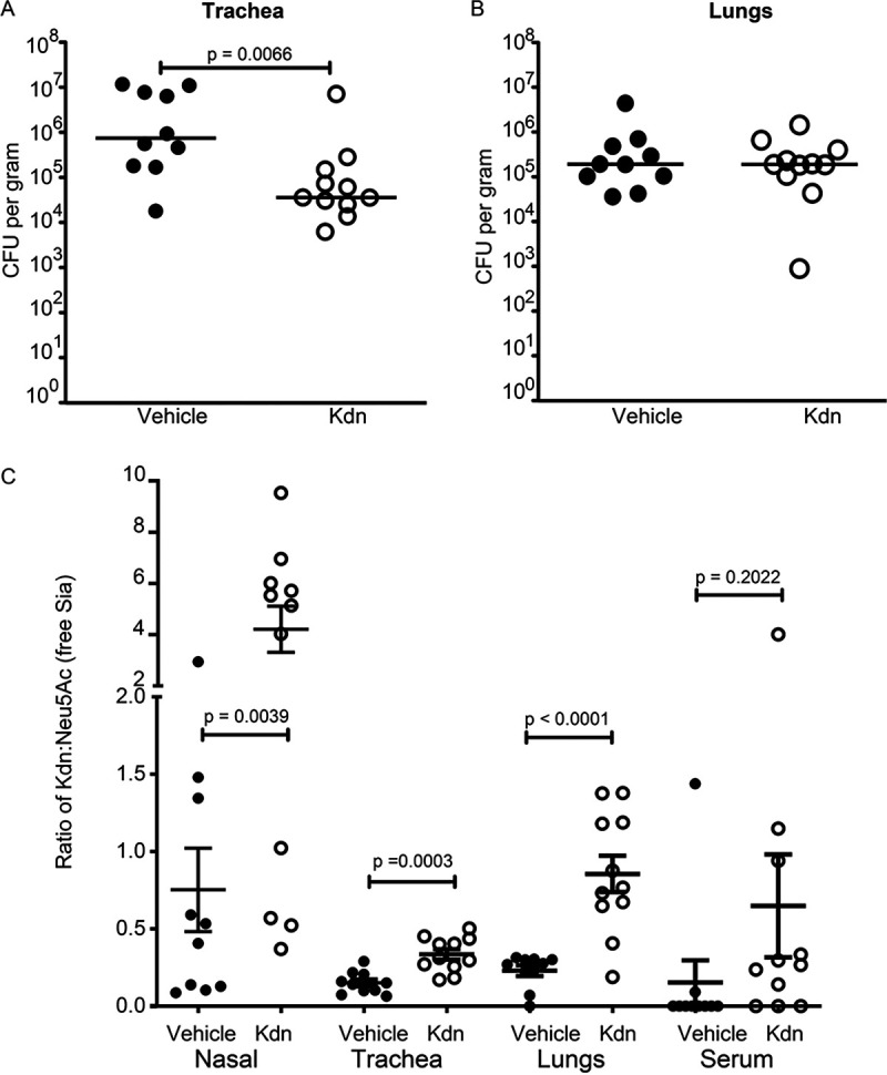
Surface expression of the common vertebrate sialic acid (Sia) N-acetylneuraminic acid (Neu5Ac) by commensal and pathogenic microbes appears structurally to represent "molecular mimicry" of host sialoglycans, facilitating multiple mechanisms of host immune evasion. In contrast, ketodeoxynonulosonic acid (Kdn) is a more ancestral Sia also present in prokaryotic glycoconjugates that are structurally quite distinct from vertebrate sialoglycans. We detected human antibodies against Kdn-terminated glycans, and sialoglycan microarray studies found these anti-Kdn antibodies to be directed against Kdn-sialoglycans structurally similar to those on human cell surface Neu5Ac-sialoglycans. Anti-Kdn-glycan antibodies appear during infancy in a pattern similar to those generated following incorporation of the nonhuman Sia N-glycolylneuraminic acid (Neu5Gc) onto the surface of nontypeable Haemophilus influenzae (NTHi), a human commensal and opportunistic pathogen. NTHi grown in the presence of free Kdn took up and incorporated the Sia into its lipooligosaccharide (LOS). Surface display of the Kdn within NTHi LOS blunted several virulence attributes of the pathogen, including Neu5Ac-mediated resistance to complement and whole blood killing, complement C3 deposition, IgM binding, and engagement of Siglec-9. Upper airway administration of Kdn reduced NTHi infection in human-like Cmah null (Neu5Gc-deficient) mice that express a Neu5Ac-rich sialome. We propose a mechanism for the induction of anti-Kdn antibodies in humans, suggesting that Kdn could be a natural and/or therapeutic "Trojan horse" that impairs colonization and virulence phenotypes of free Neu5Ac-assimilating human pathogens.IMPORTANCE All cells in vertebrates are coated with a dense array of glycans often capped with sugars called sialic acids. Sialic acids have many functions, including serving as a signal for recognition of "self" cells by the immune system, thereby guiding an appropriate immune response against foreign "nonself" and/or damaged cells. Several pathogenic bacteria have evolved mechanisms to cloak themselves with sialic acids and evade immune responses. Here we explore a type of sialic acid called "Kdn" (ketodeoxynonulosonic acid) that has not received much attention in the past and compare and contrast how it interacts with the immune system. Our results show potential for the use of Kdn as a natural intervention against pathogenic bacteria that take up and coat themselves with external sialic acid from the environment.

Keywords: CMAH; Kdn; Neu5Ac; antibody; bacterial pathogenesis; glycobiology; molecular mimicry; nontypeable Haemophilus influenzae (NTHi); sialic acid.


Figures

FIG 1
FIG 1
Structural evidence of molecular mimicry in Neu5Ac-containing, but not Kdn-containing, glycans in prokaryotes. (A) The occurrence of Neu5Ac- and Kdn-containing glycan structures in prokaryotes was determined by searching the open access database csdb.glycoscience.ru. The percent quantification is based on data compiled from 432 different Neu5Ac-containing and 55 Kdn-containing prokaryotic structures currently listed in the database. While Neu5Ac is present mainly as the terminal residue on glycan structures, Kdn is mostly an internal residue in the prokaryotic glycans (see Files S1 and S2 in the supplemental material). The black columns represent Sia as the terminal residue in glycan; white columns represent internal residue flanked by glycans. (B) Schematic representation of the common monosaccharides containing Neu5Ac (left) and Kdn (right) epitopes in lipooligosaccharide or capsular polysaccharide as obtained from the database. The pictorial symbols used are in accordance with the Symbol Nomenclature for Graphical Representation of Glycans (SNFG) (135) and are shown in the figure. The dotted line represents glycosidic linkages with the remaining glycoconjugate structure.
FIG 2
FIG 2
Antigenic specificity of the human anti-Kdn glycan antibodies. Heatmap showing the relative intensity of the fluorophore-conjugated anti-human IgG antibodies binding with the individual glycan on the microarray. Experiment was performed using commercially available pooled human IVIG (last column) as well as individual (n = 24) sera. Numbers (S-18 through S-81) on the top represent individual healthy human sera. (A) Each block represents the mean intensity of the antibody binding to the specific asialo- and sialoglycan at four independent spots in the microarray. Glycans are grouped by their nonsialylated (asialo-), Neu5Ac-, Neu5Gc-, and Kdn-terminating structures. The complete list of glycan structures represented is provided in panel B. Each column represents the relative binding preference of IgG antibodies in the corresponding serum toward the specific glycan epitopes. The color code of the heatmap is indicated, and the corresponding glycan in each row matched with the list in panel B.
FIG 3
FIG 3
Appearance of anti-Kdn IgG and IgM antibodies in the first year of human life. Binding of human sera to Kdn- and Neu5Gc-containing glycan epitopes was determined by sialoglycan microarray binding assay. Sera were obtained from cord blood (n = 15) and the child at 3, 6, and 12 months old and from 9 out of the 15 matched mothers in their third trimester of pregnancy. Control represents sera from blood from healthy human donors (both male and female) (n = 24). The average relative fluorescence unit (RFU) in arbitrary units (A.U.) against terminal Kdn (A and C) or Neu5Gc (B and D) containing glycan epitopes with underlying structures similar to vertebrate glycoconjugates are shown. Each dot represents the mean RFU values against all the Kdn (A and C)- or Neu5Gc (B and D)-containing glycans, respectively, for individual serum. (A to D) Relative abundance of the human IgG (A and B) and IgM antibodies (C and D) against the corresponding sialoglycoconjugates. Statistical significance was evaluated using one-way ANOVA with Tukey’s multiple comparison test. The mean ± standard error of the mean (SEM) (error bars) values and the adjusted P values are shown in the figure.
FIG 4
FIG 4
Exogenous Kdn is taken up and displayed by NTHi on its LOS. Surface sialylation of NTHi 2019 grown in the presence or absence of Neu5Ac or Kdn was determined using plant lectin, ECA (A) and mouse monoclonal IgM antibody 3F11 (B) in a flow cytometry-based assay. The bacteria were incubated with biotinylated lectin at 37°C or with mouse IgM antibody (Ab) 3F11 at room temperature (RT) for 30 min and then probed with fluorophore-conjugated secondary antibodies for analysis via flow cytometry. The binding epitopes of each of the reagents are shown within the panel using the symbols in accordance with SNFG nomenclature. “None” indicates the bacteria grown in the absence of Sia, while “Neu5Ac” or “Kdn” indicates the presence of the corresponding Sia in growth media. ECA binding with unsialylated bacteria done in the presence of 100 μM free lactose (none with lac) was used as a control to show the inhibition of ECA binding, since lactose is a competitive inhibitor of the lectin. Each dot represents the mean fluorescence intensity of an independent biological experiment (n = 8 for ECA; n = 5 for 3F11). Fluorescence values were expressed relative to that seen with the unsialylated samples in the same experiment to normalize for day-to-day variation. Statistical significance was determined using one-way ANOVA with Tukey’s multiple comparison test. Bars represent the mean (SEM) for each assay (A and B), and all the adjusted P values are shown in the figure. (C) Bacterial LOS was purified by hot phenol-water extraction from NTHi 2019 grown in the presence or absence of one or both Sias. Representative profiles of HPLC analysis following acid hydrolysis and DMB derivatization of purified LOS are shown. The fluorescent intensity peaks of the individual α-keto acid are identified in the figure. Kdo represents 3-deoxy-d-manno-oct-2-ulosonic acid, a key component of Gram-negative bacterial LOS and serves as the internal control in each of the LOS preparations. The table below the graph shows the bacterial cultures used to purify the corresponding LOS whose profile is shown. The presence (+) or absence (-) of the Sia in growth media of the bacteria is indicated.
FIG 5
FIG 5
Diverse NTHi strains incorporate exogenously provided Kdn. Six NTHi strains were grown in the presence of either Neu5Ac or Kdn and the bacterial LOS was purified by hot phenol-water extraction. HPLC analysis of acid-hydrolyzed and DMB-derivatized LOS shows fluorescent intensity peaks corresponding to Neu5Ac or Kdn in NTHi 375 (A), 486 (B), Rd (C), 1003 (D), PittGG (E) and R2846 (F). The individual Sia and Kdo as the internal control of the LOS preparations are identified as in Fig. 4C. The table shows the growth condition of bacterial cultures used to purify the LOS with (+) or without (−) the corresponding Sia added.
FIG 6
FIG 6
NTHi utilizes the same machineries for Kdn uptake and degradation as those for Neu5Ac. (A) Presence of Sia on the surface of wild-type (WT) NTHi 2019 and lyase deletion mutant 2019ΔnanA was determined using ECA binding. The bacterial treatment and data analysis are the same as described in the legend to Fig. 4. (B) HPLC profiles of LOS purified from NTHi 2019 or 2019ΔnanA grown in the presence or absence of 100 µM Neu5Ac or Kdn. The relative fluorophore intensity of the α-keto acid in arbitrary units (A.U.) present in the corresponding LOS is indicated on the y axis. Kdo represents an internal control as in Fig. 4C. The table below the graph shows the bacterial culture condition used to purify the corresponding LOS. (C) ECA binding assay with NTHi 2019 and the transporter deletion mutant 2019ΔsiaT grown in the presence or absence of Neu5Ac or Kdn. The dots are the same as described in the legend to Fig. 4. Statistical significance was determined using Student t test. All the values were normalized to the values for the unsialylated samples for the individual experiment to avoid day-to-day variation. Mean with SEM from independent experiments (n = 4 for panel A; n = 6 for panel C) was calculated. “None” indicates the bacteria grown in the absence of Sia, while “Neu5Ac” or “Kdn” indicates the presence of the corresponding Sia in growth media. Adjusted P values are shown.
FIG 7
FIG 7
Unlike Neu5Ac, Kdn incorporation does not provide protection against serum and whole blood killing. (A) NTHi 2019 was grown with or without Neu5Ac or Kdn and incubated in the presence of 10% pooled normal human sera at 37°C for 30 min. Percent survival was determined from CFU counts of bacteria recovered following serum incubation relative to the inoculum. The amounts of Sia added during bacterial growth are shown on the x axis. (B) Bactericidal effect of hirudin-anticoagulated whole blood collected from healthy human donors is shown (n = 4). Each dot in the figures represents the corresponding value in each experiment. Mean with SEM (n = 3 for serum killing; n = 4 for whole blood killing) and all the adjusted P values are shown. Statistical significance was determined using one-way ANOVA with Tukey’s multiple comparison test (A) and Bartlett’s test (B).
FIG 8
FIG 8
Incorporated Kdn does not abolish complement C3 and IgM antibody deposition and does not substitute for Neu5Ac in interaction with Siglecs. Bacteria grown in the presence of Neu5Ac or Kdn were incubated with 10% pooled normal human sera for 5 min at 37°C and IgM antibody (A) and complement factor C3 (B) deposition were determined using the corresponding antibody staining in flow cytometry. For IgM deposition, the bacteria were incubated with heat-inactivated sera. Each dot represents an independent experiment (n = 5 for panel A and n = 8 for panel B). The data were normalized to the values for unsialylated bacteria to eliminate the daily variation. Statistical significance was determined using one-way ANOVA with Tukey’s multiple comparison test. Mean with SEM and adjusted P values are shown in the figure. (C) Heatmap showing the binding of Neu5Ac- and Kdn-containing glycan epitopes to purified recombinant Fc chimeric proteins of human Siglec-5, -9, and -14. The map represents the binding affinities of each protein toward the corresponding glycans determined from the glycan array binding experiment. The complete list of the glycan used are shown in File S3 in the supplemental material. Glycans with lactose underlying structure are indicated by an asterisk. Each block corresponds to a distinct glycan with terminal Sia as indicated in the left. The color of each block represents the mean binding intensity done in technical quadruplet. Color code is indicated below. (D) Representative flow cytometry profiles showing Siglec-9 binding with NTHi 2019 grown in the presence or absence of Neu5Ac or Kdn in two independent biological experiments (experiments 1 and 2). Figure legend shows the Sia added to the bacterial growth media. “None” indicates the bacteria grown in the absence of Sia, while “Neu5Ac” or “Kdn” indicates the presence of the corresponding Sia in growth media.
FIG 9
FIG 9
Presence of free sialic acid correlates with in vivo infection of NTHi. (A to C) Free sialic acid content of nasal wash (A), trachea (B), and lung (C) of Cmah null (open circle) and wild-type (closed circle) mice were determined by HPLC analysis of DMB-derivatized samples of tissue collected 24 h after intranasal infection with NTHi 2019. Statistical significance was determined by one-way ANOVA and Tukey’s multiple comparison test. (D to F) Bacterial load postinfection was determined by the CFU counts of the bacteria from nasal wash (D), trachea (E) and lung (F) homogenates (same samples as used for free sialic acid analysis) that were plated on Haemophilus isolation agar. Data represent the results from two independent experiments, with each symbol representing the value for an individual mouse (n = 9). Statistical significance was determined using the nonparametric Mann-Whitney test. Adjusted P values are shown in the figure.
FIG 10
FIG 10
Free Kdn administration reduces NTHi infection in Cmah null mice. Following NTHi 2019 infection, free Kdn was administered every 6 h intranasally to the Cmah null mice. (A and B) Twenty-four hours postinfection, the mice were sacrificed and bacterial load of trachea (A) and lungs (B) homogenates was determined. Control group of infected mice denoted as “vehicle” were administered with HBSS (used as the solvent for Kdn) at the same corresponding time points as the test condition. Statistical significance was determined using the nonparametric Mann-Whitney test. (C) HPLC analysis of the tissue homogenates from the infected mice was performed to determine the abundance of free Kdn relative to Neu5Ac. Statistical significance was determined using the Student’s t test. Mean ± SEM are shown. Data represent the results from two independent experiments, and each symbol represents the value for an individual mouse. Adjusted P values are shown in the figure.

Similar articles

Cited by

References

    1. Varki A, Schnaar RL, Schauer R. 2017. Sialic acids and other nonulosonic acids In Varki A, Cummings RD, Esko JD, Stanley P, Hart GW, Aebi M, Darvill AG, Kinoshita T, Packer NH, Prestegard JH, Schnaar RL, Seeberger PH (ed), Essentials of glycobiology. Cold Spring Harbor Laboratory Press, Cold Spring Harbor, NY. - PubMed
    1. Vimr ER 2013. Unified theory of bacterial sialometabolism: how and why bacteria metabolize host sialic acids. ISRN Microbiol 2013:816713. doi:10.1155/2013/816713. - DOI - PMC - PubMed
    1. Nadano D, Iwasaki M, Endo S, Kitajima K, Inoue S, Inoue Y. 1986. A naturally occurring deaminated neuraminic acid, 3-deoxy-D-glycero-D-galacto-nonulosonic acid (KDN). Its unique occurrence at the nonreducing ends of oligosialyl chains in polysialoglycoprotein of rainbow trout eggs. J Biol Chem 261:11550–11557. - PubMed
    1. Warren L 1963. The distribution of sialic acids in nature. Comp Biochem Physiol 10:153–171. doi:10.1016/0010-406X(63)90238-X. - DOI - PubMed
    1. Mamedov T, Yusibov V. 2011. Green algae Chlamydomonas reinhardtii possess endogenous sialylated N-glycans. FEBS Open Bio 1:15–22. doi:10.1016/j.fob.2011.10.003. - DOI - PMC - PubMed

Publication types

MeSH terms