. 2021 Jan 19;12(1):e03227-20.
doi: 10.1128/mBio.03227-20.

5-Aminosalicylic Acid Ameliorates Colitis and Checks Dysbiotic Escherichia coli Expansion by Activating PPAR-γ Signaling in the Intestinal Epithelium

Affiliations

5-Aminosalicylic Acid Ameliorates Colitis and Checks Dysbiotic Escherichia coli Expansion by Activating PPAR-γ Signaling in the Intestinal Epithelium

Stephanie A Cevallos et al. mBio. .

Abstract

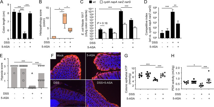
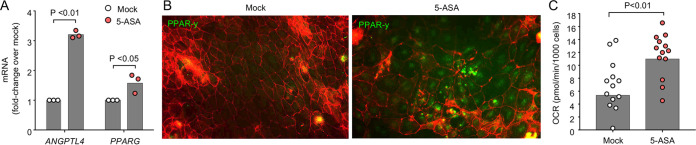
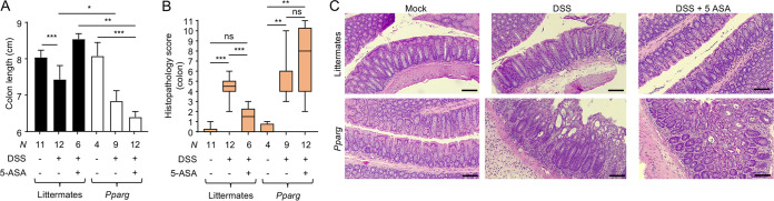
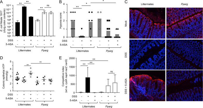
5-Aminosalicylic acid (5-ASA), a peroxisome proliferator-activated receptor gamma (PPAR-γ) agonist, is a widely used first-line medication for the treatment of ulcerative colitis, but its anti-inflammatory mechanism is not fully resolved. Here, we show that 5-ASA ameliorates colitis in dextran sulfate sodium (DSS)-treated mice by activating PPAR-γ signaling in the intestinal epithelium. DSS-induced colitis was associated with a loss of epithelial hypoxia and a respiration-dependent luminal expansion of Escherichia coli, which could be ameliorated by treatment with 5-ASA. However, 5-ASA was no longer able to reduce inflammation, restore epithelial hypoxia, or blunt an expansion of E. coli in DSS-treated mice that lacked Pparg expression specifically in the intestinal epithelium. These data suggest that the anti-inflammatory activity of 5-ASA requires activation of epithelial PPAR-γ signaling, thus pointing to the intestinal epithelium as a potential target for therapeutic intervention in ulcerative colitis.IMPORTANCE An expansion of Enterobacterales in the fecal microbiota is a microbial signature of dysbiosis that is linked to many noncommunicable diseases, including ulcerative colitis. Here, we used Escherichia coli, a representative of the Enterobacterales, to show that its dysbiotic expansion during colitis can be remediated by modulating host epithelial metabolism. Dextran sulfate sodium (DSS)-induced colitis reduced mitochondrial activity in the colonic epithelium, thereby increasing the amount of oxygen available to fuel an E. coli expansion through aerobic respiration. Activation of epithelial peroxisome proliferator-activated receptor gamma (PPAR-γ) signaling with 5-aminosalicylic acid (5-ASA) was sufficient to restore mitochondrial activity and blunt a dysbiotic E. coli expansion. These data identify the host's epithelial metabolism as a potential treatment target to remediate microbial signatures of dysbiosis, such as a dysbiotic E. coli expansion in the fecal microbiota.

Keywords: Escherichia coli; dysbiosis; gut inflammation; inflammatory bowel disease; microbial communities.


Figures

FIG 1
FIG 1
5-ASA blocks E. coli expansion during DSS-induced colitis. Groups of male C57BL/6J mice (N is indicated in panel E) receiving conventional chow (5-ASA, –) or chow supplemented with 5-ASA (5-ASA, +) and drinking water supplemented with DSS (DSS, +) or no drinking water supplementation (DSS, –) were engrafted with E. coli strains (a 1:1 mixture of the wild type [wt] and cydA napA narG narZ mutant). (A) Colon length was determined during necropsy. (B) A veterinary pathologist scored histopathological changes in blinded sections of the colon. (C) Numbers of CFU of E. coli Nissle 1917 recovered from colon contents. (D) Competitive index (ratio of the E. coli Nissle 1917 wild type to an isogenic cydA napA narG narZ mutant) in colon contents was determined. (E and F) Binding of pimonidazole was detected using Hypoxyprobe-1 primary antibody and a Cy3-conjugated goat anti-mouse secondary antibody (red fluorescence) in the sections of the proximal colon that were counterstained with DAPI nuclear stain (blue fluorescence). (E) Pimonidazole staining was quantified by scoring blinded sections of the proximal colon. (F) Representative images are shown. (G and H) Colonocytes were isolated from the colonic mucosa to measure cytosolic concentrations of ATP (G) or pyruvate dehydrogenase (PDH) activity (H). *, P < 0.05; **, P < 0.01; ***, P < 0.001.
FIG 2
FIG 2
5-ASA stimulates oxygen consumption in CaCo-2 cells. CaCo-2 cells were mock treated or treated with 5-ASA. (A) The transcript levels of the indicated genes were determined by quantitative real-time PCR. (B) The synthesis of PPAR-γ was detected by fluorescence microcopy using an anti-PPAR-γ antibody (green fluorescence) and staining of actin, with phalloidin as a counterstain (red fluorescence). (C) The oxygen consumption rate (OCR) of CaCo-2 cells was determined using an Agilent Seahorse XFe96 analyzer. (A and C) Each dot represents data from one well.
FIG 3
FIG 3
5-ASA ameliorates DSS-induced colitis by activating epithelial PPAR-γ signaling. Groups of Ppargfl/fl Villincre/− mice that lack PPAR-γ signaling in the intestinal epithelium (Pparg) and littermate Ppargfl/fl Villin−/− control mice (Littermates) received conventional chow (5-ASA, –) or chow supplemented with 5-ASA (5-ASA, +) and drinking water supplemented with DSS (DSS, +) or no drinking water supplementation (DSS, –). (A) Colon length was determined during necropsy. (B and C) Blinded sections of the colon from each animal were evaluated by a veterinary pathologist. (B) Histopathology score. The boxes in the whisker blot represent the first to third quartiles, and the mean value of the gross pathology scores is indicated by a line. (C) Representative images of colonic sections for each group are shown. The scale bars represent 300 μm. *, P < 0.05; **, P < 0.01; ***, P < 0.001; ns, differences were not significant.
FIG 4
FIG 4
5-ASA restores epithelial hypoxia and ameliorates dysbiotic E. coli expansion by activating epithelial PPAR-γ signaling. Groups (N is indicated in panel B) of Ppargfl/fl Villincre/− mice that lack PPAR-γ signaling in the intestinal epithelium (Pparg) and littermate Ppargfl/fl Villin−/− control mice (Littermates) receiving conventional chow (5-ASA, –) or chow supplemented with 5-ASA (5-ASA, +) and drinking water supplemented with DSS (DSS, +) or no drinking water supplementation (DSS, –) were engrafted with E. coli strains (a 1:1 mixture of the wild type and cydA napA narG narZ mutant). (A) Numbers of CFU of E. coli Nissle 1917 recovered from colon contents. (B and C) Binding of pimonidazole was detected using Hypoxyprobe-1 primary antibody and a Cy3-conjugated goat anti-mouse secondary antibody (red fluorescence) in the sections of proximal colon that were counterstained with DAPI nuclear stain (blue fluorescence). (B) Pimonidazole staining was quantified by scoring blinded sections of the proximal colon. (C) Representative images are shown. (D) Colonocytes were isolated from the colonic mucosa to measure cytosolic concentrations of ATP. (E) Competitive indexes (ratio of the E. coli Nissle 1917 wild type to an isogenic cydA napA narG narZ mutant) in colon contents were determined. *, P < 0.05; **, P < 0.01; ***, P < 0.001; ns, P > 0.05.

References

    1. Podolsky DK 2002. Inflammatory bowel disease. N Engl J Med 347:417–429. doi:10.1056/NEJMra020831. - DOI - PubMed
    1. Human Microbiome Project Consortium. 2012. Structure, function and diversity of the healthy human microbiome. Nature 486:207–214. doi:10.1038/nature11234. - DOI - PMC - PubMed
    1. Adeolu M, Alnajar S, Naushad S, Gupta RS. 2016. Genome-based phylogeny and taxonomy of the 'Enterobacteriales': proposal for Enterobacterales ord. nov. divided into the families Enterobacteriaceae, Erwiniaceae fam. nov., Pectobacteriaceae fam. nov., Yersiniaceae fam. nov., Hafniaceae fam. nov., Morganellaceae fam. nov., and Budviciaceae fam. nov. Int J Syst Evol Microbiol 66:5575–5599. doi:10.1099/ijsem.0.001485. - DOI - PubMed
    1. Rigottier-Gois L 2013. Dysbiosis in inflammatory bowel diseases: the oxygen hypothesis. ISME J 7:1256–1261. doi:10.1038/ismej.2013.80. - DOI - PMC - PubMed
    1. Shin NR, Whon TW, Bae JW. 2015. Proteobacteria: microbial signature of dysbiosis in gut microbiota. Trends Biotechnol 33:496–503. doi:10.1016/j.tibtech.2015.06.011. - DOI - PubMed

Publication types

MeSH terms