. 2021 Jan 19;10(1):12.
doi: 10.1038/s41389-020-00300-z.

PI3K/Akt pathway and Nanog maintain cancer stem cells in sarcomas

Affiliations

PI3K/Akt pathway and Nanog maintain cancer stem cells in sarcomas

Changhwan Yoon et al. Oncogenesis. .

Retraction in

Abstract

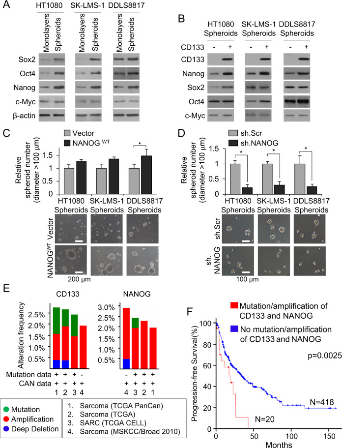
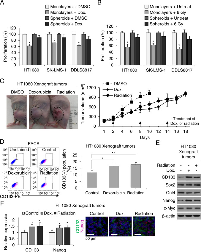
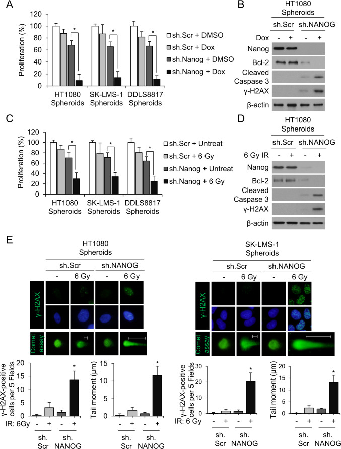
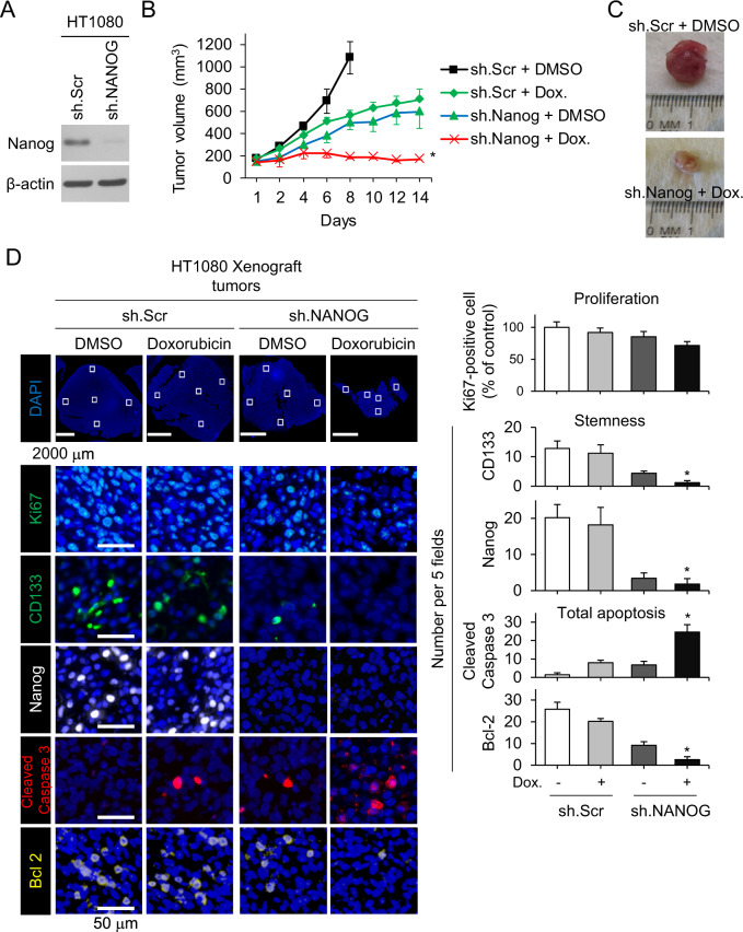
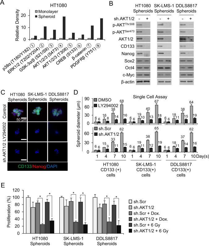
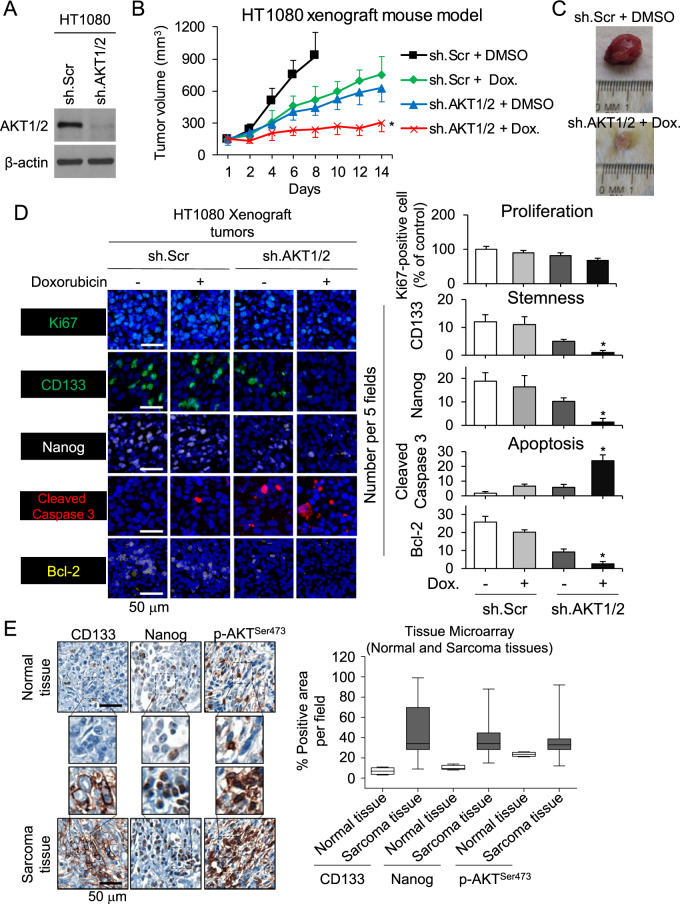
The self-renewal transcription factor Nanog and the phosphoinositide 3-kinase (PI3K)-Akt pathway are known to be essential for maintenance of mesenchymal stem cells. We evaluated their contribution to the maintenance of CD133(+) cancer stem-like cells (CSCs) and spheroid-forming cells in patient-derived cell lines from three human sarcoma subtypes: HT1080 fibrosarcoma, SK-LMS-1 leiomyosarcoma, and DDLS8817 dedifferentiated liposarcoma. Levels of Nanog and activated Akt were significantly higher in sarcoma cells grown as spheroids or sorted for CD133 expression to enrich for CSCs. shRNA knockdown of Nanog decreased spheroid formation 10- to 14-fold, and reversed resistance to both doxorubicin and radiation in vitro and in H1080 flank xenografts. In the HT1080 xenograft model, doxorubicin and Nanog knockdown reduced tumor growth by 34% and 45%, respectively, and the combination reduced tumor growth by 74%. Using a human phospho-kinase antibody array, Akt1/2 signaling, known to regulate Nanog, was found to be highly activated in sarcoma spheroid cells compared with monolayer cells. Pharmacologic inhibition of Akt using LY294002 and Akt1/2 knockdown using shRNA in sarcoma CSCs decreased Nanog expression and spheroid formation and reversed chemotherapy resistance. Akt1/2 inhibition combined with doxorubicin treatment of HT1080 flank xenografts reduced tumor growth by 73%. Finally, in a human sarcoma tumor microarray, expression of CD133, Nanog, and phospho-Akt were 1.8- to 6.8-fold higher in tumor tissue compared with normal tissue. Together, these results indicate that the Akt1/2-Nanog pathway is critical for maintenance of sarcoma CSCs and spheroid-forming cells, supporting further exploration of this pathway as a therapeutic target in sarcoma.


Conflict of interest statement

The authors declare that they have no conflict of interest.

Figures

Fig. 1
Fig. 1. Nanog promotes stemness in human sarcoma cells.
A, B Western blot for self-renewal proteins Sox2, Oct4, Nanog, and c-Myc in human sarcoma cell lines grown as A monolayers and as spheroids and B after separation of spheroid cells into CD133+ and CD133− fractions. β-Actin, loading control. C, D Spheroid formation following lentiviral transduction with C Nanog or vector and D Nanog shRNA (sh.NANOG) or scramble control shRNA (sh.Scr). Scale bar, 100, 200 µm. *p < 0.05 compared to control. E Frequency and types of CD133 and Nanog alterations in sarcoma samples according to previous genomic studies from TCGA, Liu and colleagues, and University of Tokyo. F Kaplan–Meier curves of progression-free survival of sarcoma patients following potentially curative resection, stratified by the presence or absence of CD133 and Nanog mutation/amplification. Graphs in E, F generated using cBioPortal (http://www.https://cbioportal.mskcc.org).
Fig. 2
Fig. 2. CSCs contribute to sarcomas’ resistance to chemo- and radiotherapy.
A, B Proliferation of monolayers and spheroids following treatment with doxorubicin (Dox) or radiation therapy. C Growth curve of HT1080 fibrosarcoma xenografts in athymic nude mice treated with Dox or 6 Gy radiation. D Fluorescence-activated cell sorting (FACS) analysis for CD133+ cells in untreated control and Dox- or 6-Gy-treated tumors. E Western blot of tumor lysates for CD133, Sox2, Oct4, Nanog, and c-Myc. β-Actin is the loading control. F Immunofluorescence of tumors for CD133 (green) and Nanog (red) with DAPI (blue) staining in athymic nude mice treated with doxorubicin or 6 Gy radiation. Scale bar, 50 µm. *p < 0.05, **p < 0.01 compared to control.
Fig. 3
Fig. 3. Nanog inhibition reverses chemo- and radiotherapy resistance.
AD Proliferation of sarcoma cells grown as spheroids (A, C) and western blot for Nanog, Bcl-2, cleaved caspase-3, and γ-H2AX (B, D) following treatment with sh.NANOG or sh.Scr and doxorubicin (Dox) or 6 Gy radiotherapy. β-Actin, loading control. *p < 0.01 compared to control. E Immunofluorescence of sarcoma spheroid cells (HT1080, SK-LM-1). Top panel shows γ-H2AX in green. Middle panel shows γ-H2AX in green and nuclei in blue (DAPI). Bottom panel shows Comet assay in green. All photos are of different cells under specified conditions. Graphs show quantification of γ-H2AX positive staining and mean tail moment. Nuclei were stained with DAPI (4′,6-diamidino-2-phenylindole; blue).
Fig. 4
Fig. 4. Nanog knockdown restores sensitivity to chemotherapy in mouse xenograft models.
A Western blot of HT1080 cells transduced with sh.NANOG lentivirus for Nanog and β-actin. B Growth of HT1080 fibrosarcoma xenografts in athymic nude mice treated with sh.NANOG or sh.Scr and PBS or doxorubicin (Dox). C Photos of representative tumors. D Immunohistochemical analysis of tumors for Ki67 (green), CD133 (green), Nanog (white), cleaved caspase-3 (red), and Bcl-2 (yellow). Scale bar, 50 µm. *p < 0.01 compared to control.
Fig. 5
Fig. 5. PI3K/Akt signaling regulates Nanog expression.
A Activated proteins in HT1080 monolayers and spheroids identified by human phospho-kinase array kit. B Western blot for indicated proteins following transduction with sh.Akt1/2 or sh.Scr in sarcoma spheroid cells. C CD133 (green) and Nanog (red) immunofluorescence with and DAPI (blue) counterstaining following treatment with PI3K inhibitor LY294002 or sh.Akt1/2 in spheroid cells. D Diameter of spheroids grown from single CD133+ cell cells showing following treatment with LY294002 or sh.Akt1/2 vs, DMSO or sh.Scr, respectively. E Proliferation of spheroid cells following treatment with sh.Akt1/2 and doxorubicin or 6 Gy radiation. β-Actin, loading control. Scale bar, 50 µm. *p < 0.01 compared to control.
Fig. 6
Fig. 6. Combined Akt1/2 inhibition and chemotherapy inhibits stemness and tumor growth in vivo.
A Western blot confirmation of Akt1/2 knockdown by shRNA in HT1080 cells. B Growth of HT1080 fibrosarcoma xenografts stably transduced with sh.Scr or sh.Akt1/2 in athymic nude mice treated and PBS or doxorubicin. C Photos of representative tumors. D Immunohistochemical analysis of tumors for proliferation using Ki67 (green), stemness using CD133 (green) and Nanog (white), and apoptosis using cleaved caspase-3 (red) and Bcl-2 (yellow). E Immunohistochemical staining of commercially available tissue array slide containing 79 human sarcomas and 4 human normal tissues for CD133, Nanog, and p-Akt1/2 (S473). β-Actin, loading control. Scale bar, 50 µm. *p < 0.01 compared to control.

References

    1. Siegel RL, Miller KD, Jemal A. Cancer statistics, 2019. CA Cancer J. Clin. 2019;69:7–34. - PubMed
    1. Billingsley KG, et al. Pulmonary metastases from soft tissue sarcoma: analysis of patterns of diseases and postmetastasis survival. Ann. Surg. 1999;229:602–610. - PMC - PubMed
    1. Van Glabbeke M, et al. Prognostic factors for the outcome of chemotherapy in advanced soft tissue sarcoma: an analysis of 2,185 patients treated with anthracycline-containing first-line regimens—a European Organization for Research and Treatment of Cancer Soft Tissue and Bone Sarcoma Group Study. J. Clin. Oncol. 1999;17:150–157. - PubMed
    1. Allegra A, et al. The cancer stem cell hypothesis: a guide to potential molecular targets. Cancer Invest. 2014;32:470–495. - PubMed
    1. Izumiya M, et al. Chemoresistance is associated with cancer stem cell-like properties and epithelial-to-mesenchymal transition in pancreatic cancer cells. Anticancer Res. 2012;32:3847–3853. - PubMed

Publication types