Review
. 2021 Jan 20;6(1):24.
doi: 10.1038/s41392-020-00422-1.

Interplay between cofactors and transcription factors in hematopoiesis and hematological malignancies

Affiliations
Review

Interplay between cofactors and transcription factors in hematopoiesis and hematological malignancies

Zi Wang et al. Signal Transduct Target Ther. .

Abstract

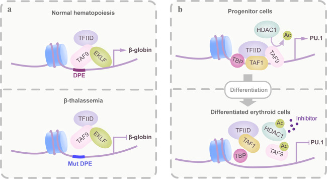
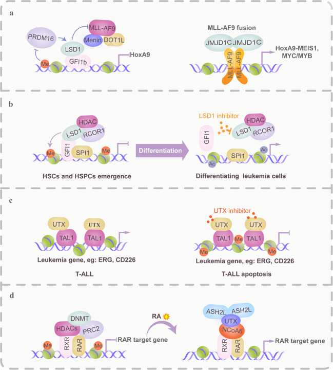
Hematopoiesis requires finely tuned regulation of gene expression at each stage of development. The regulation of gene transcription involves not only individual transcription factors (TFs) but also transcription complexes (TCs) composed of transcription factor(s) and multisubunit cofactors. In their normal compositions, TCs orchestrate lineage-specific patterns of gene expression and ensure the production of the correct proportions of individual cell lineages during hematopoiesis. The integration of posttranslational and conformational modifications in the chromatin landscape, nucleosomes, histones and interacting components via the cofactor-TF interplay is critical to optimal TF activity. Mutations or translocations of cofactor genes are expected to alter cofactor-TF interactions, which may be causative for the pathogenesis of various hematologic disorders. Blocking TF oncogenic activity in hematologic disorders through targeting cofactors in aberrant complexes has been an exciting therapeutic strategy. In this review, we summarize the current knowledge regarding the models and functions of cofactor-TF interplay in physiological hematopoiesis and highlight their implications in the etiology of hematological malignancies. This review presents a deep insight into the physiological and pathological implications of transcription machinery in the blood system.


Conflict of interest statement

The authors declare no competing interests.

Figures

Fig. 1
Fig. 1
Modes of gene transcriptional regulation by TAF-containing TCs. A deeper insight into the functional importance of TAFs as cofactors that connect transcription complexes with DNA. In addition, other cofactors within one transcription complex could indirectly affect gene transcription via regulating TAFs. a As a component of TFIID, TAF9 associated with the transcription factor EKLF provides a platform for the recruitment of TFIID transcription complexes, particularly at promoters that contain the initiator (INI) and DPE box (i.e., β-globin promoter). In disease conditions, β-thalassemia-causing mutations across the binding region disrupt the formation of the transcription complex and thus generally inhibit transcription efficiency. b The DNA-binding ability and activity of TAF9 are determined by its acetylation level. During erythropoiesis, TAF9 acetylation gradually increases due to a decrease in HDAC1-associated deacetylase activity, which subsequently results in the disassociation of the TFIID complex and transcriptional repression
Fig. 2
Fig. 2
Modes of Mediator-dependent enhancer regulation controlling both normal and malignant hematopoiesis. Generally, Mediator functions as a large coactivator complex that recruits enhancer-localized TFs to the promoter, where some members of the Mediator module cooperate with additional transcriptional cofactors to maintain active enhancers of essential hematopoietic genes. Gain or loss of function mutations of MEDs have been identified to lead to malignant hematopoiesis. To date, no drugs have been developed to target MEDs or their mutation sites. Additionally, bromodomain and extraterminal (BET) proteins, such as BRD4, regulate their downstream target genes, at least in part, by interacting with the Mediator complex and have been suggested as targets of bromodomain inhibitors to treat MED-mediated malignancies. a An enhancer-specific role of Med12 in preserving H3K27Ac levels for maintaining the active state of hematopoietic enhancers, such as the c-kit gene, via cooperation with P300. Deletion of Med12 causes H3K27Ac loss at enhancers of essential HSC genes, failure of hematopoietic-specific transcriptional programs and apoptosis in HSCs. b BRD4 and Mediator can mutually stabilize one another’s occupancy at cis elements of leukemic genes (AML) bound by acetylated nucleosomes, which promotes the transcriptional elongation of Pol II by facilitating P-TEFb recruitment. The BET bromodomain inhibitor JQ1 causes a dramatic release of Mediator from the leukemia genome, leading to transcriptional suppression of leukemic genes
Fig. 3
Fig. 3
Chromatin remodeling complexes (CRCs) are implicated in malignant hematopoiesis via their interplay with TFs. In many cases, CRCs are indispensable to the oncogenic functions of leukemia-associated and fusion TFs due to their multisubunit properties and/or recruitment of other transcriptional complexes, such as the polycomb repressive complex. In addition to remodeling histone–DNA interactions, epigenetic subunits confer to CRCs the ability to modulate regulatory region activity in the genome by affecting histone modifications or DNA methylation levels. Based on this knowledge, the following two treatment strategies have been suggested: (1) release or transition of CRC occupancy by changing DNA methylation levels or targeting oncogenic TFs. (2) Targeting the interactions between CRCs and oncogenic TFs. a The BRG1–SWI/SNF complex together with BRD4 promotes superenhancer (BRD4-dependent MYC enhancer) activity of the MYC gene. After treatment with the CBFβ-SMMHC fusion protein inhibitor, the BRG1–SWI/SNF complex occupancy is replaced by the RUNX1–PRC-repressive complex, which inhibits the MYC transcriptional program. b In AML, the binding of the Smarca5/SNF2H-CCCTC-binding factor (CTCF) complex at the PU.1 gene is blocked due to DNA methylation. Upon treatment by AZA-mediated DNA demethylation, the Smarca5/SNF2H complex is recruited to the enhancer of the PU.1 gene and blocks its expression. c In APL, the PML-RARα fusion binds and recruits NuRD, the PRC2 complex and DNA methyltransferase 3a (DNMT3a) to the tumor suppressor gene RARβ2, which in turn leads to chromatin compaction and consequent promoter silencing. d The oncogenic TF SALL4 promotes leukemogenesis, at least in part, by repressing PTEN gene expression through recruitment of the NuRD/HDAC complex. Disrupting the SALL4 and NuRD/HDAC complex interaction could reverse the repression of the PTEN gene and reduce tumor cell viability
Fig. 4
Fig. 4
Aberrant recruitment of HAT/HDAC-containing complexes is a general mechanism of leukemogenesis. Chromatin accessibility plays a critical role in regulating cell type-specific gene expression during hematopoiesis but has also been suggested to be abnormally regulated during leukemogenesis. Oncogenic TF or fusion protein binding was found at accessible chromatin regions with aberrant recruitment of the coactivator (CoA) or corepressor (CoR) complexes that affected histone modification patterns, altered the chromatin structure and thereby facilitated DNA accessibility. Furthermore, in some cases, HATs/HDACs are critical to the optimal oncogenic activity of leukemia TFs or fusion proteins via regulating their acetylation levels. Therefore, disrupting the balanced interplay of the epigenetic environment and oncogenic TFs or fusion proteins might be used as a therapeutic strategy. a Upper panel, P300-containing CoA complexes contribute to the self-renewal and leukemogenesis of HSPCs through acetylating the AML1-ETO fusion, which promotes its transcriptional activation and histone H3. b The AML1-ETO fusion recruits CoR complexes consisting of the nuclear receptor corepressor (N-CoR), HDAC1, SIN3 transcription regulator family member A (mSin3A), and DNA methyltransferase 1 (DNMT1), which repress gene transcription by enzymatic deacetylation of histones, DNA methylation, and creation of a repressive chromatin structure. c In APL blast cells, the PML-RARα fusion forms oligodimers and binds DNA through recruitment of the CoR–HDAC complex, which leads to deacetylation of histones and H3K27/H3K9 methylation and subsequently produces a condensed chromatin structure that represses the transcription of target genes. While all-trans retinoic acid (RA) or arsenic trioxide (ATO) mediates degradation of PML-RARα, which is replaced by the RARα/RXR heterodimer, and converts the CoR-HDAC into a CoA–HAT complex that reactivates gene transcription and restores differentiation
Fig. 5
Fig. 5
HMT/HDT-based regulatory mechanisms in TCs are implicated in the pathogenesis and treatment of malignant hematopoiesis. HMTs and HDTs are recurrently mutated or aberrantly expressed in a variety of hematological malignancies. Their intrinsic activities on histone and nonhistone proteins within transcriptional complexes are critical for epigenetic control in normal and malignant hematopoiesis and are amenable to drug intervention or involved in drug therapeutic effects. a Left panel in mixed lineage leukemia (MLL)-AF9 fusion leukemia. MLL-AF9 recruits DOT1L, a histone 3 lysine 79 methyltransferase (H3K79me1/me2/me3), which leads to hematopoietic transformation via H3K79 dimethylation that causes aberrant transcription of genes such as HOXA9 and MEIS1. PRDM16 (a histone H3K4 methyltransferase), an antagonist to DOT1L, activates Gfi1b-mediated gene transcription, which in turn downregulates the HOXA gene cluster. However, PRDM16 expression is always silenced by DNA methylation in MLL-AF9 leukemia. Right panel, similar to DOT1L, the H3K9me2/me1 demethylase JMJD1C contributes to MLL-AF9 leukemia maintenance by affecting MYB, MYC, and HOXA9-MEIS1 gene expression programs, suggesting that individual genes can be regulated by different kinds of HMTs and/or HDT-containing TCs. b Growth factor independence 1 (GFI1) is critical to the initiation of the endothelial-to-hematopoietic transition (EHT) due to its recruitment of the LSD1–CoREST repressive complex to epigenetically silence the endothelial program and allow the emergence of HSCs. Disruption or separation of the GFI1–LSD1 repressive complex by LSD1 inhibitors is considered to induce differentiation in certain subtypes of AML. c UTX, a coactivator of TAL1, is essential to leukemia maintenance in TAL1-positive cells due to its promotion of an open chromatin configuration at target gene sites by H3K27me3 demethylation. A therapy based on UTX inhibition is efficient at inducing cell death through downregulation of the TAL1 leukemic gene expression program. d UTX is a critical mediator of RA-induced differentiation in leukemic cells. RA treatment leads to the coordinated removal of repressive marks, the displacement of polycomb group proteins, and the deposition of activating marks. RA promotes the activation of RAR target genes by recruiting NCoA6, UTX, and ASH2L, concomitant with the demethylation of H3K27 and trimethylation of H3K4
Fig. 6
Fig. 6
The targeted domains/sites used to design cofactor inhibitors. Abnormal epigenetic changes are amenable to pharmacological intervention. The emergence of cofactors as oncology targets has spurred significant drug discovery efforts with the goal of identifying small-molecule inhibitors that target their enzymatic sites, binding pockets, and protein/TF interactions for therapeutic applications. Binding sites and representative drugs for each kind of cofactor are shown

References

    1. Wade JT, Struhl K. The transition from transcriptional initiation to elongation. Curr. Opin. Genet. Dev. 2008;18:130–136. doi: 10.1016/j.gde.2007.12.008. - DOI - PMC - PubMed
    1. Wright KJ, Marr MT, II, Tjian R. TAF4 nucleates a core subcomplex of TFIID and mediates activated transcription from a TATA-less promoter. Proc. Natl. Acad. Sci. USA. 2006;103:12347–12352. doi: 10.1073/pnas.0605499103. - DOI - PMC - PubMed
    1. Papadopoulos P, et al. TAF10 interacts with the GATA1 transcription factor and controls mouse erythropoiesis. Mol. Cell. Biol. 2015;35:2103–2118. doi: 10.1128/MCB.01370-14. - DOI - PMC - PubMed
    1. Sengupta T, Cohet N, Morle F, Bieker JJ. Distinct modes of gene regulation by a cell-specific transcriptional activator. Proc. Natl. Acad. Sci. USA. 2009;106:4213–4218. doi: 10.1073/pnas.0808347106. - DOI - PMC - PubMed
    1. Jian W, Yan B, Huang S, Qiu Y. Histone deacetylase 1 activates PU.1 gene transcription through regulating TAF9 deacetylation and transcription factor IID assembly. FASEB J. 2017;31:4104–4116. doi: 10.1096/fj.201700022R. - DOI - PMC - PubMed

Publication types

LinkOut - more resources