. 2021 Jan 19;12(1):449.
doi: 10.1038/s41467-020-20675-2.

Crystal structure of steroid reductase SRD5A reveals conserved steroid reduction mechanism

Affiliations

Crystal structure of steroid reductase SRD5A reveals conserved steroid reduction mechanism

Yufei Han et al. Nat Commun. .

Abstract

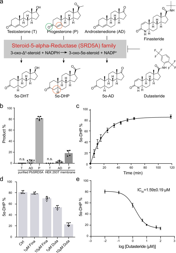
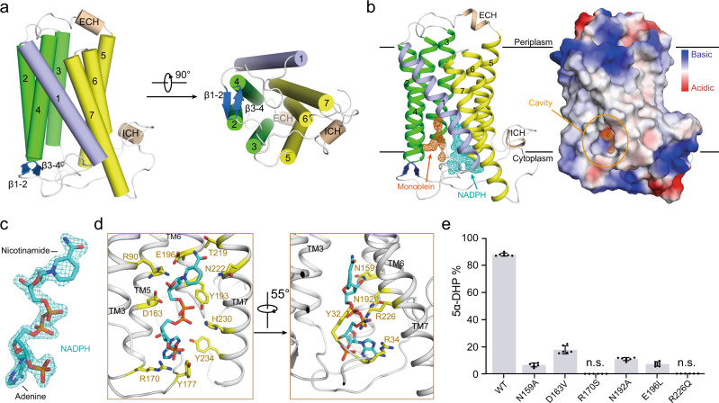
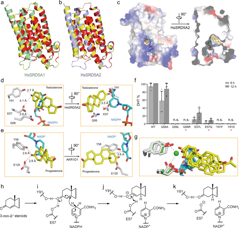
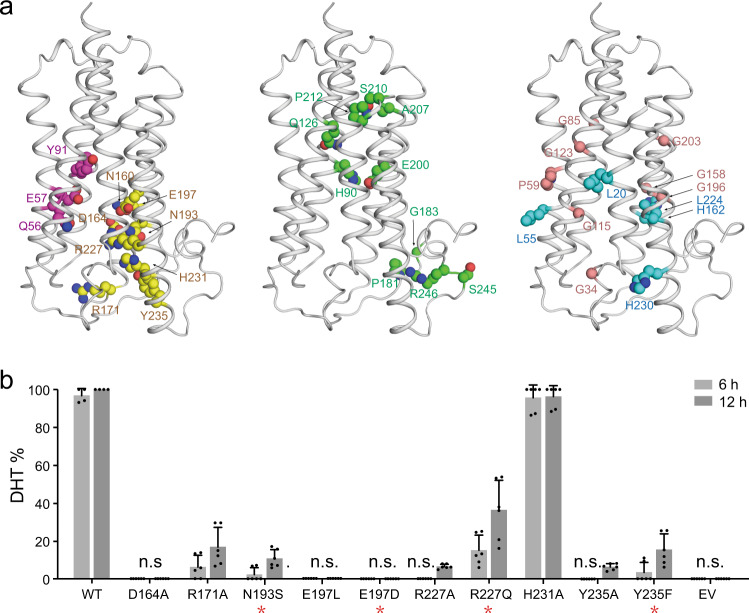
Steroid hormones are essential in stress response, immune system regulation, and reproduction in mammals. Steroids with 3-oxo-Δ4 structure, such as testosterone or progesterone, are catalyzed by steroid 5α-reductases (SRD5As) to generate their corresponding 3-oxo-5α steroids, which are essential for multiple physiological and pathological processes. SRD5A2 is already a target of clinically relevant drugs. However, the detailed mechanism of SRD5A-mediated reduction remains elusive. Here we report the crystal structure of PbSRD5A from Proteobacteria bacterium, a homolog of both SRD5A1 and SRD5A2, in complex with the cofactor NADPH at 2.0 Å resolution. PbSRD5A exists as a monomer comprised of seven transmembrane segments (TMs). The TM1-4 enclose a hydrophobic substrate binding cavity, whereas TM5-7 coordinate cofactor NADPH through extensive hydrogen bonds network. Homology-based structural models of HsSRD5A1 and -2, together with biochemical characterization, define the substrate binding pocket of SRD5As, explain the properties of disease-related mutants and provide an important framework for further understanding of the mechanism of NADPH mediated steroids 3-oxo-Δ4 reduction. Based on these analyses, the design of therapeutic molecules targeting SRD5As with improved specificity and therapeutic efficacy would be possible.


Conflict of interest statement

The authors declare no competing interests.

Figures

Fig. 1
Fig. 1. Steroid 5α-reductase activity of PbSRD5A.
a Steroid 5α-reductase family proteins can reduce Δ4 carbon–carbon double bound to form α configuration at C5 of products. These reactions can be inhibited by finasteride and dutasteride specifically. b Testosterone (T), 4-androstene-3,17-dione (AD), and progesterone (P) metabolism in vitro and in HEK 293 T cell. Purified PbSRD5A and 293 T cells transiently transfected with PbSRD5A were treated with [3H]-labeled T, AD, and P for 1 h and 12 h, respectively. n.s. means no signal. c Time curve of reduction reaction from progesterone to 5α-DHP by PbSRD5A. d Inhibition of PbSRD5A by finasteride (Fina) and dutasteride (Duta) in vitro, both with the concentration of 1 μM and 10 μM, respectively. e Dutasteride inhibits the activity of PbSRD5A in vitro, with an IC50 value of 1.59 ± 0.19 μM (logIC50 = 0.20 ± 0.05). be The percentages of products were measured using high-performance liquid chromatography (HPLC) with detected radioactivity. Data are mean±s.d. derived from technically independent experiments in duplicate. Each experiment was reproduced three times on separate occasions with similar results.
Fig. 2
Fig. 2. The crystal structure of PbSRD5A and coordination of NADPH.
a The overall structure of PbSRD5A. Two perpendicular views are shown. The seven transmembrane segments are divided into TM1 (lightblue), TM2-4 (green), and TM5-7 (yellow). The extracellular and intracellular short alpha helices (ECH and ICH) are colored wheat. The short antiparallel beta strands are colored blue. The loop regions are colored white. b 2Fo-Fc map for monoolein (orange mesh) and NADPH (cyan mesh) are both contoured at 1.0σ. The two black lines show the approximate location of the lipid bilayer. The cavity is circled in an electrostatic surface representation. c NADPH fits into the density map shown in b. Nicotinamide and adenine groups are labeled. d Coordination of NADPH by polar residues of PbSRD5A. The residues that are hydrogen-bonded to NADPH are shown in yellow sticks. e Functional validation of residues coordinating NADPH in PbSRD5A. Enzyme activities were measured by the percentage of 5α-DHP reduced from [3H]-labeled progesterone in vitro within 1 h detected by HPLC. Data are mean±s.d. derived from technically independent experiments in duplicate. Each experiment was reproduced three times on separate occasions with similar results.
Fig. 3
Fig. 3. HsSRD5A1 and -2 modeling, substrate docking, and functional characterization.
a HsSRD5A1 homology model was generated on the basis of PbSRD5A structure and represented in palegreen. Invariant and conserved residues are colored in red and yellow, respectively. b HsSRD5A2 homology model was generated on the basis of PbSRD5A structure and represented in lightblue. c The docking pose of testosterone in HsSRD5A2 structural model. The semi-transparent electrostatic surface of HsSRD5A2 is shown. Testosterone is shown as yellow stick. NADPH is colored cyan. d The coordination of conserved Q-E-Y motif with testosterone in HsSRD5A2 docking model. Two perpendicular views are shown. e The coordination of conserved Q-E-Y motif in AKR1D1-progesterone complex structure. f Biochemical characterization of the Q-E-Y motif of HsSRD5A2. All variants were transient expressed in HEK293T cells for 24 h. The cells were treated with [3H]-labeled T for 6 or 12 h. The collected and homogenized cell membranes were treated with [3H]-labeled T. HsSRD5A2 activities were measured by the percentage of DHT detected by HPLC. Data are mean ± s.d. derived from technically independent experiments in duplicate. Each experiment was reproduced three times on separate occasions with similar results. Red stars (*) indicate the variants in patients. g The water molecule, coordinated by the Q-E-Y motif in PbSRD5A structure, was shown in green sphere. The water molecule occupied the space for the C-3 carbonyl oxygen in the docking results shown in Fig. 3d and Supplementary Fig. 7d, f. h Key structural features of 3-oxo-Δ4 steroids. i The tyrosine (Y91 in HsSRD5A2) and glutamate (E57 in HsSRD5A2) may form hydrogen bonds to the carbonyl oxygen of substrates. Y91, as a super acidic hydrogen bond donor, helps to activate α, β-unsaturated ketone moiety of substrates. j The proposed mechanism of reduction involves a two-step reduction by the enzyme and NADPH. In the first step, NADPH coordinates on α face of the substrate and adds a hydride to C5, leading to a selective reduction at C5 to form an enolate ion intermediate. k Second, the resonance-stabilized enolate ion is protonated by E57 at C4 and thus releasing a saturated ketone product and an NADP+.
Fig. 4
Fig. 4. Disease-related loss-of-function mutations in HsSRD5A2 model.
a 32 disease-related loss-of-function mutations were classified into five categories. Catalytic site residues are in magenta, NADPH-binding residues are in yellow, structural destabilizing mutations are in green, wheat, or cyan based on residue properties. b Biochemical analysis of disease-related HsSRD5A2 variants. All variants were transient expressed in HEK293T cells for 24 h. The cells were treated with [3H]-labeled T for 6 or 12 h. The collected and homogenized cell membranes were treated with [3H]-labeled T. HsSRD5A2 activities were measured by the percentage of DHT detected by HPLC. Data are mean±s.d. derived from technically independent experiments in duplicate. Each experiment was reproduced three times on separate occasions with similar results. Red stars (*) below variant labels indicate the variants in patients.

Similar articles

Cited by

References

    1. Miller WL, Auchus RJ. The molecular biology, biochemistry, and physiology of human steroidogenesis and its disorders. Endocr. Rev. 2011;32:81–151. doi: 10.1210/er.2010-0013. - DOI - PMC - PubMed
    1. Payne AH, Hales DB. Overview of steroidogenic enzymes in the pathway from cholesterol to active steroid hormones. Endocr. Rev. 2004;25:947–970. doi: 10.1210/er.2003-0030. - DOI - PubMed
    1. Sakiani S, Olsen NJ, Kovacs WJ. Gonadal steroids and humoral immunity. Nat. Rev. Endocrinol. 2013;9:56–62. doi: 10.1038/nrendo.2012.206. - DOI - PubMed
    1. Belelli D, Lambert JJ. Neurosteroids: endogenous regulators of the GABA(A) receptor. Nat. Rev. Neurosci. 2005;6:565–575. doi: 10.1038/nrn1703. - DOI - PubMed
    1. Schiffer L, et al. Human steroid biosynthesis, metabolism and excretion are differentially reflected by serum and urine steroid metabolomes: a comprehensive review. J. Steroid Biochem Mol. Biol. 2019;194:105439. doi: 10.1016/j.jsbmb.2019.105439. - DOI - PMC - PubMed

Publication types

MeSH terms

LinkOut - more resources