. 2021 Jan 19;11(1):1807.
doi: 10.1038/s41598-020-80536-2.

Lactobacillus rhamnosus attenuates bone loss and maintains bone health by skewing Treg-Th17 cell balance in Ovx mice

Affiliations

Lactobacillus rhamnosus attenuates bone loss and maintains bone health by skewing Treg-Th17 cell balance in Ovx mice

Leena Sapra et al. Sci Rep. .

Abstract

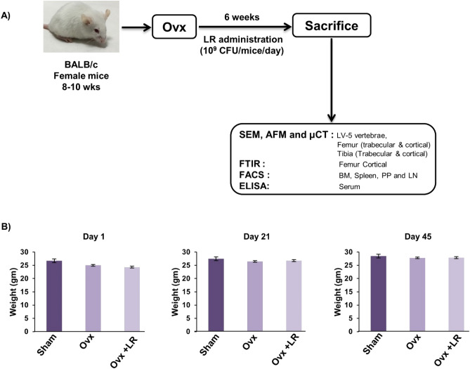
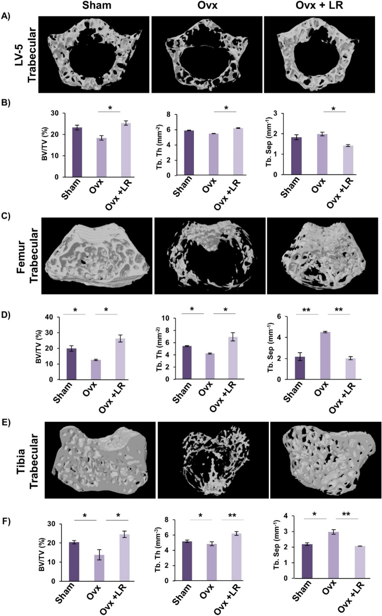
Osteoporosis is a systemic-skeletal disorder characterized by enhanced fragility of bones leading to increased rates of fractures and morbidity in large number of populations. Probiotics are known to be involved in management of various-inflammatory diseases including osteoporosis. But no study till date had delineated the immunomodulatory potential of Lactobacillus rhamnosus (LR) in bone-health. In the present study, we examined the effect of probiotic-LR on bone-health in ovariectomy (Ovx) induced postmenopausal mice model. In the present study, we for the first time report that LR inhibits osteoclastogenesis and modulates differentiation of Treg-Th17 cells under in vitro conditions. We further observed that LR attenuates bone loss under in vivo conditions in Ovx mice. Both the cortical and trabecular bone-content of Ovx+LR treated group was significantly higher than Ovx-group. Remarkably, the percentage of osteoclastogenic CD4+Rorγt+Th17 cells at distinct immunological sites such as BM, spleen, LN and PP were significantly reduced, whereas the percentage of anti-osteoclastogenic CD4+Foxp3+Tregs and CD8+Foxp3+Tregs were significantly enhanced in LR-treated group thereby resulting in inhibition of bone loss. The osteoprotective role of LR was further supported by serum cytokine data with a significant reduction in osteoclastogenic cytokines (IL-6, IL-17 and TNF-α) along with enhancement in anti-osteoclastogenic cytokines (IL-4, IL-10, IFN-γ) in LR treated-group. Altogether, the present study for the first time establishes the osteoprotective role of LR on bone health, thus highlighting the immunomodulatory potential of LR in the treatment and management of various bone related diseases including osteoporosis.


Conflict of interest statement

The authors declare no competing interests.

Figures

Figure 1
Figure 1
LR-CM inhibits osteoclastogenesis in a dose dependent manner: Osteoclasts differentiation was induced in Bone Marrow Macrophages (BMMs) with M-CSF (30 ng/ml) and RANKL (100 ng/ml) with or without Lactobacillus rhamnosus—conditioned media (LR-CM) at different ratios of 1:10 and 1:1 for 6 days. Giant multinucleated cells were stained with TRAP and cells with ≥ 3 nuclei were considered as mature osteoclasts. (A) Photomicrographs at different magnifications (10×, 20× and 40×) were taken. (B) Number of TRAP positive cells. (C) Number of TRAP positive cells with more than 3 nuclei. (D) Area of osteoclasts. The above images are indicative of one independent experiment and similar results were obtained in three different independent experiments. Statistical significance was considered as p ≤ 0.05 (*p ≤ 0.05, **p ≤ 0.01, ***p ≤ 0.001) with respect to indicated groups.
Figure 2
Figure 2
LR-CM inhibits RANKL stimulated F-actin ring formation: Bone Marrow Macrophages (BMMs) were treated with M-CSF (30 ng/ml) and RANKL (100 ng/ml) with or without Lactobacillus rhamnosus—Conditioned Media (LR-CM) at different ratios of 1:10 and 1:1 for 6 days. F-actin and nuclei were stained with FITC-conjugated phalloidin and DAPI respectively. Images were captured in fluorescence microscope (Imager.Z2 Zeiss microscope) at 10× magnification. (B) Number of F-actin rings. (C) Number of nuclei per osteoclasts. (D) Area of F-actin rings. The above images are indicative of one independent experiment and similar results were obtained in three different independent experiments. Statistical significance was considered as p ≤ 0.05 (*p ≤ 0.05, **p ≤ 0.01, ***p ≤ 0.001) with respect to indicated groups.
Figure 3
Figure 3
Experimental work plan for in vivo experiments. (A) Mice were divided into 3 groups. viz. Sham group, Ovx and Ovx+LR received LR. At the end of experiment (D45), mice were sacrificed and further studies performed (Mouse Image courtesy: Leena Sapra). (B) Effect of Lactobacillus rhamnosus (LR) on body weight; values are reported as mean ± SEM (n = 6/gp).
Figure 4
Figure 4
LR attenuates bone loss in Ovx mice. Mice were sacrificed at the end of experiment and cortical bones of all groups were collected for SEM and AFM analysis. (A) 2D SEM images. (B) 2D MATLAB analysis of SEM images. (C) 3D AFM images. (D) 2D MATLAB analysis of AFM image. The above images are indicative of one independent experiment and comparable results were obtained in two different independent experiments with n = 6 mice/group/experiment.
Figure 5
Figure 5
LR administration enhances trabecular bone microarchitecture. 3-D µCT reconstruction of LV-5 Trabecular, Femur Trabecular and Tibia Trabecular of all groups. (A) Bone micro-architecture of LV-5. (B) Histomorphometric parameters of LV-5. (C) Bone micro-architecture of femur trabecular. (D) Histomorphometric parameters of femur trabecular. (E) Bone micro-architecture of Tibia trabecular. (F) Histomorphometric parameters of Tibia trabecular. Bone volume/tissue volume ratio (BV/TV); Tb. Th., trabecular thickness; Tb. Sp., trabecular separation. The results were evaluated by ANOVA with subsequent comparisons by Student t-test for paired or nonpaired data. Values are reported as mean ± SEM. The above graphical representations are indicative of one independent experiment and similar results were obtained in two different independent experiments with n = 6. Statistical significance was considered as p ≤ 0.05 (*p ≤ 0.05, **p ≤ 0.01, ***p ≤ 0.001) with respect to indicated mice groups.
Figure 6
Figure 6
LR administration enhances cortical bone microarchitecture. 3-D u-CT reconstruction of Femur Cortical and Tibia Cortical of all groups. (A) Bone micro-architecture of Femur Cortical. (B) Histomorphometric parameters of Femur Cortical. (C) Bone micro-architecture of Tibia Cortical. (D) Histomorphometric parameters of Tibia Cortical. T. Ar., bone tissue area; T. Pm., total cross-sectional perimeter; Ct. Po., cortical porosity. The results were evaluated by ANOVA with subsequent comparisons by Student t-test for paired or nonpaired data. Values are reported as mean ± SEM. The above graphical representations are indicative of one independent experiment and similar results were obtained in two different independent experiments with n = 6. Statistical significance was considered as p ≤ 0.05 (*p ≤ 0.05, **p ≤ 0.01, ***p ≤ 0.001) with respect to indicated mice groups.
Figure 7
Figure 7
LR administration enhances bone mineral density and heterogeneity of bones. (A) Graphical representation of BMD of LV-5, trabecular and cortical regions of femur and tibial bones of all groups. (B) Graphical representation of compositional changes in bones as detected by FTIR (bone mineral/organic matrix ratio (m/m), crystallinity (XST) and carbonate to phosphate ratio (C/P). Data are reported as mean ± SEM. Similar results were obtained in two independent experiments with n = 6. Statistical significance of each parameter was assessed by ANOVA followed by paired group comparison. *p < 0.05, **p < 0.01, ***p < 0.001 compared with indicated groups.
Figure 8
Figure 8
LR intake modulates Treg-Th17 cell balance in vivo. Cells from BM, PP, spleen and LN of mice from Sham, Ovx and Ovx+LR groups were harvested and analysed by Flow cytometry for percentage of (A) CD4+Foxp3+Tregs. (B) CD8+Foxp3+Tregs. (C) CD4+Roryt+Th17 cells. (D) CD4+RANKL+T cells. Data are reported as Mean ± SEM. Similar results were obtained in two independent experiments with n = 6. Statistical significance of each parameter was assessed by ANOVA followed by paired group comparison. *p < 0.05, **p < 0.01, ***p < 0.001, ****p < 0.0001 compared with indicated groups.
Figure 9
Figure 9
LR modulates Treg and Th17 cell differentiation in vitro. (A) For Tregs differentiation, splenic naïve CD4+ T cells stimulated with anti-CD3 and anti-CD28 mAbs were incubated with TGF-β1 and IL-2 with or without LR-CM. After 4 days, cells were analysed for Foxp3 expression by FACS. (B) Average percentage of CD4+Foxp3+ cells from two independent experiments of A. (C) MFI showing the expression of Foxp3 in CD4+ T cells. (D) For Th17 differentiation, splenic naïve CD4+ T cells stimulated with anti-CD3 and anti-CD28 mAbs, incubated with TGF-β1, IL-6, IL-23 with or without LR-CM. (E) Average percentage of CD4+Roryt+ cells from three independent experiments of D. (F) MFI showing the expression of Roryt in CD4+ T cells. Data is reported as Mean ± SEM. Similar results were obtained in two independent experiments. MFI, mean fluorescence intensity. Statistical significance of each parameter was assessed by ANOVA followed by paired group comparison. *p < 0.05, **p < 0.01, ***p < 0.001, ****p < 0.0001 compared with indicated groups.
Figure 10
Figure 10
LR skews cytokines balance in Ovx mice. Osteoclastogenic and anti-osteoclastogenic cytokines were analysed in serum samples of mice by ELISA/CBA. The results were evaluated by using ANOVA with subsequent comparisons by Student t-test for paired or non-paired data, as appropriate. Values are expressed as mean ± SEM (n = 6) and similar results were obtained in two independent experiments. Statistical significance was defined as p ≤ 0.05, *p ≤ 0.05, **p < 0.01 ***p ≤ 0.001 with respect to indicated mice group.
Figure 11
Figure 11
Summary of our results: LR administration attenuates bone loss via inhibiting osteoclasts and modulating the Treg-Th17 cell balance under both in vitro and in vivo conditions. (Image illustrated using Medical Art https://smart.servier.com/).

Similar articles

Cited by

References

    1. Rachner, T.D., Khosla, S. F., Hofbauer, L.C. & Hofbauer, L.C. Osteoporosis: now and the future. The Lancet (2011). - PMC - PubMed
    1. Pisani P, et al. Major osteoporotic fragility fractures: Risk factor updates and societal impact. World J. Orthop. 2016;7:171–181. doi: 10.5312/wjo.v7.i3.171. - DOI - PMC - PubMed
    1. Srivastava RK, Dar HY, Mishra PK. Immunoporosis: Immunology of osteoporosis-role of T cells. Front. Immunol. 2018;9:657–657. doi: 10.3389/fimmu.2018.00657. - DOI - PMC - PubMed
    1. Dar HY, Lone Y, Koiri RK, Mishra PK, Srivastava RK. Microcystin-leucine arginine (MC-LR) induces bone loss and impairs bone micro-architecture by modulating host immunity in mice: Implications for bone health. Environ. Pollut. 2018;238:792–802. doi: 10.1016/j.envpol.2018.03.059. - DOI - PubMed
    1. Hadji P, et al. The epidemiology of osteoporosis–Bone evaluation study (BEST): An analysis of routine health insurance data. Deutsches Arzteblatt Int. 2013;110:52–57. - PMC - PubMed

Publication types