. 2021 Jan 19;11(1):1788.
doi: 10.1038/s41598-020-79892-w.

Mechanistic basis of breast cancer resistance protein inhibition by new indeno[1,2-b]indoles

Affiliations

Mechanistic basis of breast cancer resistance protein inhibition by new indeno[1,2-b]indoles

Diogo Henrique Kita et al. Sci Rep. .

Abstract

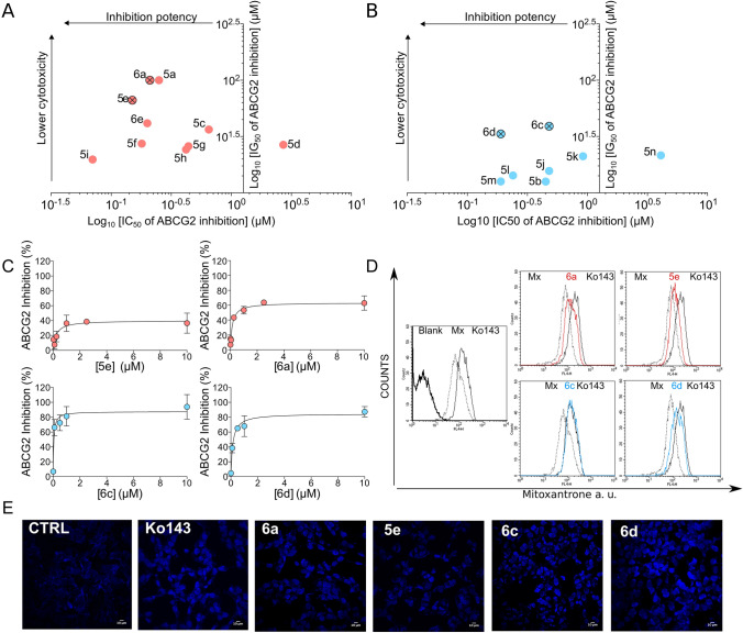
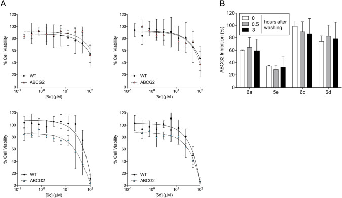
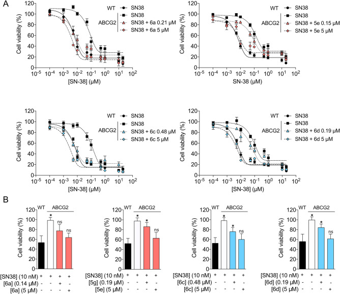
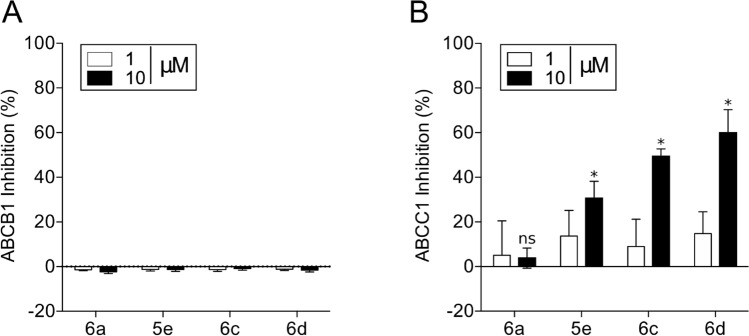
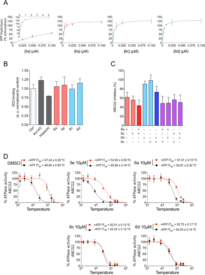
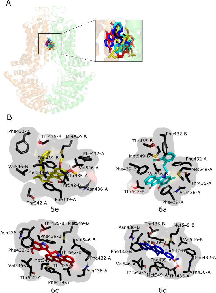
The ATP-binding cassette transporter ABCG2 mediates the efflux of several chemotherapeutic drugs, contributing to the development of multidrug resistance (MDR) in many cancers. The most promising strategy to overcome ABCG2-mediated MDR is the use of specific inhibitors. Despite many efforts, the identification of new potent and specific ABCG2 inhibitors remains urgent. In this study, a structural optimization of indeno[1,2-b]indole was performed and a new generation of 18 compounds was synthesized and tested as ABCG2 inhibitors. Most compounds showed ABCG2 inhibition with IC50 values below 0.5 µM. The ratio between cytotoxicity (IG50) and ABCG2 inhibition potency (IC50) was used to identify the best inhibitors. In addition, it was observed that some indeno[1,2-b]indole derivatives produced complete inhibition, while others only partially inhibited the transport function of ABCG2. All indeno[1,2-b]indole derivatives are not transported by ABCG2, and even the partial inhibitors are able to fully chemosensitize cancer cells overexpressing ABCG2. The high affinity of these indeno[1,2-b]indole derivatives was confirmed by the strong stimulatory effect on ABCG2 ATPase activity. These compounds did not affect the binding of conformation-sensitive antibody 5D3 binding, but stabilized the protein structure, as revealed by the thermostabilization assay. Finally, a docking study showed the indeno[1,2-b]indole derivatives share the same binding site as the substrate estrone-3-sulfate.


Conflict of interest statement

The authors declare no competing interests.

Figures

Figure 1
Figure 1
Structures of all studied ketonic and phenolic indeno[1,2-b]indole derivatives (compounds p4h, p4j, p4k and p5h were already published, ).
Figure 2
Figure 2
Inhibition potency and cytotoxicity of compounds with the best therapeutic ratios. (A) Partial inhibitors (red) and (B) complete inhibitors (blue). (C) Representative IC50 curves of the partial and complete inhibitors. (D) Representative flow cytometry histograms of mitoxantrone accumulation in HEK293-ABCG2 cells. Overlay of histograms obtained by compounds at 10 µM mitoxantrone and Ko143. (E) Representative confocal microscopy images of Hoechst 33342 (1 µM) accumulation in HEK293-ABCG2 cells, using the program ImageJ2 (URL: https://imagej.net/Fiji). Effect produced by compounds at 10 µM compared to the reference inhibitor Ko143 at 0.5 µM.
Figure 3
Figure 3
Cytotoxicity of inhibitors and washout assay. Cell viability was determined by MTT assay. (A) Cell viability of HEK293-ABCG2 cells and HEK293 (wild-type) control cells upon 72 h treatment with inhibitors at increasing concentrations (0.1–100 µM), as indicated. (B) Washout assay performed on HEK293-ABCG2 cells with the compounds at 10 µM. The white bars represent the classical experiment (30 min of concomitant incubation with compounds and mitoxantrone before analysis), grey and black bars represent pre-treatment with compounds for 30 min followed by washing procedure at 0.5 and 3 h, as described in methods. The data are the mean ± SD of three independent experiments performed in duplicate.
Figure 4
Figure 4
Sensitization of transfected and cancer cells overexpressing ABCG2 to SN-38. Cell viability was determined by MTT assay. (A) Cell viability of HEK293-ABCG2 and HEK293 (wild-type) control cells upon 72 h treatment with SN-38 at increasing concentrations, as indicated, and HEK293-ABCG2 cells upon co-treatment with SN-38 and inhibitors at either IC50 values or 5 µM. (B) Cell viability of H460MX20 and H460 control cells upon 72 h treatment with SN-38 at 10 nM, as indicated, and H460MX20 cells upon co-treatment with SN-38 and inhibitors at either IC50 values or 5 µM. The data are the mean ± SEM of three independent experiments performed in triplicate and compared using the Student's t test (2-sided) for independent samples. *p < 0.05 were considered significant for all tests.
Figure 5
Figure 5
Selectivity of indeno[1,2-b]indole derivatives toward ABCG2. (A, B) Effect of compounds at 1 and 10 µM on ABCB1 (P-gp) for rhodamine 123 transport inhibition and on ABCC1 (MRP1) for calcein-AM transport inhibition. The data are the mean ± SD of three independent experiments performed in duplicate and compared using the Student's t test (2-sided) for independent samples. *p < 0.05 were considered significant for all tests.
Figure 6
Figure 6
Studies on ABCG2 ATPase activity, binding of conformational antibody 5D3, biomodulation assay and thermostabilization assay. (A) Effect of compounds at increasing concentrations (0.001–5 µM) on basal ABCG2 ATPase activity. (B) Effect of compounds at 10 µM on the binding of conformational antibody 5D3. (C) Bimodulation assay using partial and complete inhibitors at 10 µM on ABCG2 transport activity using mitoxantrone as a substrate. (D) Thermostabilization assay with partial and complete inhibitors at saturating concentration of 10 µM. The data are the mean ± SD of three independent experiments performed in duplicate.
Figure 7
Figure 7
Docking analysis in human ABCG2 (PDB 6HCO). (A) Representative docking poses of the inhibitors overlapping the binding site of estrone-3-sulfate (E3S – black, 6c—red, 6d—blue, 6a—cyan and 5e—yellow), in stick representation. The two monomers are colored differently, orange (chain A) and green (chain B). (B) Interacting residues on the best energy score pose of the inhibitors are represented as stick models. The images were generated using the program PyMOL Molecular Graphics System, Version 1.2r3pre, Schrödinger, LLC (URL: https://pymol.org/2/).

References

    1. Gottesman MM, Lavi O, Hall MD, Gillet J-P. Toward a better understanding of the complexity of cancer drug resistance. Annu. Rev. Pharmacol. Toxicol. 2016;56:85–102. doi: 10.1146/annurev-pharmtox-010715-103111. - DOI - PubMed
    1. Robey RW, et al. Revisiting the role of ABC transporters in multidrug-resistant cancer. Nat. Rev. Cancer. 2018;18:452–464. doi: 10.1038/s41568-018-0005-8. - DOI - PMC - PubMed
    1. Szakács G, Váradi A, Özvegy-Laczka C, Sarkadi B. The role of ABC transporters in drug absorption, distribution, metabolism, excretion and toxicity (ADME-Tox) Drug Discov. Today. 2008;13:379–393. doi: 10.1016/j.drudis.2007.12.010. - DOI - PubMed
    1. Aller SG, et al. Structure of P-glycoprotein reveals a molecular basis for poly-specific drug binding. Science. 2009;323:1718–1722. doi: 10.1126/science.1168750. - DOI - PMC - PubMed
    1. Fletcher JI, Haber M, Henderson MJ, Norris MD. ABC transporters in cancer: more than just drug efflux pumps. Nat. Rev. Cancer. 2010;10:147–156. doi: 10.1038/nrc2789. - DOI - PubMed

Publication types

MeSH terms