. 2021 Jun;15(6):1782-1793.
doi: 10.1038/s41396-020-00886-7. Epub 2021 Jan 19.

Fungal foraging behaviour and hyphal space exploration in micro-structured Soil Chips

Affiliations

Fungal foraging behaviour and hyphal space exploration in micro-structured Soil Chips

Kristin Aleklett et al. ISME J. 2021 Jun.

Abstract

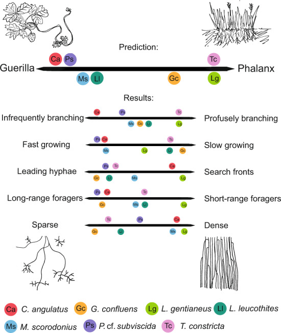
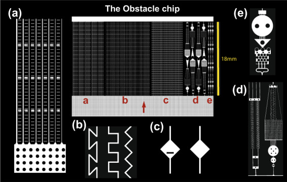
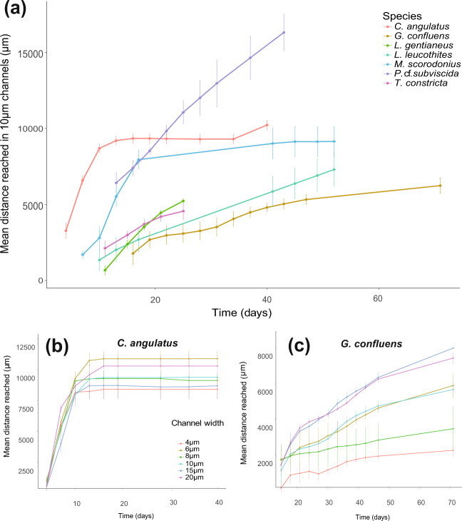
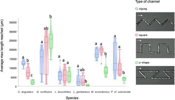
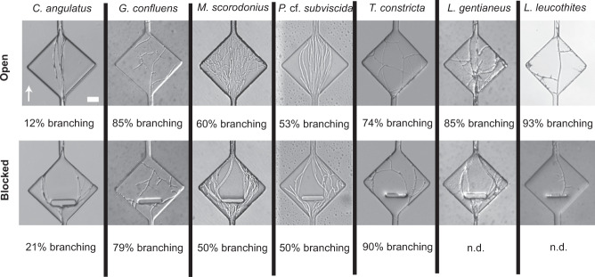
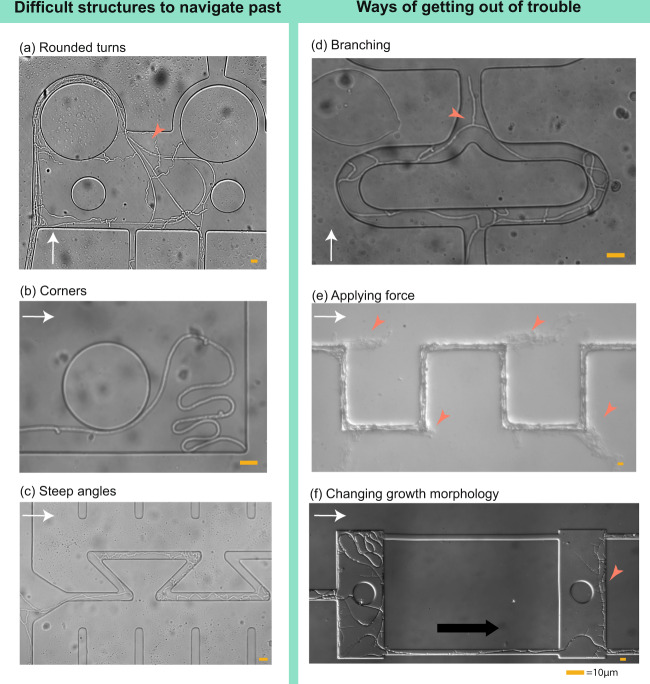
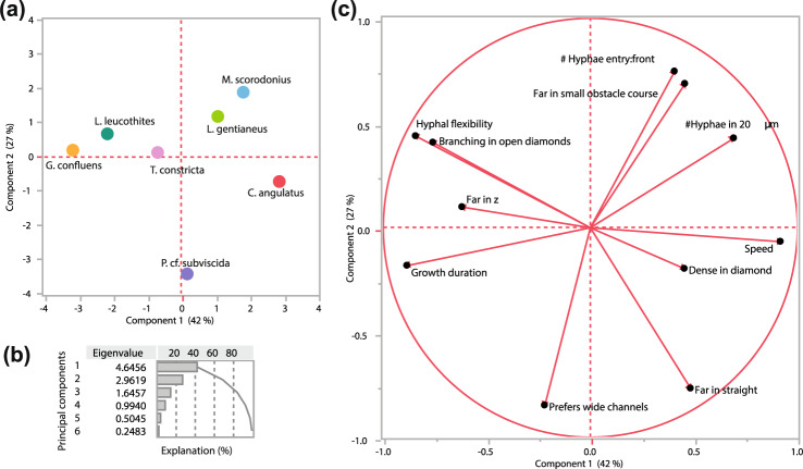
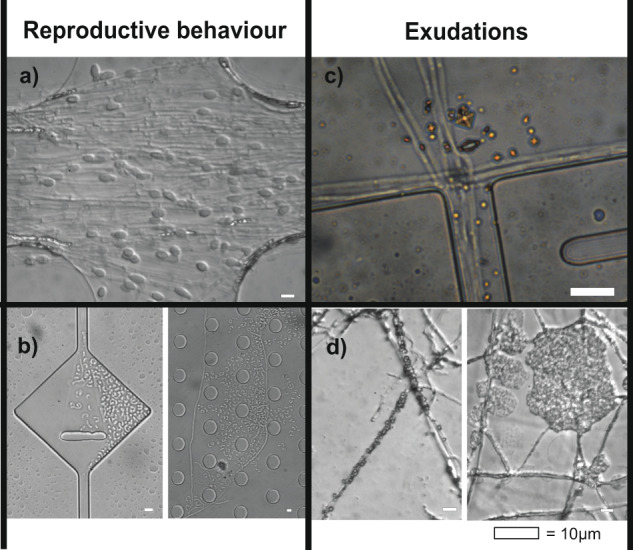
How do fungi navigate through the complex microscopic maze-like structures found in the soil? Fungal behaviour, especially at the hyphal scale, is largely unknown and challenging to study in natural habitats such as the opaque soil matrix. We monitored hyphal growth behaviour and strategies of seven Basidiomycete litter decomposing species in a micro-fabricated "Soil Chip" system that simulates principal aspects of the soil pore space and its micro-spatial heterogeneity. The hyphae were faced with micrometre constrictions, sharp turns and protruding obstacles, and the species examined were found to have profoundly different responses in terms of foraging range and persistence, spatial exploration and ability to pass obstacles. Hyphal behaviour was not predictable solely based on ecological assumptions, and our results obtained a level of trait information at the hyphal scale that cannot be fully explained using classical concepts of space exploration and exploitation such as the phalanx/guerrilla strategies. Instead, we propose a multivariate trait analysis, acknowledging the complex trade-offs and microscale strategies that fungal mycelia exhibit. Our results provide novel insights about hyphal behaviour, as well as an additional understanding of fungal habitat colonisation, their foraging strategies and niche partitioning in the soil environment.


Conflict of interest statement

The authors declare that they have no conflict of interest.

Figures

Fig. 1
Fig. 1. Summary of hyphal growth behaviour in the context of the ecological concept of phalanx- and guerrilla-type foraging.
This concept originates from plant ecology and is used to describe growth patterns of clonal plants, where Fragaria (strawberry; guerrilla, left) and Festuca (phalanx, right) are typical examples of the concept. Guerrilla-type foraging in fungi has been defined by infrequent branching, fast growth, leading hyphae, long-range foraging and sparse growth (listed to the left), whereas phalanx-type foraging has been defined by the opposite set of characteristics: frequent branching, slow growth, a front of hyphae advancing in synchrony, short-range foraging and dense growth [15, 17]. The seven examined species were hypothetically placed along the continuum of the phalanx and guerrilla division based on assumptions associated with what type of litter the different species have been found to grow on (Table S1) (above), and further placed along the continuums of their defining trait components based on the results of this study (below). Based on our results, none of the examined species fell clearly into one of the categories, or even at a comparable location within the continuum for all trait axes. Instead, we saw that species could be typical guerrilla for one trait and typical phalanx for another.
Fig. 2
Fig. 2. Schematic overview of the design of the Obstacle Chip.
The chip design is shown in the centre, surrounded by enlarged details from the different experiments (ae). The red arrow shows the hyphal growth direction. a Parallel straight channels in a series of different widths (20, 15, 10, 8, 6, 4 µm; n = 6) with each width repeated five times within the chip. Rulers were incorporated between the channels to measure how far the hyphae reached under microscope. b Channels of 10 µm width angled in a zigzag pattern with 90° corners, meandering square pattern with 90° corners or a z-shaped pattern with 135° corners, organised in a randomised order, n = 11. c Channels of 10 µm width with the repeated occurrence of 140-µm-diameter diamond-shaped openings that either was free for passage, included a 50-µm-wide and 10-µm-thick obstacle blocking the straight passage of the fungi, or a random occurrence of open and blocked openings in the same channel in randomised order, n = 12. d Larger obstacle courses with a combination of challenging structures for the fungi to navigate through. e Smaller-sized obstacle courses with more frequent repetition of obstacles.
Fig. 3
Fig. 3. Hyphal growth rates by the fungal species over time—measured in the section of the chip with straight channels of different widths (Fig. 2a).
a Comparison of the mean growth rate of the different species in colonised 10-µm-wide straight channels, n = 6. b Comparison of mean growth rates in straight channels of different width (20, 15, 10, 8, 6 and 4 µm) for C. angulatus, which did not show a significant difference in advancement between different widths (p = 0.52), c similar comparison for G. confluens that advanced significantly shorter distances in narrower channels (p < 0.0001). Error bars show ±1 SEM.
Fig. 4
Fig. 4. Distances reached by the fungal species within the differently angled channels (Fig. 2b).
Box plots show the mean final distances reached by the hyphae in channels of the same type (n = 11). Different letters indicate a significant difference of colonisation distances for each species according to ANOVA followed by Tukey-Kramer’s HSD test at p < 0.05 (Table S3). The legend contains examples of the differently angled channels colonised by P. cf. subviscida, where arrows indicate growth directions.
Fig. 5
Fig. 5. Hyphal growth responses to sudden channel openings (with or without a perpendicular obstacle) (Fig. 2c) in the different species examined.
The level of branching was recorded for colonised openings in each chip and listed as a percentage for each type of widening (open and blocked). If the opening was colonised by multiple hyphae, the occurrence of one branching hypha was enough to record it as ‘branching’. The percentage of branching was calculated per number of colonised openings. Percentage of branching could not be determined (n.d.) for blocked openings of L. gentianeus and L. leucothites since their hyphae frequently grew beneath the blockages. The arrow indicating growth direction and the scale bar of 20 µm apply to all images.
Fig. 6
Fig. 6. Fungal navigation in complex structures.
Certain structures in the chips proved notoriously difficult for fungal hyphae to navigate past (ac), and different growth strategies were applied by the fungi to increasing their foraging range past these obstacles (df). a Rounded turns confused the growth direction of hyphae (here P. cf. subviscidae) and led them to grow back towards their origin (red arrowhead). b Corner-trapped hyphae (here G. confluens). The tip was not able to navigate out of the corner, and the hyphae instead elongated behind the tip in a folding manner. c Sharp angles restricted hyphal advancement of many species (as seen in Fig. 3, here P. cf. subviscidae) and in some cases led hyphae to turn back towards their original growth direction. d Branching increased the likelihood that a newly formed tip found the passage for progression (red arrowhead). In that case, the apex switched to this tip (here P. cf. subviscidae). e Hyphae hitting solid chip parts could apply tip force. Some species (here L. gentianeus) were able to break through the bonding of the chip and continue to grow between the PDMS and the glass slide in its original direction (red arrowheads), instead of following the maze pattern. f G. confluens altered its growth morphology from thicker, flexible runner hyphae (young growth morphology, left red arrowhead) to thinner hyphae with frequent lateral short branches (old growth morphology, right red arrowhead). The white arrows indicate growth direction.
Fig. 7
Fig. 7. Principal component analysis (PCA) of the measured traits for the seven fungal species investigated (Table S2).
a Score plot for the seven species along two dimensions of PC1 and PC2. b Explanation of the variation. Cumulatively explained variation of PC1 and PC2 is 69.4%. c Loading plot of the variables included in the PC analysis. Dashed lines connect the different locations of the loading variables for distance growth in different spatial structures. Details on the measurements can be found in Table S2.
Fig. 8
Fig. 8. Observations of reproductive behaviour and exudations inside the Obstacle Chips.
a L. gentianeus forming chlamydospores in the entrance system of the chip. Chlamydospores have previously been documented in L. gentianeus [49]. b P. cf. subviscida forming monokaryotic arthroconidia from dikaryotic hyphae. P. cf. subviscida has previously been shown to produce monokaryotic arthroconidia [50, 51]. c P. cf. subviscida exudates forming copper coloured crystals (likely oxalates [52]). d L. leucothites exudates forming crystals that cluster around the hyphae.

References

    1. Ferguson BA, Dreisbach TA, Parks CG, Filip GM, Schmitt CL. Coarse-scale population structure of pathogenic Armillaria species in a mixed-conifer forest in the Blue Mountains of northeast Oregon. Can J Res. 2003;33:612–23.
    1. Fricker MD, Heaton LLM, Jones NS, Boddy L. The mycelium as a network. Microbiol Spectr. 2017;5:1–32. - PMC - PubMed
    1. Treseder KK, Lennon JT. Fungal traits that drive ecosystem dynamics on land. Microbiol Mol Biol Rev. 2015;79:243–62. - PMC - PubMed
    1. Smith SE, Read DJ. Mycorrhizal symbiosis. 3rd ed. London, UK: Academic Press; 2008.
    1. Maron JL, Marler M, Klironomos JN, Cleveland CC. Soil fungal pathogens and the relationship between plant diversity and productivity. Ecol Lett. 2011;14:36–41. - PubMed

Publication types