Review
. 2021 May;18(5):261-279.
doi: 10.1038/s41571-020-00459-9. Epub 2021 Jan 19.

Targeting cancer-promoting inflammation - have anti-inflammatory therapies come of age?

Affiliations
Review

Targeting cancer-promoting inflammation - have anti-inflammatory therapies come of age?

Jiajie Hou et al. Nat Rev Clin Oncol. 2021 May.

Abstract

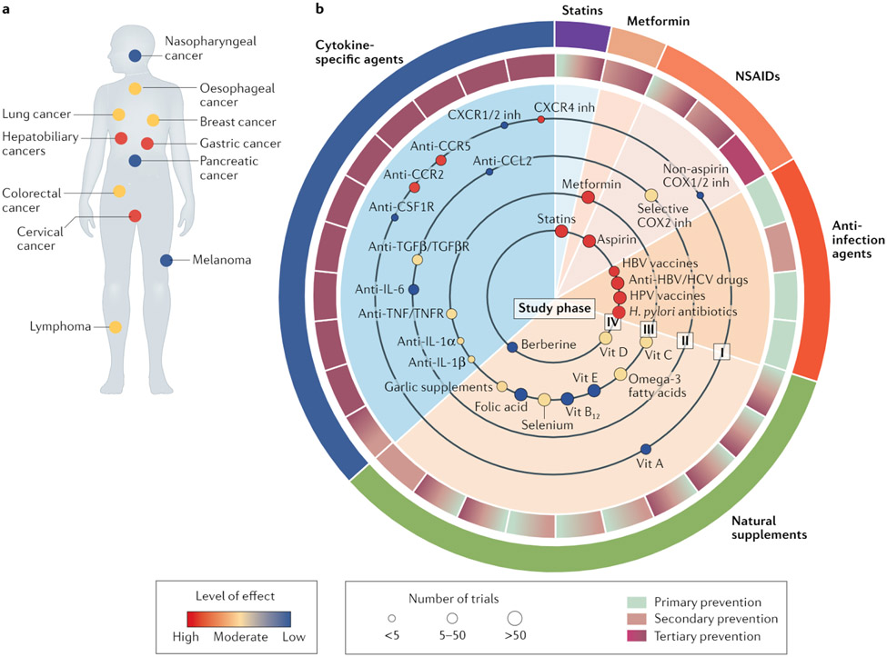
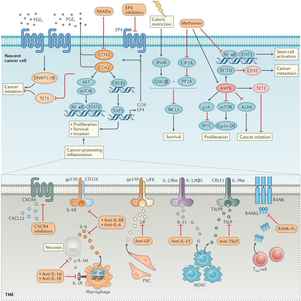
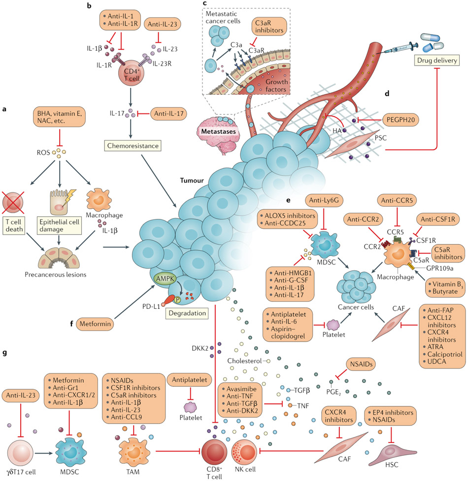
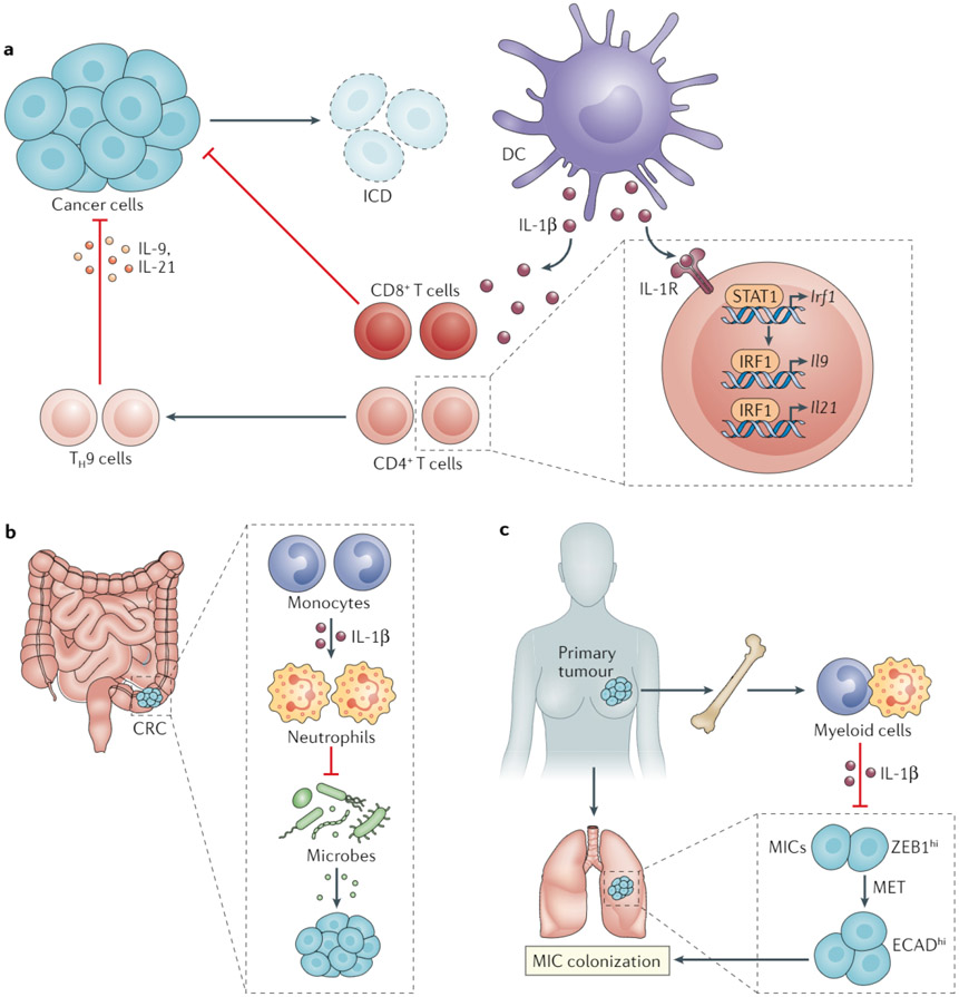
The immune system has crucial roles in cancer development and treatment. Whereas adaptive immunity can prevent or constrain cancer through immunosurveillance, innate immunity and inflammation often promote tumorigenesis and malignant progression of nascent cancer. The past decade has witnessed the translation of knowledge derived from preclinical studies of antitumour immunity into clinically effective, approved immunotherapies for cancer. By contrast, the successful implementation of treatments that target cancer-associated inflammation is still awaited. Anti-inflammatory agents have the potential to not only prevent or delay cancer onset but also to improve the efficacy of conventional therapeutics and next-generation immunotherapies. Herein, we review the current clinical advances and experimental findings supporting the utility of an anti-inflammatory approach to the treatment of solid malignancies. Gaining a better mechanistic understanding of the mode of action of anti-inflammatory agents and designing more effective treatment combinations would advance the clinical application of this therapeutic approach.


Figures

Fig. 1 ∣
Fig. 1 ∣. Evidence grading for anti-inflammatory agents in cancer prevention.
a ∣ Statistics of anti-inflammatory treatments assigned to patients with different cancer types. Each small circle represents a cancer type for which anti-inflammatory treatments have been examined. The colour of the circles reflects the level of effect defined based on the number of positive versus negative prospective trials registered in the ClinicalTrials.gov database. b ∣ Anti-inflammatory treatments using various anti-infective agents or modalities, nonsteroidal anti-inflammatory drugs (NSAIDs), statins, metformin, cytokine-specific drugs, and natural supplements are illustrated according to levels of evidence and efficacy in the management of cancer. The arcs in the outer circle surrounding the central pie chart indicate the various categories of therapeutic agents. The shading in each portion of the inner circle surrounding the pie chart represents the preventive applications in which each respective agent (or class of agent) shown within the body of the pie chart has been evaluated: primary prevention to deduce the aetiological role of the therapeutic target, secondary prevention to mitigate development of the disease in at-risk populations or tertiary prevention for the treatment of cancer following diagnosis. Each small circle in the inner sectors of the pie chart reflects the clinical evidence for each agent, indicating the level of effect (circle colour), the number of clinical studies performed (circle size) and the phase of clinical testing (as indicated by the ‘study phase’ designation on each large circle within the pie chart). Ongoing trials are summarized in Supplementary Table 1. CCR, CC-chemokine receptor; COX, cyclooxygenase; CSF1R, colony stimulating factor 1 receptor; CXCR, CXC-chemokine receptor; HBV, hepatitis B virus; HCV, hepatitis C virus; HPV, human papillomavirus; inh, inhibitor; TGFβ, transforming growth factor-β; TGFβR, TGFβ receptor; TNF, tumour necrosis factor; TNFR, TNF receptor; Vit, vitamin.
Fig. 2 ∣
Fig. 2 ∣. Impact of anti-inflammatory agents on oncogenic pathways.
Cyclooxygenase 1 (COX1) and COX2, which are the targets of nonsteroidal anti-inflammatory drugs (NSAIDs), can activate AKT, mTOR and NF-κB to support cancer cell survival and proliferation, either directly or via the production of prostaglandin E2(PGE2). PGE2 produced in a COX2-dependent manner can bind to the PGE2 receptor EP4 and induce intracellular signal transduction. Whereas COX2 functions to downregulate the expression of DNA demethylase TET1, EP4 signalling upregulates the expression of DNA (cytosine 5)-methyltransferase 1 and/or 3B (DNMT1/3B). The altered expression of both of these epigenetic regulators results in silencing of tumour suppressor genes and thus promotes cancer initiation, which could potentially be prevented through treatment with the NSAID COX2 inhibitor celecoxib. PGE2-EP4 signalling can also activate a positive feedforward loop for tumour initiation and promotion involving upregulation of the transcriptional coactivator YAP1, which in turn upregulates the expression of COX enzymes and EP4. NSAIDs or EP4 antagonists might effectively disrupt this inflammation-driven process. The anticancer effects of metformin largely depend on the activation of AMPK, which negatively regulates downstream signalling cascades involved in cancer initiation. In addition, AMPK activation confers a tumour-suppressive epigenome via the stabilization of TET2 and/or degradation of the histone-lysine N-methyltransferase EZH2. In an AMPK-independent fashion, metformin attenuates an NF-κB-mediated inflammatory response required for stem cell function and can activate a protein phosphatase 2A (PP2A) pathway to exert control over cell survival via inhibition of the antiapoptotic protein MCL1. The latter mechanism is otherwise ineffective for cancer prevention in mice without GSK3β activation through caloric restriction. Various cytokines present in the inflammatory tumour microenvironment (TME), including IL-6, IL-23, leukaemia inhibitory factor (LIF), receptor activator of nuclear factor-κB ligand (RANKL), thymic stromal lymphopoietin (TSLP) and CXC-chemokine ligand 12 (CXCL12), can induce pro-tumorigenic signals via cognate receptors expressed by cancer cells themselves. Therefore, inhibitory antibodies or other antagonists targeting these cytokines or their receptors might have anticancer effects. These cytokines are derived from distinct immune cells, including macrophages, myeloid-derived suppressor cells (MDSCs) and regulatory T (Treg) cells or stromal cells, such as pancreatic stellate cells (PSCs), in a context-dependent manner. For instance, IL-6 can be produced abundantly by macrophages in response to necrotic tumour cells and IL-1α. Hence, the blockade of IL-1α signalling might prevent downstream tumorigenic events. B56δ, PP2A B subunit isoform B56δ; CIP2A, cancerous inhibitor of protein phosphatase 2A; CREB1, cAMP-responsive element-binding protein 1; CXCR4, CXC-chemokine receptor 4; STAT3, signal transducer and activator of transcription 3.
Fig. 3 ∣
Fig. 3 ∣. Approaches to resolving cancer-associated inflammation and normalizing antitumour immunity.
a ∣ Reactive oxygen species (ROS) can cause epithelial cell damage, immune cell death, unresolved inflammation and subsequent precancerous lesions. ROS and their effects can be counteracted by antioxidants, such as butylated hydroxyanisole (BHA), vitamin E and N-acetylcysteine (NAC). b ∣ IL-17 has a pivotal role in inflammation-driven cancer initiation as well as in angiogenesis and chemotherapy resistance. IL-17 is generally produced by CD4+ T cells in response to IL-23 or IL-1β. Accordingly, antagonistic antibodies targeting IL-17, IL-23 or IL-1β receptor (IL-1R) have substantial therapeutic anticancer effects in mice-. c ∣ Leptomeningeal metastatic cells can secret complement component 3 (C3a), which activates the C3a receptor (C3aR) expressed on the choroid plexus epithelium and thereby enables circulating growth factors to enter the leptomeningeal space via disruption of the blood–brain barrier. C3aR antagonism might therefore interrupt the nutrition supply to metastatic cells in the cerebrospinal fluid. d ∣ The glycosaminoglycan hyaluronan (HA) is a central component of the extracellular matrix, fostering tissue stiffness and restricting drug perfusion. A PEGylated form of PH20 hyaluronidase (PEGPH20), which can degrade HA in the tumour microenvironment, is capable of enhancing chemotherapeutic efficacy. e ∣ Tumour-associated macrophages (TAMs), neutrophils or myeloid-derived suppressor cells (MDSCs), cancer-associated fibroblasts (CAFs) and platelets directly communicate with cancer cells and supply them with various pro-tumorigenic signals, which can be interrupted by targeting the corresponding inflammatory stimulants or their receptors. f ∣ Tumour cells often express the inhibitory immune-checkpoint protein PD-L1 to avoid elimination by CD8+ T cells; AMPK can catalyse the phosphorylation and thus promote the degradation of PD-L1, and this process can be activated by metformin. g ∣ Moreover, cholesterol, prostaglandin E2 (PGE2), tumour necrosis factor (TNF), transforming growth factor-β (TGFβ) and oncoproteins such as Dickkopf-related protein 2 (DKK2) present in the tumour-associated inflammatory milieu can directly suppress the function of CD8+ T cells and natural killer (NK) cells; therefore, the cholesterol acyltransferase inhibitor avasimibe, nonsteroidal anti-inflammatory drugs (NSAIDs), PGE2 receptor EP4 antagonists, and cytokine-specific antibodies or antagonists might reinforce the antitumour effects of these cytotoxic lymphocytes. TAMs, MDSCs, CAFs (or hepatic stellate cells (HSCs)), platelets and other cell types that coordinate the inflammatory responses within the tumour microenvironment also frequently produce factors that are suppressive to effector lymphocytes. Other pro-inflammatory cells, such as IL-17-producting γδT (γδT17) cells, can further strengthen the immunosuppressive phenotype of MDSCs. Anti-inflammatory strategies for depleting or reprogramming these cells might restore the cancer–immunity cycle. ALOX5, arachidonate 5-lipoxygenase; ATRA, all-trans retinoic acid; CCDC25, coiled-coil domain containing protein 25; CCL, CC-chemokine ligand; CCR, CC-chemokine receptor; CSF1R, colony-stimulating factor 1 receptor; CXCL, CXC-chemokine ligand; CXCR, CXC-chemokine receptor; C5aR, complement component 5a receptor; FAR fibroblast activation protein; G-CSF, granulocyte colony-stimulating factor; GPR109A, G protein-coupled receptor 109A; HMGB1, high mobility group box1; Ly6G, lymphocyte antigen 6G (also known as Gr1); UDCA, ursodeoxycholic acid.
Fig. 4 ∣
Fig. 4 ∣. Potential perils of anti-IL-1β therapy for cancer.
The discovery of important roles for IL-1 in promoting antitumour immunity and suppressing cancer-promoting inflammation warrants careful consideration in approaches to targeting this cytokine for cancer therapy. a ∣ Chemotherapy-induced immunogenic cell death (ICD) of cancer cells activates dendritic cells (DCs) and thereby results in the production of IL-1β, which is mandatory for the cross-priming of antitumour CD8+ T cells. IL-1β also dictates the anticancer effect of T helper 9 (TH9) cells by upregulating IL-9 and IL-21 expression in these CD4+ T cells via signal transducer and activator of transcription 1 (STAT1) and interferon regulatory factor 1 (IRF1). b ∣ In a mouse model of colorectal cancer (CRC), monocyte-derived IL-1β imparts an anti-inflammatory phenotype in neutrophils, which attenuate intestinal dysbiosis and cancer progression. c ∣ In models of breast cancer, primary tumours elicit a systemic inflammatory response that includes the expansion of bone marrow and circulating myeloid cells and the production of IL-1β by these myeloid cells suppresses metastatic colonization by preventing mesenchymal-to-epithelial transition (MET) of metastasis-initiating cells (MICs). ECAD, E-cadherin; ZEB1, zinc finger E-box binding homeobox 1.

References

    1. Greten FR & Grivennikov SI Inflammation and cancer: triggers, mechanisms, and consequences. Immunity 51, 27–41 (2019). - PMC - PubMed
    1. Grivennikov SI, Greten FR & Karin M Immunity, inflammation, and cancer. Cell 140, 883–899 (2010). - PMC - PubMed
    1. Balkwill F & Mantovani A Inflammation and cancer: back to Virchow? Lancet 357, 539–545 (2001). - PubMed
    1. Diakos CI, Charles KA, McMillan DC & Clarke SJ Cancer-related inflammation and treatment effectiveness. Lancet Oncol. 15, e493–e503 (2014). - PubMed
    1. Yang JD et al. A global view of hepatocellular carcinoma: trends, risk, prevention and management. Nat. Rev. Gastroenterol. Hepatol 16, 589–604 (2019). - PMC - PubMed

Publication types

MeSH terms

Substances