Review
. 2021 Apr;17(4):195-206.
doi: 10.1038/s41574-020-00451-4. Epub 2021 Jan 19.

Current understanding of the molecular and cellular pathology of diabetic retinopathy

Affiliations
Review

Current understanding of the molecular and cellular pathology of diabetic retinopathy

David A Antonetti et al. Nat Rev Endocrinol. 2021 Apr.

Erratum in

Abstract

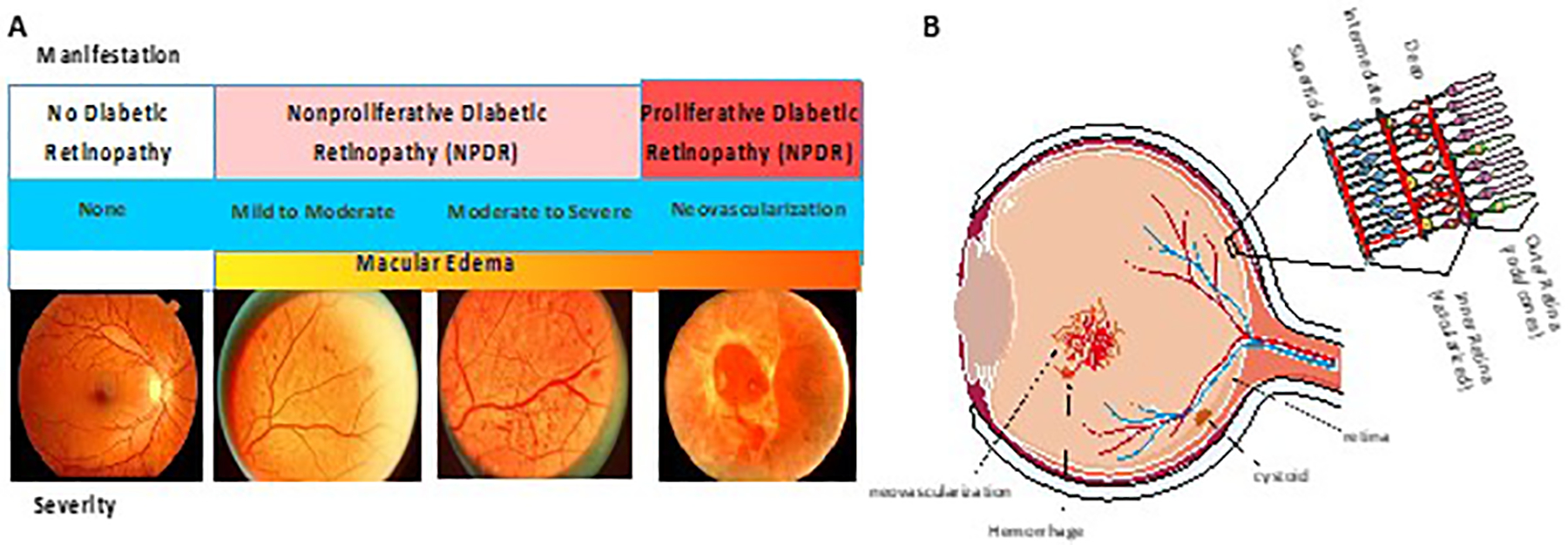
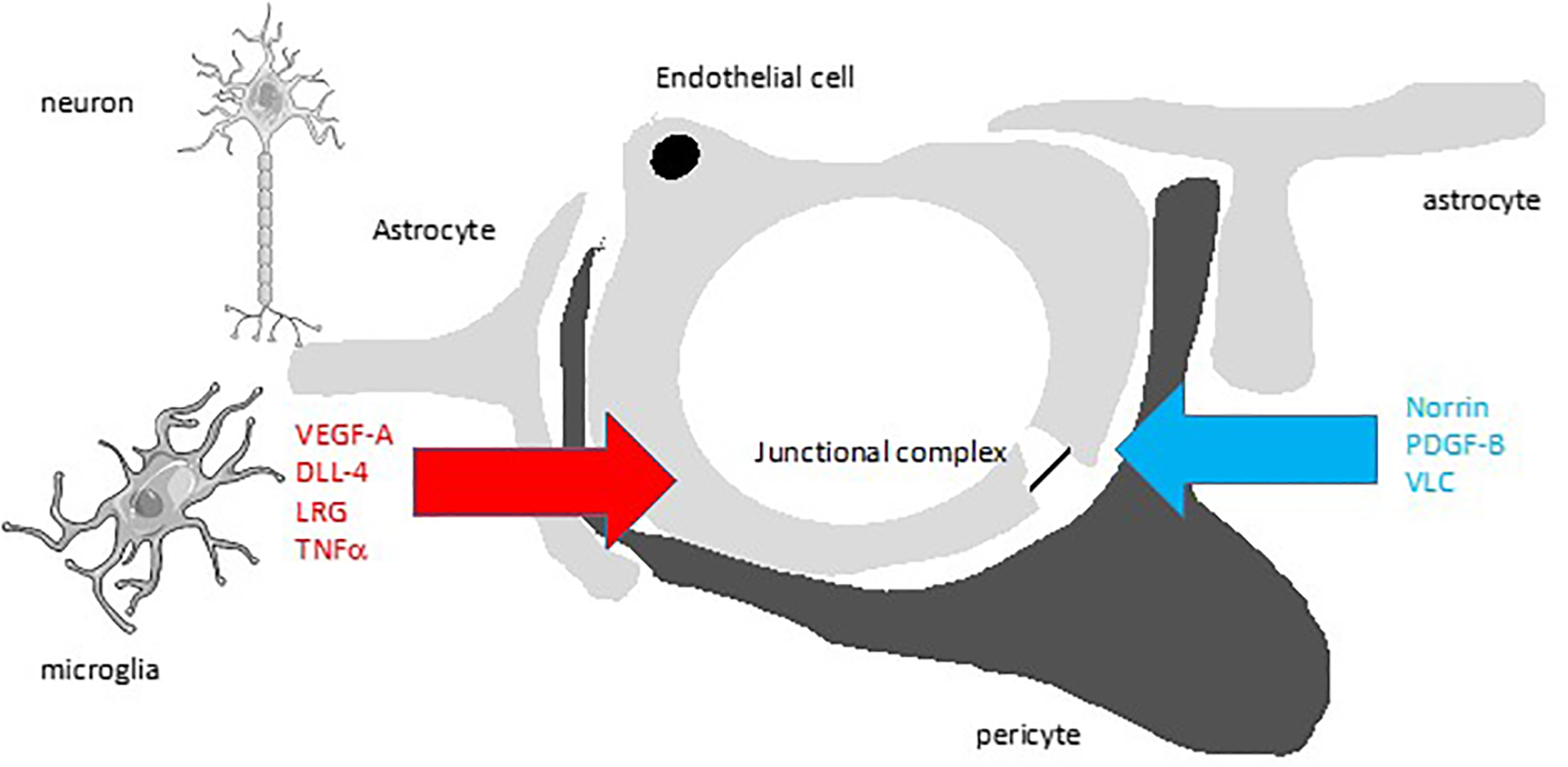
Diabetes mellitus has profound effects on multiple organ systems; however, the loss of vision caused by diabetic retinopathy might be one of the most impactful in a patient's life. The retina is a highly metabolically active tissue that requires a complex interaction of cells, spanning light sensing photoreceptors to neurons that transfer the electrochemical signal to the brain with support by glia and vascular tissue. Neuronal function depends on a complex inter-dependency of retinal cells that includes the formation of a blood-retinal barrier. This dynamic system is negatively affected by diabetes mellitus, which alters normal cell-cell interactions and leads to profound vascular abnormalities, loss of the blood-retinal barrier and impaired neuronal function. Understanding the normal cell signalling interactions and how they are altered by diabetes mellitus has already led to novel therapies that have improved visual outcomes in many patients. Research highlighted in this Review has led to a new understanding of retinal pathophysiology during diabetes mellitus and has uncovered potential new therapeutic avenues to treat this debilitating disease.


Figures

Figure 1.
Figure 1.. Diabetic Retinopathy (DR) Manifests with Multiple Pathologies.
A. Patients with diabetes may have no readily observable alterations to the retina as observed by fundus photography. Alternatively, microvascular abnormalities, hemorrhages, microaneurysms and venous beading reveal evidence of disease process that may range from mild to severe and occur in patients with non-proliferative DR (NPDR). Patients with proliferative DR (PDR) have neovascularization in the retina that may lead to retinal detachment. Diabetic macular edema (DME) may occur in both NPDR or PDR. (Adapted from) B. Schematic diagram of a cross section of the eye. Vessel leak, neovascularization and cystoid formation due to DME are indicated. Cross section of retina indicating organization of ganglion cells and bipolar cells in the inner retina versus rods and cones in the outer retina. Blood vessels in the inner retina make the inner blood-retinal barrier and the retinal pigment epithelium (RPE) makes the outer blood-retinal barrier.
Figure 2.
Figure 2.. The Neurovascular Unit (NVU) and Cytokine Signaling in Diabetic Retinopathy (DR).
A) Proper retinal functions require an intimate relationship of the retinal blood vessels in the inner retina with neurons, glia (astrocytes and Müller cells), and pericytes. Glia provide norrin signaling required for BRB formation. Endothelial cells recruit pericytes by PDGF-B signaling and pericytes promote BRB by an unknown mechanism. B) In DR, glia have increased aquaporin and Kir4.1 channels contributing to swelling and now produce vasoactive substances such as VEGF-A and associated DLL-4, ANGPTL4 and LRG that promote permeability, angiogenesis or both. Loss of pericytes leads to hyper-responsiveness of endothelial cells to VEGF signaling. Further, inflammatory cytokines such as TNFα, IL1β and CCL2 among many others, are produced by microglia and other retinal cells as well as adherent inflammatory cells. In addition, hyperglycemia induces direct endothelial dysfunction through change in redox state (NAD(P)H and ROS). Not shown, RPE also undergo dysfunction with increased cytokine production. Collectively these changes disrupt the neurovascular unit and alter normal retinal function.

Similar articles

Cited by

References

    1. Ogurtsova K et al. IDF Diabetes Atlas: Global estimates for the prevalence of diabetes for 2015 and 2040. Diabetes research and clinical practice 128, 40–50, doi:10.1016/j.diabres.2017.03.024 (2017). - DOI - PubMed
    1. Grading diabetic retinopathy from stereoscopic color fundus photographs--an extension of the modified Airlie House classification. ETDRS report number 10. Early Treatment Diabetic Retinopathy Study Research Group. Ophthalmology 98, 786–806 (1991). - PubMed
    1. Solomon SD et al. Diabetic Retinopathy: A Position Statement by the American Diabetes Association. Diabetes Care 40, 412–418, doi:10.2337/dc16-2641 (2017). - DOI - PMC - PubMed
    1. Kohner EM, Patel V & Rassam SM Role of blood flow and impaired autoregulation in the pathogenesis of diabetic retinopathy. Diabetes 44, 603–607, doi:10.2337/diab.44.6.603 (1995). - DOI - PubMed
    1. Roy S, Ha J, Trudeau K & Beglova E Vascular basement membrane thickening in diabetic retinopathy. Curr Eye Res 35, 1045–1056, doi:10.3109/02713683.2010.514659 (2010). - DOI - PubMed