. 2021 Jan 11:14:49-58.
doi: 10.2147/IDR.S286187. eCollection 2021.

PspA Diversity, Serotype Distribution and Antimicrobial Resistance of Invasive Pneumococcal Isolates from Paediatric Patients in Shenzhen, China

Affiliations

PspA Diversity, Serotype Distribution and Antimicrobial Resistance of Invasive Pneumococcal Isolates from Paediatric Patients in Shenzhen, China

Hanfang Jiang et al. Infect Drug Resist. .

Abstract

Introduction: To determine the phenotypes and genotypes of invasive Streptococcus pneumoniae (S. pneumoniae), 108 strains were isolated from paediatric patients with invasive pneumococcal diseases (IPDs) in Shenzhen from 2014 to 2018.

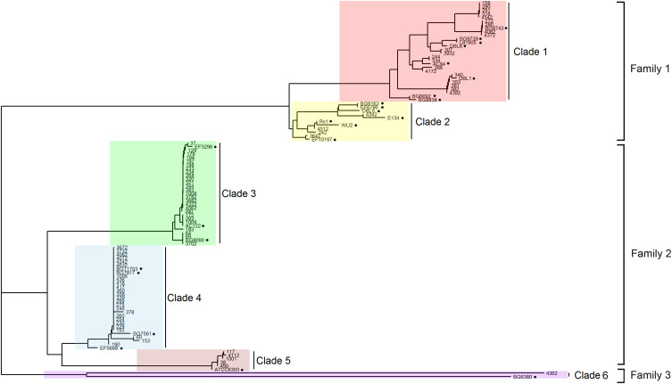
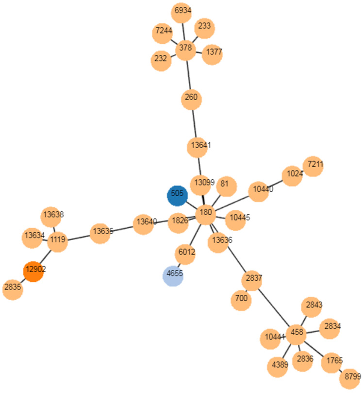
Methods: Serotype profiles were defined by multiplex PCR of the capsule gene. Pneumococcal surface protein A (PspA) classification was performed through pspA gene sequencing. Antimicrobial resistance was examined by broth microdilution. Multilocus sequence typing (MLST) was determined based on next-generation sequencing data.

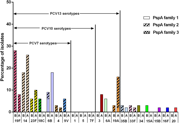
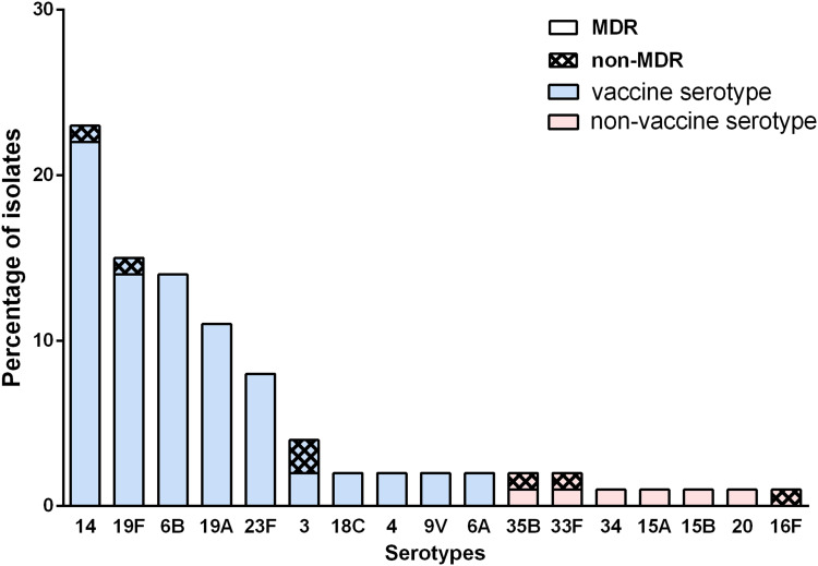
Results: Eighty-one S. pneumoniae of 17 serotypes were finally collected. The coverage of the 13-conjugated polysaccharide vaccine (PCV13) was 88.9%. After the introduction of PCV13, the nonvaccine serotypes were added by serotypes 15b, 16F and 20. Vaccine serotype 3 increased by four serious cases. The pspA family 1 and pspA family 2 are predominant. The multiple drug resistance rate is 91.3%. None of the nonmeningitis isolates were resistant to penicillin, while 98.8% of all the isolates were resistant to erythromycin.

Discussion: This work characterizes the molecular epidemiology of invasive S. pneumoniae in Shenzhen. Continued surveillance of serotype distribution and antimicrobial susceptibility is necessary to alert antibiotic-resistant nonvaccine serotypes and highly virulent serotypes.

Keywords: PspA family; Streptococcus pneumoniae; antimicrobial resistance; invasive pneumococcal disease; serotype.


Conflict of interest statement

The authors declare that there is no conflict of interest regarding the publication of this paper.

Figures

Figure 1
Figure 1
PspA families and clades of Shenzhen`s isolates. PspA clades 1–6 were highlighted by red, yellow, green, blue, pink and purple respectively. The black solid circles indicate the 24 strains reported previously, which were identified using their names.
Figure 2
Figure 2
Serotype distribution before and after PCV13 introduction. B represents before introduction of PCV13, and A represents after introduction of PCV13. Each serotype is colored specifically. The height of the column indicates the composition ratio of the serotype in either period. The proportion of PspA family 1, 2 and 3 are expressed by different filling patterns.
Figure 3
Figure 3
The multiple drug resistance rate of prevalent serotypes in Shenzhen. Vaccine serotypes refers to the serotypes covered by PCVs, which are colored by blue. The non-vaccine serotypes were not covered by PCVs and are colored by red. The proportion of MDR and non-MDR are distinguished by different filling patterns.
Figure 4
Figure 4
The goeBURST analysis of serotype 3. PHYLOViZ was used to identify the relationship of clonal complexes (CCs) of serotype 3 globally. Each node represents a ST. STs found in Shenzhen are colored with dark blue (ST 505), sky blue (ST 4655) and orange (ST 12902), respectively. STs colored with yellow are from foreign countries.

Similar articles

Cited by

References

    1. Kadioglu A, Weiser JN, Paton JC, Andrew PW. The role of Streptococcus pneumoniae virulence factors in host respiratory colonization and disease. Nat Rev Microbiol. 2008;6(4):288–301. doi:10.1038/nrmicro1871 - DOI - PubMed
    1. Subramanian K, Henriques-Normark B, Normark S. Emerging concepts in the pathogenesis of the Streptococcus pneumoniae: from nasopharyngeal colonizer to intracellular pathogen. Cell Microbiol. 2019;21(11):e13077. doi:10.1111/cmi.13077 - DOI - PMC - PubMed
    1. Engholm DH, Kilian M, Goodsell DS, et al. A visual review of the human pathogen Streptococcus pneumoniae. FEMS Microbiol Rev. 2017;41(6):854–879. doi:10.1093/femsre/fux037 - DOI - PubMed
    1. Li Q, Li Y, Yi Q, et al. Prognostic roles of time to positivity of blood culture in children with Streptococcus pneumoniae bacteremia. Eur J Clin Microbiol Infect Dis. 2019;38(3):457–465. doi:10.1007/s10096-018-03443-5 - DOI - PubMed
    1. Paton JC, Trappetti C. Streptococcus pneumoniae capsular polysaccharide. Microbiol Spectr. 2019;7(2). doi:10.1128/microbiolspec.GPP3-0019-2018 - DOI - PMC - PubMed

LinkOut - more resources