Review
. 2021 Jan 14:14:139-152.
doi: 10.2147/DMSO.S292991. eCollection 2021.

Application of High-Resolution Ultrasound on Diagnosing Diabetic Peripheral Neuropathy

Affiliations
Review

Application of High-Resolution Ultrasound on Diagnosing Diabetic Peripheral Neuropathy

Hailun Huang et al. Diabetes Metab Syndr Obes. .

Abstract

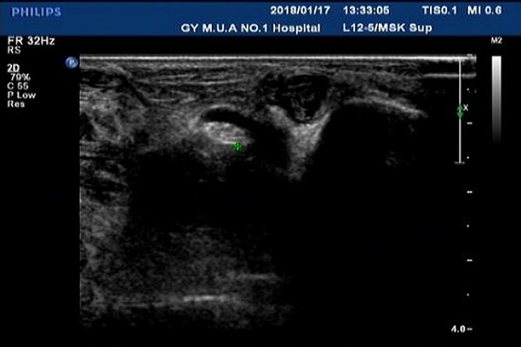
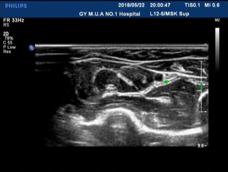
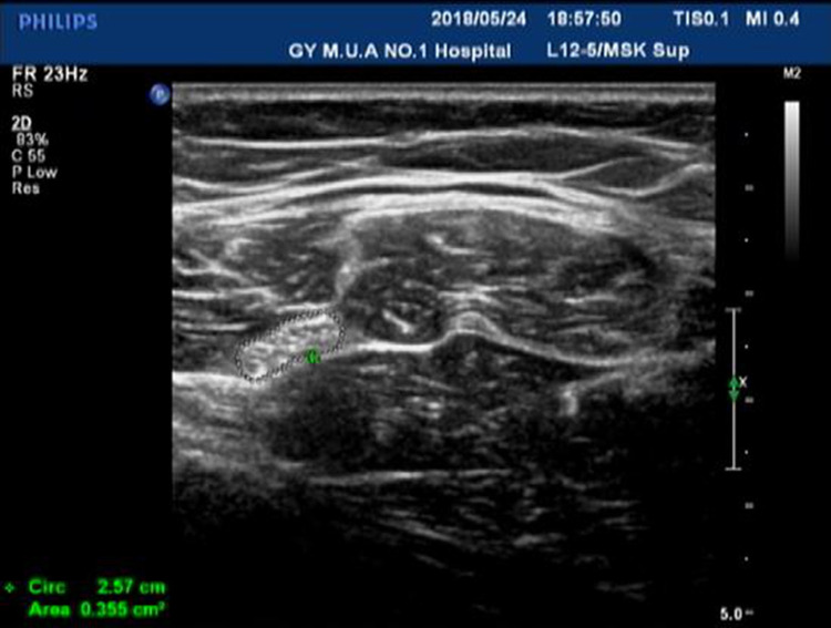
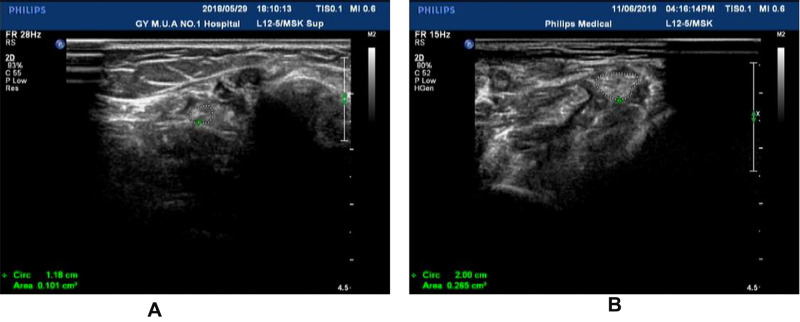
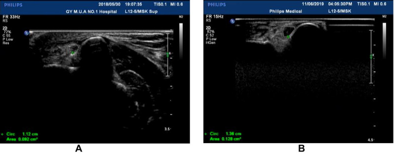
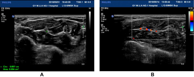
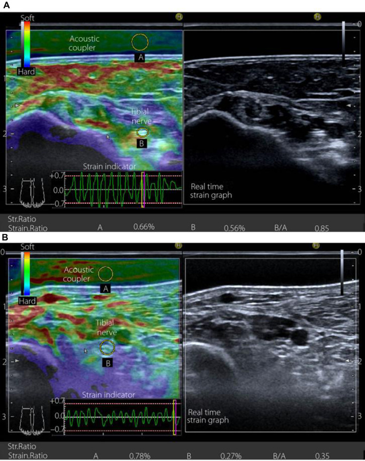
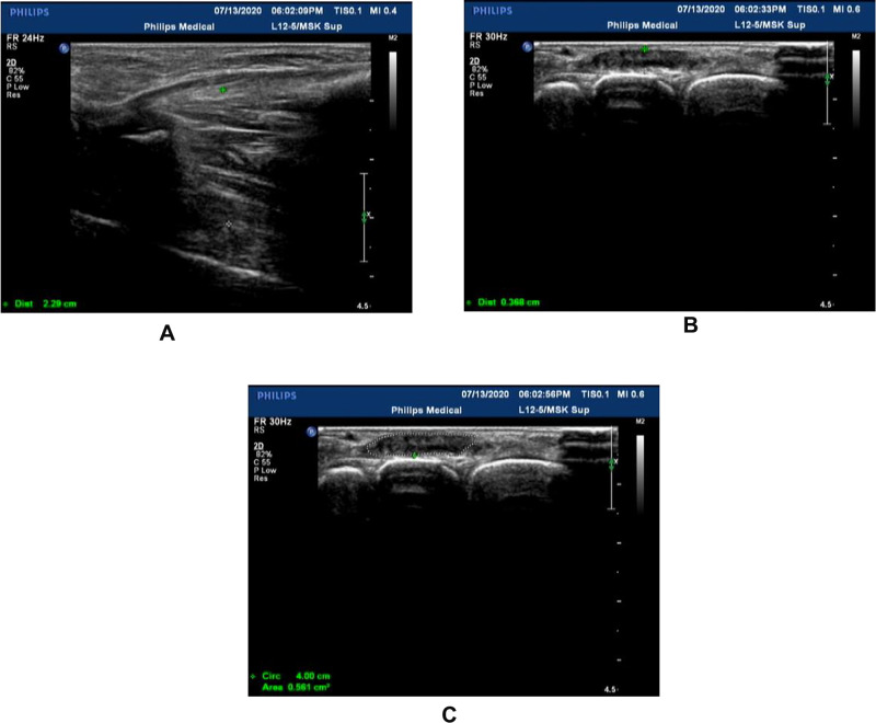
Diabetic peripheral neuropathy (DPN) is a common complication of diabetes mellitus (DM). The typical manifestation is a length-dependent "glove and sock" sensation. At present, diagnosis is mainly dependent on clinical manifestations. Since the pathogenesis is not clear, there are no effective treatment measures. Management consists mainly of glucose control, peripheral nerve nutrition, and other measures to delay the progress of the disease; early diagnosis is therefore crucial to improving prognosis and quality of life for patients with DPN. Due to the lack of obvious symptoms in 50% of patients and the low sensitivity of neuro-electrophysiology to small fibers, the missed diagnosis rate is high. High-resolution ultrasound (HRU), as a convenient noninvasive tool, has been proven by many studies to have excellent clinical value in diagnosing DPN. With the development of related new technology, HRU shows promise for the screening, diagnosing, and follow-up of DPN, which could serve as a biomarker and provide new diagnostic insights. In this paper, we review the ability of HRU to detect nerve cross-sectional area and blood flow, and echo and other image changes, and in showing the characteristics of peripheral nerve morphological changes in patients with DPN. We also explore the application of two other recent technological developments-shear wave elastography (SWE) and ultrasound scoring systems-in improving the diagnostic efficiency of HRU in peripheral neuropathy.

Keywords: diabetes; diabetic peripheral neuropathy; high-resolution ultrasound diagnosis; muscle ultrasound.


Conflict of interest statement

There are no conflicts of interest for this work.

Figures

Figure 1
Figure 1
The right posterior tibial nerve of normal people shows “honeycomb” in cross section.
Figure 2
Figure 2
The right ulnar nerve (under forearm) of normal people shows “honeycomb” in cross section.
Figure 3
Figure 3
The left sciatic nerve (inferior margin of gluteus maximus) of normal people shows” honeycomb” on the transverse axis.
Figure 4
Figure 4
(A) The right posterior tibial nerve (white circle) in the malleolus medialis of normal people shows “ honeycomb” in cross section. (B) The right posterior tibial nerve (white circle) in the malleolus medialis of patients with DPN. The development was blurred and the cross-sectional area of the nerve was significantly larger than the normal nerve with “honeycomb” disappeared.
Figure 5
Figure 5
(A) The right ulnar nerve (in the carpal canal) of normal people shows “ honeycomb” in cross section. (B) The echo in the carpal canal of the right ulnar nerve was significantly lower than that of the normal nerve besides enlarged cross-sectional area.
Figure 6
Figure 6
(A) Median nerve in forearm from healthy people. (B) The median nerve in the forearm of DPN patients, the blood flow signals of peripheral nerve and surrounding tissues were abundant.
Figure 7
Figure 7
Transverse sectional image of the tibial nerve in (A) a 48‐year‐old male control participant and (B) a 52‐year‐old male patient with type 2 diabetes at stage III neuropathy, visualized by high‐resolution ultrasonography using an 18.0‐MHz linear array probe (HIVISION Ascendus; Hitachi Medical, Tokyo, Japan) attached with an acoustic coupler (EZU‐TECPL1; Hitachi‐Aloka Medical, Tokyo, Japan). A translucent color‐coded image represents the relative stiffness of tissues. Mild compression and decompression using a probe attached with an acoustic coupler were repeated on the tibial nerve. Representative sonoelastographic images were chosen from images stored as cine loops. The tibial nerve and region of the acoustic coupler located directly above the tibial nerve are shown with a circle. The elastograms were constructed automatically. The elasticity of the tibial nerve was assessed as the tibial nerve:acoustic coupler strain ratio. The measurements were repeated three times and averaged. Reproduced from Ishibashi F, Taniguchi M, Kojima R, Kawasaki A, Kosaka A, Uetake H, Elasticity of the tibial nerve assessed by sonoelastography was reduced before the development of neuropathy and further deterioration associated with the severity of neuropathy in patients with type 2 diabetes. J Diabetes Investig. 7(3):404–412. Copyright © [2015], John Wiley and Sons.
Figure 8
Figure 8
(A) Thickness of the first and second metatarsal interosseous muscles in the left dorsum of diabetic peripheral neuropathy (between green * and white *); (B) Thickness of extensor digitorum brevis of  the left foot in patients with diabetic peripheral neuropathy (between green * and white *); (C) Cross-sectional area of extensor digitorum brevis of theleft foot in patients with diabetic peripheral neuropathy (white circle).

References

    1. Kessler JM, de la Lama M, Umans HR, et al. High-frequency sonography of the volar digital nerves of the hand. Muscle Nerve. 2012;45(2):222–226. doi:10.1002/mus.22277 - DOI - PubMed
    1. Fornage BD, Rifkin MD. Ultrasound examination of the hand and foot. Radiol Clin North Am. 1988;26(1):109–129. - PubMed
    1. Suk JI, Walker FO, Cartwright MS. Ultrasonography of peripheral nerves. Curr Neurol Neurosci Rep. 2013;13(2):328. doi:10.1007/s11910-012-0328-x - DOI - PMC - PubMed
    1. Huang Y, Hu B, Zhu J. Study on the use of quantitative ultrasound evaluation of diabetic neuropathy in the rat sciatic nerve. Australas Phys Eng Sci Med. 2016;39(4):997–1005. doi:10.1007/s13246-016-0448-8 - DOI - PubMed
    1. Tan CY, Arumugam T, Razali SNO, Yahya MA, Goh KJ, Shahrizaila N. Nerve ultrasound can distinguish chronic inflammatory demyelinating polyneuropathy from demyelinating diabetic sensorimotor polyneuropathy. J Clin Neurosci. 2018;57:198–201. doi:10.1016/j.jocn.2018.08.031 - DOI - PubMed

LinkOut - more resources