. 2021 Jan 5:11:524833.
doi: 10.3389/fphys.2020.524833. eCollection 2020.

Effects of Immunization With the Soil-Derived Bacterium Mycobacterium vaccae on Stress Coping Behaviors and Cognitive Performance in a "Two Hit" Stressor Model

Affiliations

Effects of Immunization With the Soil-Derived Bacterium Mycobacterium vaccae on Stress Coping Behaviors and Cognitive Performance in a "Two Hit" Stressor Model

Christine L Foxx et al. Front Physiol. .

Abstract

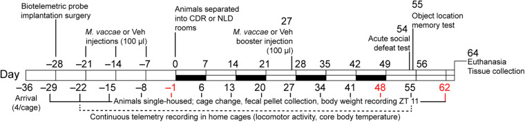
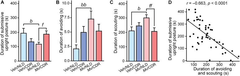
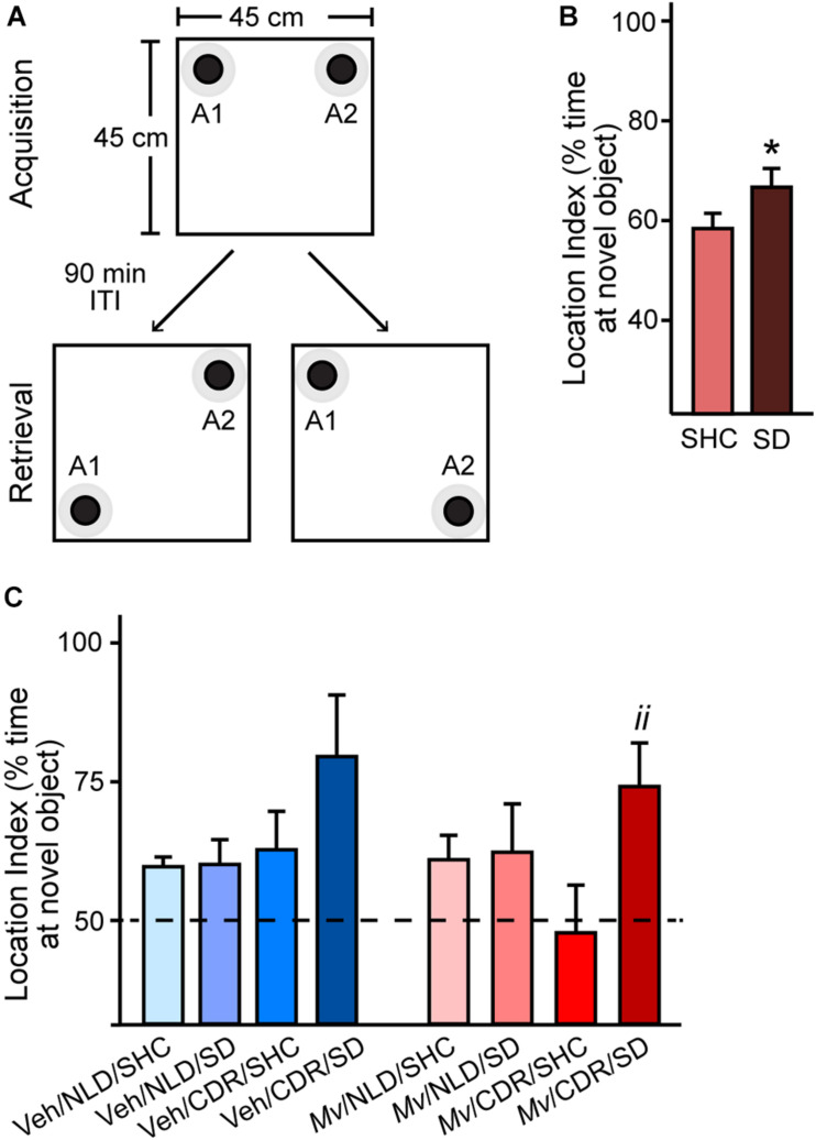
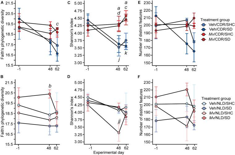
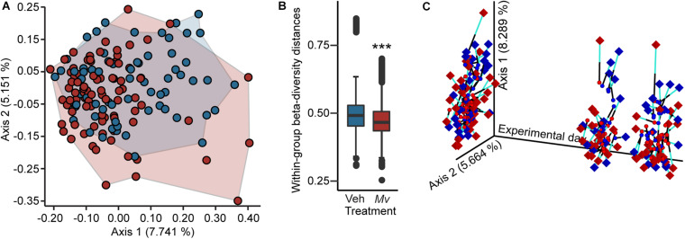
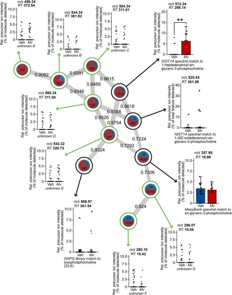
Previous studies demonstrate that Mycobacterium vaccae NCTC 11659 (M. vaccae), a soil-derived bacterium with anti-inflammatory and immunoregulatory properties, is a potentially useful countermeasure against negative outcomes to stressors. Here we used male C57BL/6NCrl mice to determine if repeated immunization with M. vaccae is an effective countermeasure in a "two hit" stress exposure model of chronic disruption of rhythms (CDR) followed by acute social defeat (SD). On day -28, mice received implants of biotelemetric recording devices to monitor 24-h rhythms of locomotor activity. Mice were subsequently treated with a heat-killed preparation of M. vaccae (0.1 mg, administered subcutaneously on days -21, -14, -7, and 27) or borate-buffered saline vehicle. Mice were then exposed to 8 consecutive weeks of either stable normal 12:12 h light:dark (LD) conditions or CDR, consisting of 12-h reversals of the LD cycle every 7 days (days 0-56). Finally, mice were exposed to either a 10-min SD or a home cage control condition on day 54. All mice were exposed to object location memory testing 24 h following SD. The gut microbiome and metabolome were assessed in fecal samples collected on days -1, 48, and 62 using 16S rRNA gene sequence and LC-MS/MS spectral data, respectively; the plasma metabolome was additionally measured on day 64. Among mice exposed to normal LD conditions, immunization with M. vaccae induced a shift toward a more proactive behavioral coping response to SD as measured by increases in scouting and avoiding an approaching male CD-1 aggressor, and decreases in submissive upright defensive postures. In the object location memory test, exposure to SD increased cognitive function in CDR mice previously immunized with M. vaccae. Immunization with M. vaccae stabilized the gut microbiome, attenuating CDR-induced reductions in alpha diversity and decreasing within-group measures of beta diversity. Immunization with M. vaccae also increased the relative abundance of 1-heptadecanoyl-sn-glycero-3-phosphocholine, a lysophospholipid, in plasma. Together, these data support the hypothesis that immunization with M. vaccae stabilizes the gut microbiome, induces a shift toward a more proactive response to stress exposure, and promotes stress resilience.

Keywords: cognition; diurnal; locomotor activity; metabolome; microbiome; microbiome-gut-brain axis; microbiota; stress resilience.


Conflict of interest statement

CL serves on the Scientific Advisory Board of Immodulon Therapeutics, Ltd., is cofounder and Chief Scientific Officer of Mycobacteria Therapeutics Corporation, serves as an unpaid scientific consultant with Aurum Switzerland AG and serves at a member of the faculty of the Integrative Psychiatry Institute, Boulder, Colorado, United States. KPW has received research support from the National Institutes of Health, the Pac-12 Conference, and SomaLogic, Inc. outside of this work; consulting fees from or served as a paid member of scientific advisory boards for the Sleep Disorders Research Advisory Board – National Heart, Lung and Blood Institute and CurAegis Technologies, Circadian Therapeutics, Ltd.; and has received speaker/educational/travel consultant honorarium fees from the American Academy of Sleep Medicine, American College of Chest Physicians, American College of Sports Medicine, American Diabetes Association, Associated Professional Sleep Societies, Kellogg Company, and The European Association for the Study of Obesity. The remaining authors declare that the research was conducted in the absence of any commercial or financial relationships that could be construed as a potential conflict of interest.

Figures

FIGURE 1
FIGURE 1
Diagrammatic illustration of the experimental protocol. White bars indicate normal 12-h light:dark cycling with lights on at 0600 MDT or 0700 h MST, black bars indicate light cycle reversal periods for the chronic disruption of rhythms (CDR) group. CDR, chronic disruption of rhythms; MDT, Mountain Daylight Time; MST, Mountain Standard Time; M. vaccae, Mycobacterium vaccae NCTC 11659; NLD, normal light:dark condition; Vehicle, borate-buffered saline vehicle; ZT, Zeitgeber time.
FIGURE 2
FIGURE 2
Behavioral responses of experimental mice during the 10-min social defeat test. Individual panels represent means + standard errors of the means of mice from seven independently tested cohorts of mice; (A) duration of submissive upright posture, (B) duration of avoiding behavior, and (C) duration of scouting behavior in s. (D) Shows the relationship between the duration of avoiding and scouting (s) and the duration of submissive upright posture (s); circles indicate individual data points. Post hoc pairwise comparisons indicated by bp < 0.05 and bbp < 0.01, Veh/NLD against Mv/NLD group; fp < 0.05 and ffp < 0.01, Mv/NLD against Mv/CDR group. CDR, chronic disruption of rhythms; Mv, Mycobacterium vaccae NCTC 11659; NLD, normal light:dark cycle; Veh, borate-buffered saline vehicle. For sample sizes of each group, see Supplementary File S1.
FIGURE 3
FIGURE 3
Object location memory test configuration and location indices. (A) Arena configuration for the pre-test phase, where black bottlecaps of equal dimensions and coloration (objects A1 and A2; black circles) with defined contact zones (gray circles) were placed in adjacent corners, 2.5 cm away from the walls of the 45 cm × 45 cm × 40 cm arena. Each individual mouse was placed in the center of the arena and permitted to freely explore the arena for 5 min during the pre-test Acquisition phase. The mouse was then removed from the testing arena and returned to its home cage for a 90-min inter-trial interval (ITI) while the testing arena and objects A1 and A2 were cleaned with 70% ethanol and paper towels. The arena was then reconfigured for the test phase (Retrieval) such that either object A1 (lower left) or A2 (lower right) was displaced from its original position, diagonally positioned from the other object in non-adjacent corners. After the 90-min ITI, the mouse was then placed back in the testing arena. Location index data were obtained via Noldus Ethovision XT for (B) mice exposed to social defeat (SD) or left undisturbed in single-housed home cage control (SHC) conditions and (C) for all treatment groups. Data represent means + standard errors of the means of mice from seven independently tested cohorts of mice. Dashed line represents equal exploration of the displaced object and the unmoved object; bars above this threshold indicate preferential exploration of the displaced object. Pairwise post hoc comparisons shown: *p < 0.05, SD against SHC, and iip < 0.01, Mv/CDR/SD against Mv/CDR/SHC. CDR, chronic disruption of rhythms; Mv, Mycobacterium vaccae NCTC 11659; NLD, normal light:dark condition; SD, social defeat; SHC, single-housed home cage control condition; Veh, borate-buffered saline vehicle. For sample sizes of each group at each time point, see Supplementary File S1.
FIGURE 4
FIGURE 4
Within-subject alpha diversity of samples shown separately for chronic disruption of rhythms (CDR) conditions and normal light:dark conditions (NLD), as measured by (A,B) Faith’s phylogenetic diversity, (C,D) Shannon’s index, and (E,F) number of distinct features over time. Data represent treatment group means ± standard errors of the means of alpha diversity in fecal samples from days –1, 48, and 62 of mice in five independently tested cohorts of mice. Analyses were performed on 16 small subunit ribosomal RNA (16S rRNA) gene V4 amplicon data at a rarefaction depth of 15,000 reads per sample. 16S rRNA, 16 small subunit ribosomal RNA; CDR, chronic disruption of rhythms; Mv, Mycobacterium vaccae NCTC 11659; NLD, normal light:dark conditions; SD, social defeat; SHC, single-housed home cage control condition; Veh, borate-buffered saline vehicle. For sample sizes of each group at each time point, see Supplementary File S1; for definitions of symbols used to indicate significant post hoc pairwise comparisons, see Table 4.
FIGURE 5
FIGURE 5
Beta diversity and Procrustes analyses of gut microbiomes. (A) Principal coordinates analysis (PCoA) of microbial community-wide beta diversities of mice from five independently tested cohorts of mice using Unweighted UniFrac, grouped by treatment (Veh, blue; Mv, red). PCoA axes 1 and 2 explain 12.9% of the variation. (B) Within-group beta diversity distances among individual samples (Veh, blue; Mv, red); ***p < 0.0001, Kruskal–Wallis H test. (C) Procrustes analysis plot produced from the superimposition of the PCoA of the 16 small subunit ribosomal RNA (16S rRNA) gene V4 amplicon data (circles) and the LC-MS/MS-based metabolomics dataset (diamonds). Longer lines on Procrustes plots indicate greater within-subjects dissimilarity of the microbiome and metabolome. PCoA axes 1 and 2 explain 14.0% of the variation observed in the data. For sample sizes of each group at each time point, see Supplementary File S1. 16S rRNA, 16 small subunit ribosomal RNA; Mv, Mycobacterium vaccae NCTC 11659; PCoA, principal coordinates analysis; Veh, borate-buffered saline vehicle.
FIGURE 6
FIGURE 6
Spectral network of a molecular family of lysophospholipids present in the untargeted plasma metabolomics dataset of mice from six independently tested cohorts of mice. Nodes from solvent blanks, standards, and controls were subtracted. Nodes are labeled according to treatment group origin (Veh, blue; Mv, red) if spectra came exclusively from a single group, as in unknown A, and are split into labeled frequency pie charts if spectra above the lower limit of detection came from both groups, as in unknown B. Nodes without a library hit are circled in green, identified according to precursor mass-to-charge ratio (m/z) and retention time (RT) in s, and annotated as unknowns A–G. Nodes with a library hit are circled in black and identified according to spectral database and library compound name, in addition to m/z and RT information as above. For sample sizes of each group, see Supplementary File S1. GNPS, Global Natural Products Social Molecular Networking; Mv, Mycobacterium vaccae NCTC 11659; m/z, mass-to-charge ratio; NIST, National Institutes of Standards and Technology; Rel., relative; RT, retention time; Veh, borate-buffered saline vehicle.
FIGURE 7
FIGURE 7
In situ hybridization histochemistry-based analysis of tryptophan hydroxylase 2 (Tph2) and high-affinity, low-capacity sodium-dependent serotonin transporter (Slc6a4) mRNA expression. Atlases (A,C) based on autoradiographic images of (A) Tph2, and (C) Slc6a4 mRNA expression in the midbrain and pontine raphe complex (84 μm intervals) from a representative adult male mouse in this study, used for analysis of subregions of the dorsal raphe nucleus (DR) and median raphe nucleus (MnR) with a high level of neuroanatomical resolution. The levels chosen for analysis ranged from (A) –4.160 mm bregma through (N) –5.252 mm bregma. Dashed lines delineate different subdivisions of the DR and MnR analyzed in this study, based on a stereotaxic atlas of the mouse brain (Paxinos and Franklin, 2001). Numbers in the lower left of each panel indicate the rostrocaudal coordinates relative to bregma in mm. Numbers in the upper right of each panel correspond to the x-axis values in graphical representation of the data. Scale bar, 1 mm. Effects of immunization with Mycobacterium vaccae NCTC 11659 (Mv), exposure to chronic disruption of rhythms (CDR), and exposure to social defeat (SD) on (B) Tph2 mRNA expression within the dorsal raphe nucleus, dorsal part (DRD) and (D) on Slc6a4 mRNA expression within the DRD. Data represent treatment group means + standard errors of the means of mice from six independently tested cohorts of mice at each rostrocaudal level throughout the DRD. Post hoc testing was not conducted when one or more groups contained less than 50% of the full sample size, indicated by the right y-axis in (B) and (D). (E) Tph2 and Slc6a4 mRNA expression throughout the rostrocaudal extent of the DR and MnR are highly correlated (Pearson’s r = 0.839, p < 0.0001). For sample sizes of each group, see Supplementary File S1. For definitions of symbols used to indicate significant post hoc pairwise comparisons, see Table 4. CDR, chronic disruption of rhythms; DR, dorsal raphe nucleus; DRC, dorsal raphe nucleus, caudal part; DRD, dorsal raphe nucleus, dorsal part; DRI, dorsal raphe nucleus, interfascicular part; DRV, dorsal raphe nucleus, ventral part; DRVL/VLPAG, dorsal raphe nucleus, ventrolateral part/ventrolateral periaqueductal gray; MnR, median raphe nucleus; GV, gray value; mRNA, messenger RNA; Mv, Mycobacterium vaccae NCTC 11659; SD, social defeat; SHC, single-housed home cage control condition; Slc6a4, solute carrier family 6 member 4 (high-affinity, low-capacity sodium-dependent serotonin transporter); Tph2, tryptophan hydroxylase 2.

References

    1. Abrams J. K., Johnson P. L., Hollis J. H., Lowry C. A. (2004). Anatomic and functional topography of the dorsal raphe nucleus. Ann. N. Y. Acad. Sci. 1018 46–57. 10.1196/annals.1296.005 - DOI - PubMed
    1. Adams K. L., Castanon-Cervantes O., Evans J. A., Davidson A. J. (2013). Environmental circadian disruption elevates the IL-6 response to lipopolysaccharide in blood. J. Biol. Rhythms 28 272–277. 10.1177/0748730413494561 - DOI - PMC - PubMed
    1. Amara J., Saliba Y., Hajal J., Smayra V., Bakhos J. J., Sayegh R., et al. (2019). Circadian rhythm disruption aggravates DSS-induced colitis in mice with fecal calprotectin as a marker of colitis severity. Dig. Dis. Sci. 64 3122–3133. 10.1007/s10620-019-05675-7 - DOI - PubMed
    1. Ambrée O., Ruland C., Zwanzger P., Klotz L., Baune B. T., Arolt V., et al. (2019). Social defeat modulates T helper cell percentages in stress susceptible and resilient mice. Int. J. Mol. Sci. 20:3512. 10.3390/ijms20143512 - DOI - PMC - PubMed
    1. Amir A., McDonald D., Navas-Molina J. A., Kopylova E., Morton J. T., Xu Z. Z., et al. (2017). Deblur rapidly resolves single-nucleotide community sequence patterns. mSystems 2:e00191-16. - PMC - PubMed