Gorham Stout disease: a case report from Syria
- PMID: 33469472
- PMCID: PMC7802812
- DOI: 10.1093/omcr/omaa121
Gorham Stout disease: a case report from Syria
Abstract
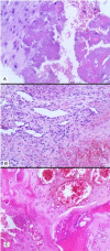
Gorham-Stout disease (GSD) is a rare entity that destroys the bone matrix resulting mainly in osteolysis, pain and pathologic fractures among a broader clinical picture. We report a case of a 60-year-old female with a sudden discovery of pathologic fractures in the pelvis and the absence of the left femoral head. On biopsy, no cellular atypia was found, instead disturbed bone formation with prominent vascularity with scattered foci of necrosis & osteolysis, which lead to the diagnosis of GSD. Possible differential diagnoses were discussed and excluded. The patient was put on Bisphosphonate that led to a relative improvement in the symptoms. This disease needs a more thorough investigation to identify the key cause, what is beyond the scope of this report.
Keywords: Gorham Stout disease; angiomatosis; lytic bone disease; osteolysis; vanishing bone disease.
© The Author(s) 2020. Published by Oxford University Press. All rights reserved. For Permissions, please email: journals.permissions@oup.com.
Figures


References
-
- Malde R, Agrawal HM, Ghosh SL, Dinshaw KA. Vanishing bone disease involving the pelvis. J Cancer Res Ther 2005;1:227–8. - PubMed
-
- Mukhopadhyay S, Chattopadhyay A, Bhattacharya R, Roy U. Gorham’s disease of mandible--a rare case presentation in pediatric patient. J Indian Soc Pedod Prev Dent 2016;34:180–4. - PubMed
-
- Dellinger MT, Garg N, Olsen BR. Viewpoints on vessels and vanishing bones in Gorham-stout disease. Bone 2014;63:47–52. - PubMed
-
- University of Washington Bone densitometry [internet]. 2011. https://courses.washington.edu/bonephys/opbmdtz.html (15 September 2019, date last accessed).
Publication types
LinkOut - more resources
Full Text Sources
Other Literature Sources

