

Preliminary studies of constructing a tissue-engineered lamellar corneal graft by culturing mesenchymal stem cells onto decellularized corneal matrix
- PMID: 33469478
- PMCID: PMC7790677
- DOI: 10.18240/ijo.2021.01.02
Preliminary studies of constructing a tissue-engineered lamellar corneal graft by culturing mesenchymal stem cells onto decellularized corneal matrix
Abstract
Aim: To construct a competent corneal lamellar substitute in order to alleviate the shortage of human corneal donor.
Methods: Rabbit mesenchymal stem cells (MSCs) were isolated from bone marrow and identified by flow cytometric, osteogenic and adipogenic induction. Xenogenic decellularized corneal matrix (XDCM) was generated from dog corneas. MSCs were seeded and cultured on XDCM to construct the tissue-engineered cornea. Post-transplantation biocompatibility of engineered corneal graft were tested by animal experiment. Rabbits were divided into two groups then underwent lamellar keratoplasty (LK) with different corneal grafts: 1) XDCM group (n=5): XDCM; 2) XDCM-MSCs groups (n=4): tissue-engineered cornea made up with XDCM and MSCs. The ocular surface recovery procedure was observed while corneal transparency, neovascularization and epithelium defection were measured and compared. In vivo on focal exam was performed 3mo postoperatively.
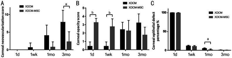
Results: Rabbit MSCs were isolated and identified. Flow cytometry demonstrated isolated cells were CD90 positive and CD34, CD45 negative. Osteogenic and adipogenic induction verified their multipotent abilities. MSC-XDCM grafts were constructed and observed. In vivo transplantation showed the neovascularization in XDCM-MSC group was much less than that in XDCM group postoperatively. Post-transplant 3-month confocal test showed less nerve regeneration and bigger cell-absent area in XDCM-MSC group.
Conclusion: This study present a novel corneal tissue-engineered graft that could reduce post-operatively neovascularization and remain transparency, meanwhile shows that co-transplantation of MSCs may help increase corneal transplantation successful rate and enlarge the source range of corneal substitute to overcome cornea donor shortage.
Keywords: acellular corneal matrix; mesenchymal stem cells; neovascularization; tissue-engineered cornea; xenogenic decellularized corneal matrix.
International Journal of Ophthalmology Press.
Figures


Similar articles
-
In vivo confocal microscopic observation of lamellar corneal transplantation in the rabbit using xenogenic acellular corneal scaffolds as a substitute.Chin Med J (Engl). 2015 Apr 5;128(7):933-40. doi: 10.4103/0366-6999.154301. Chin Med J (Engl). 2015. PMID: 25836615 Free PMC article.
-
Suppression of alkali-induced oxidative injury in the cornea by mesenchymal stem cells growing on nanofiber scaffolds and transferred onto the damaged corneal surface.Exp Eye Res. 2013 Nov;116:312-23. doi: 10.1016/j.exer.2013.10.002. Epub 2013 Oct 18. Exp Eye Res. 2013. PMID: 24145108
-
Acellular human corneal matrix sheets seeded with human adipose-derived mesenchymal stem cells integrate functionally in an experimental animal model.Exp Eye Res. 2015 Mar;132:91-100. doi: 10.1016/j.exer.2015.01.020. Epub 2015 Jan 24. Exp Eye Res. 2015. PMID: 25625506
-
Current Trends and Future Perspective of Mesenchymal Stem Cells and Exosomes in Corneal Diseases.Int J Mol Sci. 2019 Jun 12;20(12):2853. doi: 10.3390/ijms20122853. Int J Mol Sci. 2019. PMID: 31212734 Free PMC article. Review.
-
[Transplantation of corneal endothelial cells].Nippon Ganka Gakkai Zasshi. 2002 Dec;106(12):805-35; discussion 836. Nippon Ganka Gakkai Zasshi. 2002. PMID: 12610838 Review. Japanese.
Cited by
-
Chemical, Physical, and Biological Corneal Decellularization Methods: A Review of Literature.J Ophthalmol. 2024 Mar 25;2024:1191462. doi: 10.1155/2024/1191462. eCollection 2024. J Ophthalmol. 2024. PMID: 38567029 Free PMC article. Review.
References
-
- Hori J, Yamaguchi T, Keino H, Hamrah P, Maruyama K. Immune privilege in corneal transplantation. Prog Retin Eye Res. 2019;72:100758. - PubMed
-
- Hos D, Matthaei M, Bock F, Maruyama K, Notara M, Clahsen T, Hou YH, Le VNH, Salabarria AC, Horstmann J, Bachmann BO, Cursiefen C. Immune reactions after modern lamellar (DALK, DSAEK, DMEK) versus conventional penetrating corneal transplantation. Prog Retin Eye Res. 2019;73:100768. - PubMed
-
- Brunette I, Roberts CJ, Vidal F, Harissi-Dagher M, Lachaine J, Sheardown H, Durr GM, Proulx S, Griffith M. Alternatives to eye bank native tissue for corneal stromal replacement. Prog Retin Eye Res. 2017;59:97–130. - PubMed
-
- Whitcher JP, Srinivasan M, Upadhyay MP. Prevention of corneal ulceration in the developing world. Int Ophthalmol Clin. 2002;42(1):71–77. - PubMed
LinkOut - more resources
Full Text Sources
Other Literature Sources
Research Materials
Miscellaneous