

Endogenous fungal endophthalmitis: risk factors, clinical course, and visual outcome in 13 patients
- PMID: 33469490
- PMCID: PMC7790672
- DOI: 10.18240/ijo.2021.01.14
Endogenous fungal endophthalmitis: risk factors, clinical course, and visual outcome in 13 patients
Abstract
Aim: To analyze the risk factors, ophthalmological features, treatment modalities and their effect on the visual outcome in patients with endogenous fungal endophthalmitis (EFE).
Methods: Data retrieved from the medical files included age at presentation to the uveitis clinic, gender, ocular symptoms and their duration before presentation, history of fever, eye affected, anatomical diagnosis and laboratory evidence of fungal infection. Medical therapy recorded included systemic antifungal therapy and its duration, use of intravitreal antifungal agents and use of oral/intravitreal steroids. Surgical procedures and the data of ophthalmologic examination at presentation and at last follow-up were also collected.
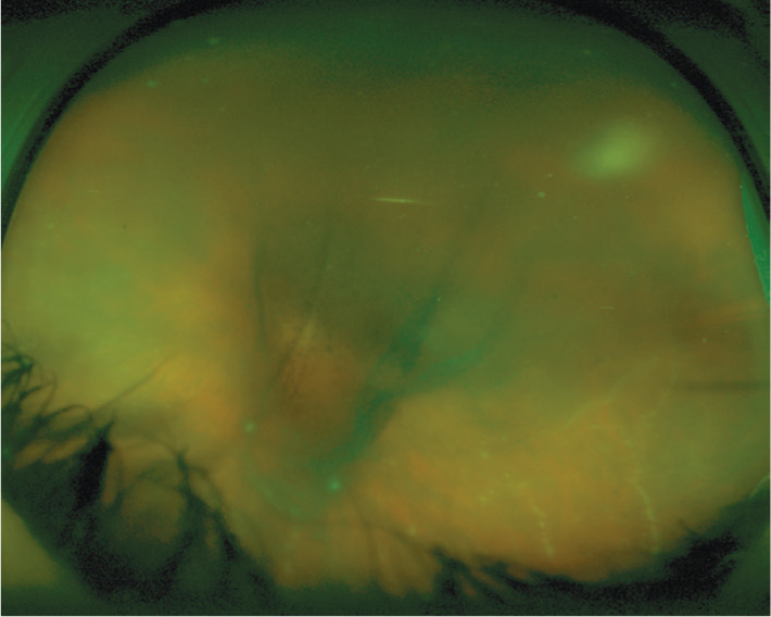
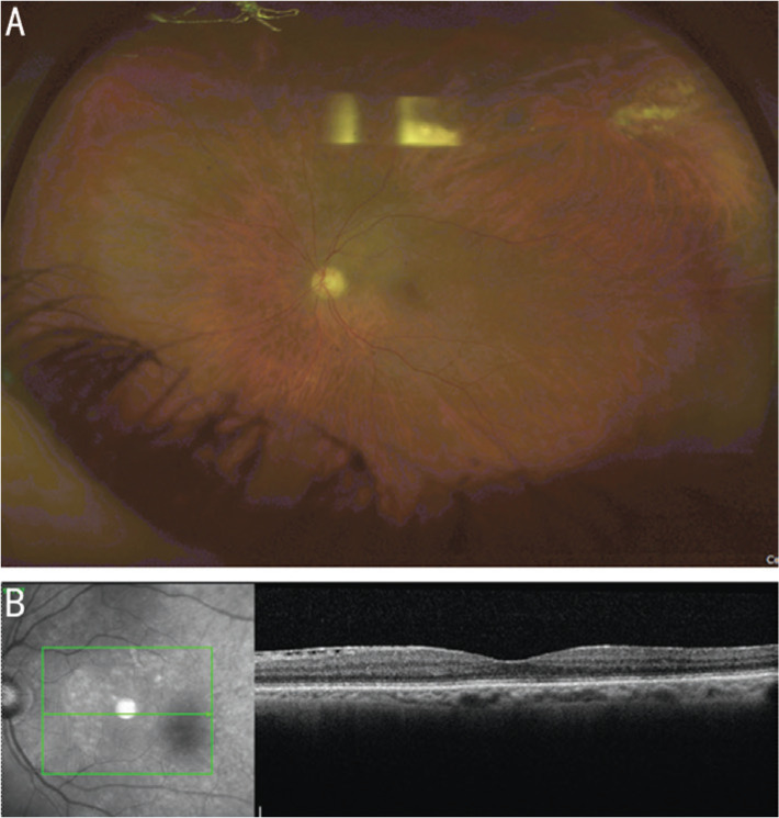
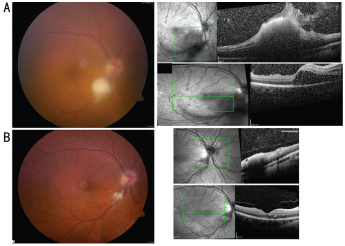
Results: Included were 13 patients (20 eyes, mean age 58y). Ten patients presented after gastrointestinal or urological interventions and two presented after organ transplantation. In one patient, there was no history of previous intervention. Diagnostic vitrectomy was performed in 16 eyes (80%) and vitreous cultures were positive in 10 of the vitrectomized eyes (62.5%). In only 4 patients (31%), blood cultures were positive. All patients received systemic antifungal therapy. Sixteen eyes (80%) received intravitreal antifungal agent with voriconazole being the most commonly used. Visual acuity (VA) improved from 0.9±0.9 at initial exam to 0.5±0.8 logMAR at last follow-up (P=0.03). A trend of greater visual improvement was noted in favor of eyes treated with oral steroids (±intravitreal dexamethasone) than eyes that were not treated with steroids. The most common complication was maculopathy. Twelve eyes (60%) showed no ocular complications.
Conclusion: High index of suspicion in patients with inciting risk factors is essential because of the low yield of blood cultures and the good general condition of patients at presentation. Visual prognosis is improved with the prompt institution of systemic and intravitreal pharmacotherapy and the immediate surgical intervention. Oral±local steroids could be considered in cases of prolonged or marked inflammatory responses in order to hasten control of inflammation and limit ocular complications.
Keywords: candida endophthalmitis; endogenous endophthalmitis; endogenous fungal endophthalmitis; endophthalmitis; fungal endophthalmitis.
International Journal of Ophthalmology Press.
Figures
References
-
- Kostick DA, Foster RE, Lowder CY, Meyers SM, McHenry MC. Endogenous endophthalmitis caused by candida albicans in a healthy woman. Am J Ophthalmol. 1992;113(5):593–595. - PubMed
-
- Valluri S, Moorthy RS, Liggett PE, Rao NA. Endogenous Aspergillus endophthalmitis in an immunocompetent individual. Int Ophthalmol. 1993;17(3):131–135. - PubMed
-
- Ishibashi Y. Proposed classification of stages of endogenous fungal endophthalmitis. Rinsho Ganka (Jpn J Clin Ophthalmol) 1993;47:845–849.
LinkOut - more resources
Full Text Sources
Other Literature Sources