Review
. 2021 Jan 5:8:615461.
doi: 10.3389/fcell.2020.615461. eCollection 2020.

Targeting Mitochondrial Impairment in Parkinson's Disease: Challenges and Opportunities

Affiliations
Review

Targeting Mitochondrial Impairment in Parkinson's Disease: Challenges and Opportunities

Jannik Prasuhn et al. Front Cell Dev Biol. .

Abstract

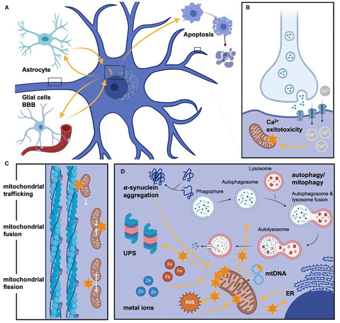
The underlying pathophysiology of Parkinson's disease is complex, but mitochondrial dysfunction has an established and prominent role. This is supported by an already large and rapidly growing body of evidence showing that the role of mitochondrial (dys)function is central and multifaceted. However, there are clear gaps in knowledge, including the dilemma of explaining why inherited mitochondriopathies do not usually present with parkinsonian symptoms. Many aspects of mitochondrial function are potential therapeutic targets, including reactive oxygen species production, mitophagy, mitochondrial biogenesis, mitochondrial dynamics and trafficking, mitochondrial metal ion homeostasis, sirtuins, and endoplasmic reticulum links with mitochondria. Potential therapeutic strategies may also incorporate exercise, microRNAs, mitochondrial transplantation, stem cell therapies, and photobiomodulation. Despite multiple studies adopting numerous treatment strategies, clinical trials to date have generally failed to show benefit. To overcome this hurdle, more accurate biomarkers of mitochondrial dysfunction are required to detect subtle beneficial effects. Furthermore, selecting study participants early in the disease course, studying them for suitable durations, and stratifying them according to genetic and neuroimaging findings may increase the likelihood of successful clinical trials. Moreover, treatments involving combined approaches will likely better address the complexity of mitochondrial dysfunction in Parkinson's disease. Therefore, selecting the right patients, at the right time, and using targeted combination treatments, may offer the best chance for development of an effective novel therapy targeting mitochondrial dysfunction in Parkinson's disease.

Keywords: Parkinson's disease; mitochondria; mitochondrial dysfunction; neurodegeneration; therapy.


Conflict of interest statement

The authors declare that the research was conducted in the absence of any commercial or financial relationships that could be construed as a potential conflict of interest.

Figures

Figure 1
Figure 1
Schematic representation of the Electron Transport Chain (ETC) and potential neuroimaging modalities to assess bioenergetic disturbances in Parkinson's disease (PD) patients. The localisation of the ETC complexes is highlighted in the right upper corner. Lactate (as a surrogate marker for mitochondrial dysfunction) can be assessed by 1H-MRSI (1). bNIRS devices can measure Cyt c oxidation state under dynamic conditions (2). ADP and ATP can either be measured under dynamic (ATP synthase activity) by 31P-MRSI magnetisation transfer (3) or static conditions (regular 31P-MRSI) (6). Complex I activity can be studied by 31P-MRSI (by measuring the NAD+/NADH ratio) or via a radiolabeled PET tracer (18F-BCPP-EF) (4). Many different approaches can be used to study in-vivo oximetry and oxygen turnover. The most common non-invasive methods are 17O-MRI (direct measurement) or a combination of BOLD and ASL imaging (indirect measurement by employing different physiological models) (5). 1H-MRSI, proton magnetic resonance spectroscopy imaging; 17O-MRI, oxygen magnetic resonance imaging; 18F-BCPP-EF, 2-tert-butyl-4-chloro-5-[6-[2-2[([18F]fluoroethoxy)-ethoxy]-pyridin-3-ylmethoxy]-2H]-pyridazin-3-one; 31P-MRSI, phosphorus magnetic resonance spectroscopy imaging; ADP, adenosine diphosphate; ASL, arterial spin labelling; ATP, adenosine triphosphate; bNIRS, broadband near-infrared spectroscopy; BOLD, blood oxygen-dependent imaging, functional MRI; CoQ, coenzyme Q; Cyt c. Cytochrome c; e, electron; FAD, flavin adenine dinucleotide (oxidised form); FADH2, flavin adenine dinucleotide (reduced form); H+, proton; H2O, water; I, complex I; NADH, ubiquinone oxidoreductase, Type I NADH dehydrogenase; II, complex II, Succinate dehydrogenase, succinate-coenzyme Q reductase; III, complex III, coenzyme Q: cytochrome c—oxidoreductase; IV, complex IV, cytochrome c oxidase; MRI, magnetic resonance imaging; n, number; NAD+, nicotinamide dinucleotide (oxidised form); NADH, nicotinamide dinucleotide (reduced form); O2, molecular oxygen; PD, Parkinson's disease; PET, positron emission tomography. Created with https://biorender.com/.
Figure 2
Figure 2
The exemplary workflow of phosphorus-magnetic resonance spectroscopy imaging (31P-MRSI) analysis and its potential role in imaging mitochondrial dysfunction in Parkinson's disease (PD) patients. (A) Depicts the voxel placement (orange bordered) for metabolic studies in-vivo. As seen here, one of the most significant limitations in 31P-MRSI studies is the relatively poor spatial resolution. The arbitrarily chosen voxel-of-interest is highlighted by orange striation. We receive a spectrum of phosphorus-containing metabolites for each voxel (see B.1), which we marked by small black arrows. a/b/g-ATP do not reflect the number of covalently bound phosphate groups (by means of AMP/ADP, and ATP) but result, e.g., from electron-mediated interaction of two (or more) nuclear spins residing on the same molecule (among other quantum physical mechanisms). Usually, the raw spectrum (see B.1) is processed by mathematical operations that incorporate prior knowledge on investigated molecules' physical properties (see B.2). In (B.3), one can see the 'unfitted' noise of the raw spectrum. To illustrate how 31P-MRSI can be applied to study mitochondrial dysfunction in PD, two exemplary patients were investigated here (see C): One patient carrying two wild type (wt) alleles for the Parkin allele and, in contrast, one patient carrying two homozygous loss-of-function mutations (Parkin−/−). The spectral integral of investigated metabolites (here b-ATP and Pi) is used for the (semi-)quantification of metabolite content. Commonly, metabolites are normalised (as a ratio to Pi) to account for the overall phosphorus amount in human brain tissue (which might be influenced by, e.g., nutritional intake). -/-, homozygous Parkin mutation carrier; 31P-MRSI, phosphorus magnetic resonance spectroscopy imaging; a/b/g-ATP, alpha/beta/gamma-adenosine triphosphate; ADP, adenosine diphosphate; AMP, adenosine monophosphate; arb. unit, arbitrary unit; NAD, nicotinamide adenine dinucleotide; PD, Parkinson's disease; Pi, inorganic phosphate; ppm, parts per million; wt, wild type.
Figure 3
Figure 3
Mitochondrial pathways involved in PD pathogenesis. Different impaired pathways in mitochondrial dysfunction are schematically highlighted but not exhaustively listed. (A) Provides an overview of neurons' histopathological orientation and otherwise active cell types (left upper corner: glial cells, left lower corner: pericytes, which form -mong others- the blood-brain barrier, right upper corner: schematic representation of neuronal cell death via apoptosis). Orange arrows are indicating the direct and indirect influence on various factors on mitochondrial dysfunction, which are additionally highlighted by orange stars to stress severe impairment (e.g., the bioenergetic relevance of glial cells for neurons). (B–D) Representing more in-depth insights on molecular aspects of mitochondrial dysfunction. (B) Illustrates the increased Ca2+ influx, which leads to mitochondrial impairment mediated by glutamate-excitotoxicity. (C) Displays (represented by a magnified axonal section) alterations of mitochondrial dynamics by impaired mitochondrial trafficking, fusion, or fission. (D) Depicts schematically (especially concerning the size of involved cellular components) the commonly shared pathomechanisms of mitochondrial dysfunction. Again, we highlighted the direct and indirect influence on the mitochondria with orange arrows. Even though not all pathomechanisms are named here, the lysosome/autophagosome pathway's involvement and the ubiquitin-proteasome clearance of dysfunctional mitochondria are stated. The influence of toxic cellular compounds is stressed by protein aggregation, Zn and Fe dyshomeostasis, and ROS. In addition, alterations of mtDNA are highlighted. Even though many altered pathways are interconnectedly leading to mitochondrial dysfunction, we only listed the respective influence on mitochondria and, for clarity, not on each other. BBB, blood brain barrier; Ca2+, calcium; ER, endoplasmic reticulum; Fe, iron; mtDNA, mitochondrial DNA; ROS, reactive oxygen species; UPS, ubiquitin-proteasome system; Zn, zinc. Created with https://biorender.com/.

References

    1. Abdelkader N. F., Safar M. M., Salem H. A. (2016). Ursodeoxycholic acid ameliorates apoptotic cascade in the rotenone model of parkinson's disease: modulation of mitochondrial perturbations. Mol. Neurobiol. 53, 810–817. 10.1007/s12035-014-9043-8 - DOI - PubMed
    1. Aman Y., Ryan B., Torsetnes S. B., Knapskog A. B., Watne L. O., Mcewan W. A., et al. . (2020). Enhancing mitophagy as a therapeutic approach for neurodegenerative diseases. Int. Rev. Neurobiol. 155, 169–202. 10.1016/bs.irn.2020.02.008 - DOI - PubMed
    1. Angelova P. R., Abramov A. Y. (2018). Role of mitochondrial ROS in the brain: from physiology to neurodegeneration. FEBS Lett. 592, 692–702. 10.1002/1873-3468.12964 - DOI - PubMed
    1. Anis E., Zafeer M. F., Firdaus F., Islam S. N., Anees Khan A., Ali A., et al. . (2020). Ferulic acid reinstates mitochondrial dynamics through PGC1alpha expression modulation in 6-hydroxydopamine lesioned rats. Phytother. Res. 34, 214–226. 10.1002/ptr.6523 - DOI - PubMed
    1. Ascherio A., Schwarzschild M. A. (2016). The epidemiology of Parkinson's disease: risk factors and prevention. Lancet Neurol. 15, 1257–1272. 10.1016/S1474-4422(16)30230-7 - DOI - PubMed