[Preprint]. 2021 Jan 18:2021.01.17.427024.
doi: 10.1101/2021.01.17.427024.

Tropism of SARS-CoV-2 for Developing Human Cortical Astrocytes

Affiliations

Tropism of SARS-CoV-2 for Developing Human Cortical Astrocytes

Madeline G Andrews et al. bioRxiv. .

Update in

  • Tropism of SARS-CoV-2 for human cortical astrocytes.
    Andrews MG, Mukhtar T, Eze UC, Simoneau CR, Ross J, Parikshak N, Wang S, Zhou L, Koontz M, Velmeshev D, Siebert CV, Gemenes KM, Tabata T, Perez Y, Wang L, Mostajo-Radji MA, de Majo M, Donohue KC, Shin D, Salma J, Pollen AA, Nowakowski TJ, Ullian E, Kumar GR, Winkler EA, Crouch EE, Ott M, Kriegstein AR. Andrews MG, et al. Proc Natl Acad Sci U S A. 2022 Jul 26;119(30):e2122236119. doi: 10.1073/pnas.2122236119. Epub 2022 Jul 12. Proc Natl Acad Sci U S A. 2022. PMID: 35858406 Free PMC article.

Abstract

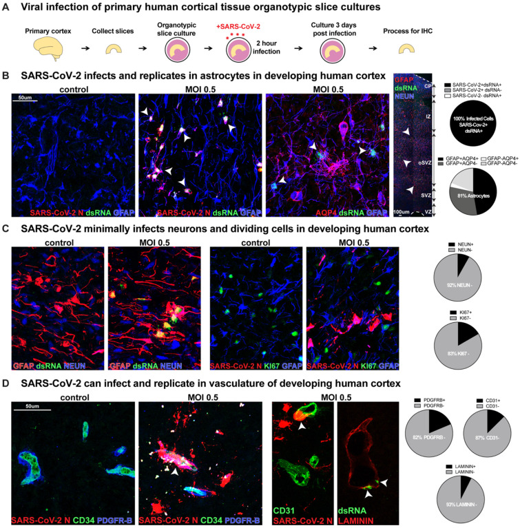
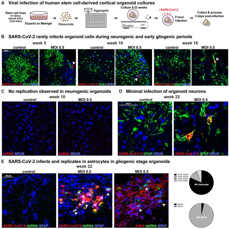
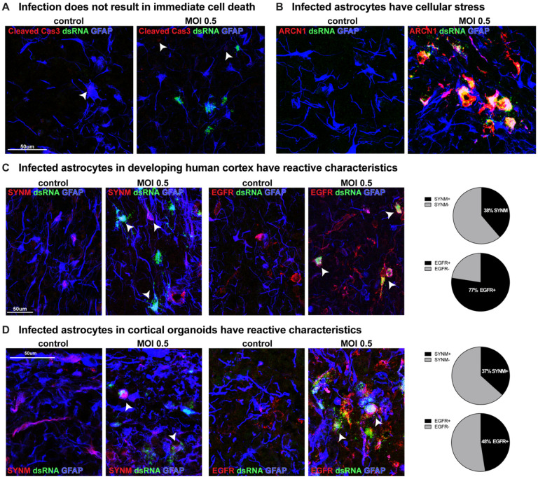
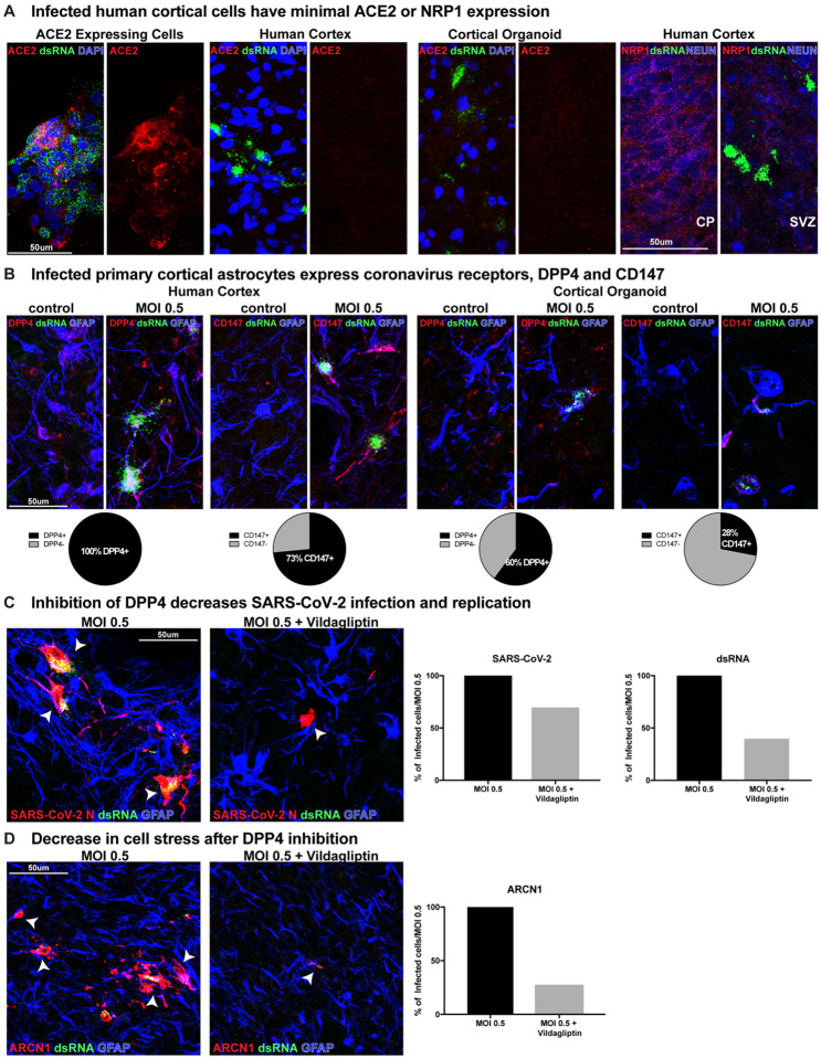
The severe acute respiratory syndrome coronavirus 2 (SARS-CoV-2) readily infects a variety of cell types impacting the function of vital organ systems, with particularly severe impact on respiratory function. It proves fatal for one percent of those infected. Neurological symptoms, which range in severity, accompany a significant proportion of COVID-19 cases, indicating a potential vulnerability of neural cell types. To assess whether human cortical cells can be directly infected by SARS-CoV-2, we utilized primary human cortical tissue and stem cell-derived cortical organoids. We find significant and predominant infection in cortical astrocytes in both primary and organoid cultures, with minimal infection of other cortical populations. Infected astrocytes had a corresponding increase in reactivity characteristics, growth factor signaling, and cellular stress. Although human cortical cells, including astrocytes, have minimal ACE2 expression, we find high levels of alternative coronavirus receptors in infected astrocytes, including DPP4 and CD147. Inhibition of DPP4 reduced infection and decreased expression of the cell stress marker, ARCN1. We find tropism of SARS-CoV-2 for human astrocytes mediated by DPP4, resulting in reactive gliosis-type injury.


Figures

Figure 1.
Figure 1.. SARS-CoV-2 Infects Astrocytes in Developing Human Cortex
A) Experimental paradigm for viral infection of human cortical tissue. Primary cortical tissue was acutely sectioned and cultured at the air-liquid interface. The next day tissue was infected with SARS-CoV-2 at MOI 0.5 and cultured for 72 hours before being collected and processed. B) SARS-CoV-2 infects GFAP+AQP4+ astrocyte cells in the developing human cortex, which are predominantly located in the subventricular zone (SVZ), where >81% of cells assayed expressed markers of astrocytes. 100% of infected cells expressed both SARS-CoV-2+ nucleocapsid (N) antibody and double-stranded (ds)RNA antibody. White arrowheads indicate dsRNA+GFAP+ infected astrocytes (dsRNA+SARS-CoV-2 N+ n=31 cells, GFAP+AQP4+ n=74 cells across two biological samples and four technical replicates). C) Few other neural types were infected, as indicated by co-labeling of SARS-CoV-2 N or dsRNA, where <8% of cells assayed expressed NEUN, a marker of cortical neurons, and <17% expressed KI67, a marker of dividing cells (NEUN n=613 cells, KI67 n=186 cells across two biological samples and four technical replicates). D) Vascular cell types can also be infected where 7% of LAMININ+ blood vessels, 13% of CD31+ endothelial cells and 18% of PDGFR-β+ mural cells have infection. White arrowheads indicate infected vascular cells (Laminin n=269 cells, CD31 n=247 cells, PDGFRB=225 cells across two biological samples and four technical replicates).
Figure 2.
Figure 2.. SARS-CoV-2 Infects Astrocytes in Cortical Organoids
A) Viral infection of cortical organoids. Human stem cells from several lines were aggregated and differentiated toward cortical identity in suspension. After 5, 10, 16 or 22 weeks of differentiation, organoids were exposed to SARS-CoV-2 for 2 hours, media was replaced and then collected 72 hours later. B) After 5, 10, or 16 weeks organoids only indicated rare infection (white arrowheads). At five and 10 weeks, SARS-CoV-2 N+ cells did not co-express SOX2, NEUN or GFAP indicating infected cells are not cortical progenitors, neurons or astrocytes and may instead be an off-target population. However, after 16 weeks, in one stem cell line a few GFAP+ astrocytes were infected. C) Although rare cells are infected at neurogenic stages, as indicated by coronavirus N antibody presence, there was no observed viral replication with dsRNA at these timepoints. D) At late gliogenic stages, by week 22 infection was readily observed in GFAP astrocytes but not NEUN+ neurons. E) 94% of infected cells stained positive for markers of astrocytes GFAP or AQP4, while only 4% are NEUN+ neurons. White arrowheads indicate SARS-CoV-2+ dsRNA+ GFAP+ AQP4+ astrocytes (GFAP+AQP4+ n=169 cells, NEUN n=143 cells).
Figure 3.
Figure 3.. SARS-CoV-2 Infection Increases Cell Stress and Reactivity in Cortical Astrocytes
A) After SAR-CoV-2 infection, there is no immediate increase in cell death in infected cells, as indicated by Cleaved Caspase 3 staining. B) Infected cells have an increase in cell stress, as indicated by the ER stress marker, ARCN1. C) Approximately one-third of infected astrocytes in primary cortical tissue express the reactive marker SYNM and three-quarters have the receptor for growth factor signaling, EGFR (SYNM n=62 cells, EGFR n=142 cells). D) The same proportion of infected organoid cells express SYNM and about one-half express EGFR (SYNM n=172 cells, EGFR n=143 cells).
Figure 4.
Figure 4.. Coronavirus Receptors, DPP4 and CD147, but not ACE2 are Expressed in Developing Human Cortex
A) ACE2 expressing cells are readily infected by SARS-CoV-2, where they have abundant ACE2 and dsRNA presence. Primary cortical tissue and cortical organoids do not have observable ACE2 protein in cortical tissue or infected cells. NRP1 is present in cortical neurons, but not in infected cells. B) Infected cortical astrocytes in the SVZ of primary cortex abundantly express coronavirus receptors DPP4 and CD147, where 100% of cells assayed have DPP4 and 73% have CD147. In cortical organoids, 60% are DPP4+ and 28% are CD147+ (Primary: DPP4 n=61cells, CD147 n=83 cells; Organoid: DPP4 n=296 cells, CD147 n=239 cells) C) Inhibition of DPP4 by Vildagliptin results in a 30% decrease in the number of SARS-CoV-2 N+ cells and a 70% reduction in dsRNA+ cells. SARS-CoV-2 N+ dsRNA+ cells are indicated by white arrowheads (SARS-CoV-2 MOI 0.5 n=1273 cells, MOI 0.5+Vildagliptin n=879 cells, dsRNA MOI 0.5 n=571 cells, MOI 0.5+Vildagliptin n=227 cells across two biological samples and three technical replicates). D) ARCN1 is broadly expressed in SARS-CoV-2 infected samples, which is reduced by 70% after DPP4 inhibition by Vildagliptin. ARCN1+ dsRNA+ cells indicated by white arrowheads (MOI 0.5 n=1224 cells, MOI 0.5+ Vildagliptin n=331 cells across two biological samples and three technical replicates).

Similar articles

Cited by

References

    1. Varatharaj A., Thomas N., Ellul M. A., Davies N. W. S., Pollak T. A., Tenorio E. L., Sultan M., Easton A., Breen G., Zandi M., Coles J. P., Manji H., Al-Shahi Salman R., Menon D. K., Nicholson T. R., Benjamin L. A., Carson A., Smith C., Turner M. R., Solomon T., Kneen R., Pett S. L., Galea I., Thomas R. H., Michael B. D., CoroNerve Study Group, Neurological and neuropsychiatric complications of COVID-19 in 153 patients: a UK-wide surveillance study. Lancet Psychiatry. 7, 875–882 (2020). - PMC - PubMed
    1. Solomon I. H., Normandin E., Bhattacharyya S., Mukerji S. S., Keller K., Ali A. S., Adams G., Hornick J. L., Padera R. F. Jr, Sabeti P., Neuropathological Features of Covid-19. N. Engl. J. Med. 383, 989–992 (2020). - PMC - PubMed
    1. Wang Q., Xu R., Volkow N. D., Increased risk of COVID-19 infection and mortality in people with mental disorders: analysis from electronic health records in the United States. World Psychiatry (2020), doi:10.1002/wps.20806. - DOI - PMC - PubMed
    1. Meinhardt J., Radke J., Dittmayer C., Franz J., Thomas C., Mothes R., Laue M., Schneider J., Brünink S., Greuel S., Lehmann M., Hassan O., Aschman T., Schumann E., Chua R. L., Conrad C., Eils R., Stenzel W., Windgassen M., Rößler L., Goebel H.-H., Gelderblom H. R., Martin H., Nitsche A., Schulz-Schaeffer W. J., Hakroush S., Winkler M. S., Tampe B., Scheibe F., Körtvélyessy P., Reinhold D., Siegmund B., Kühl A. A., Elezkurtaj S., Horst D., Oesterhelweg L., Tsokos M., Ingold-Heppner B., Stadelmann C., Drosten C., Corman V. M., Radbruch H., Heppner F. L., Olfactory transmucosal SARS-CoV-2 invasion as a port of central nervous system entry in individuals with COVID-19. Nat. Neurosci. (2020), doi:10.1038/s41593-020-00758-5. - DOI - PubMed
    1. Placantonakis D. G., Aguero-Rosenfeld M., Flaifel A., Colavito J., Inglima K., Zagzag D., Snuderl M., Louie E., Frontera J. A., Lewis A., SARS-CoV-2 Is Not Detected in the Cerebrospinal Fluid of Encephalopathic COVID-19 Patients. Front. Neurol. 11, 587384 (2020). - PMC - PubMed

Publication types

LinkOut - more resources