

Lethal multiple colon necrosis and perforation due to fulminant amoebic colitis: a surgical case report and literature review
- PMID: 33469722
- PMCID: PMC7815445
- DOI: 10.1186/s40792-020-01095-2
Lethal multiple colon necrosis and perforation due to fulminant amoebic colitis: a surgical case report and literature review
Abstract
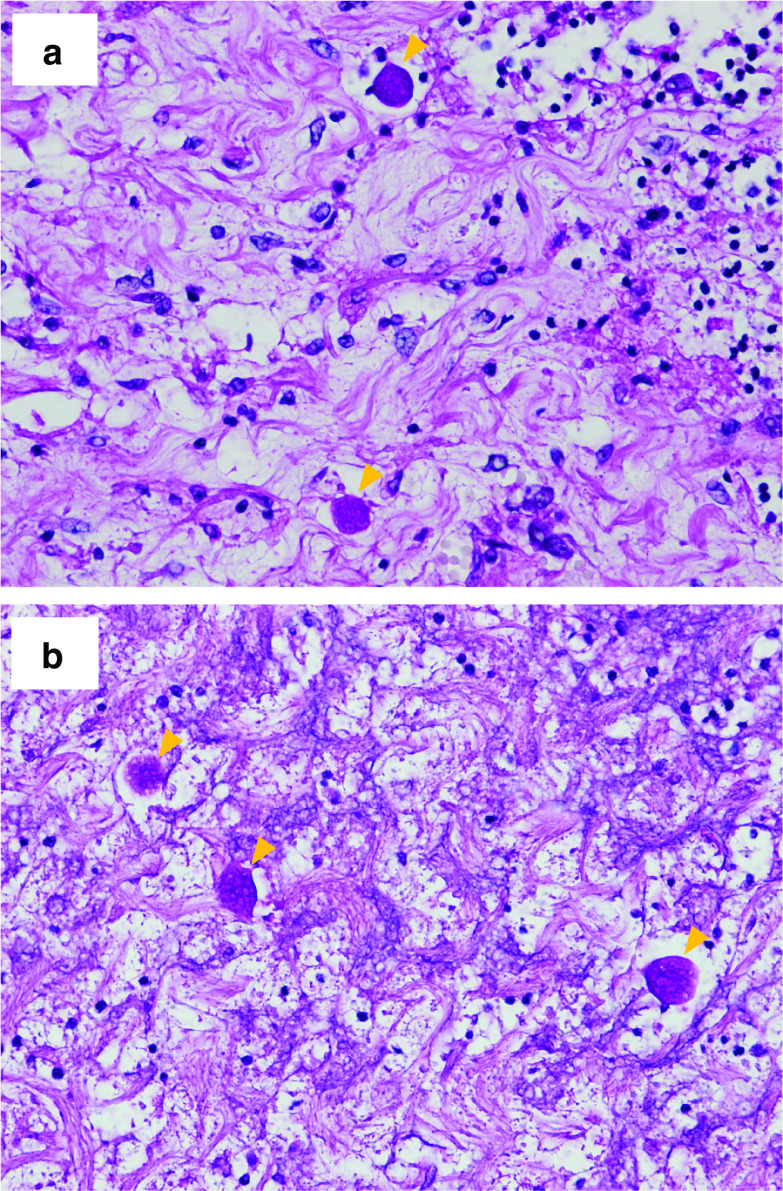
Background: Amoebiasis caused by the protozoan species Entamoeba histolytica rarely develops into fulminant amoebic colitis (FAC), but when it does, it shows an aggressive clinical course including colonic perforation, necrotizing colitis, and high mortality. Surgical treatment for FAC patients should be carried out urgently. However, even after surgery, the mortality rate can be 40-50%. Although FAC is one of the most unfavorable surgical diseases with a poor prognosis, there are a few reports on the perioperative diagnosis and management of FAC based on autopsy findings. We herein report the surgical case of a 64-year-old man who developed multiple colon necrosis and perforation due to FAC. A detailed autopsy revealed FAC as the cause of death. Additionally, we reviewed the existing literature on FAC patients who underwent surgery and followed their perioperative diagnosis and management.
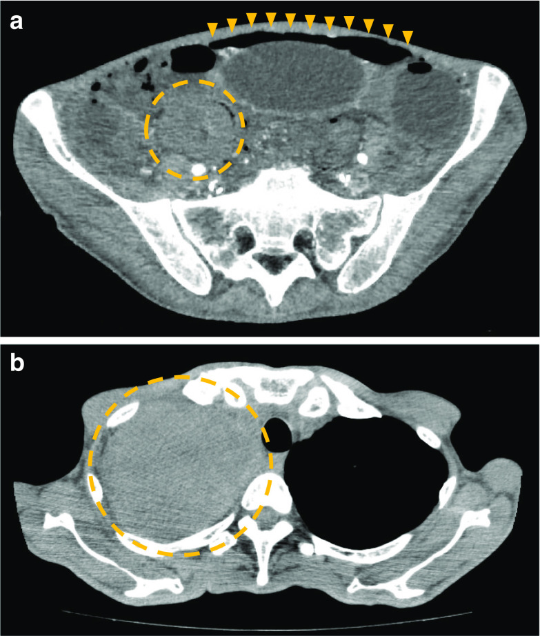
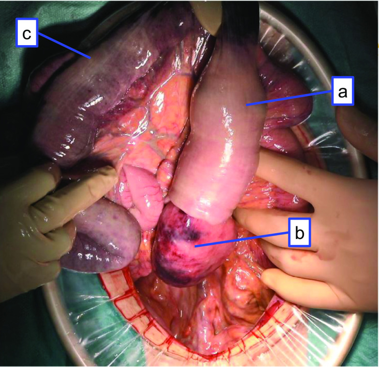
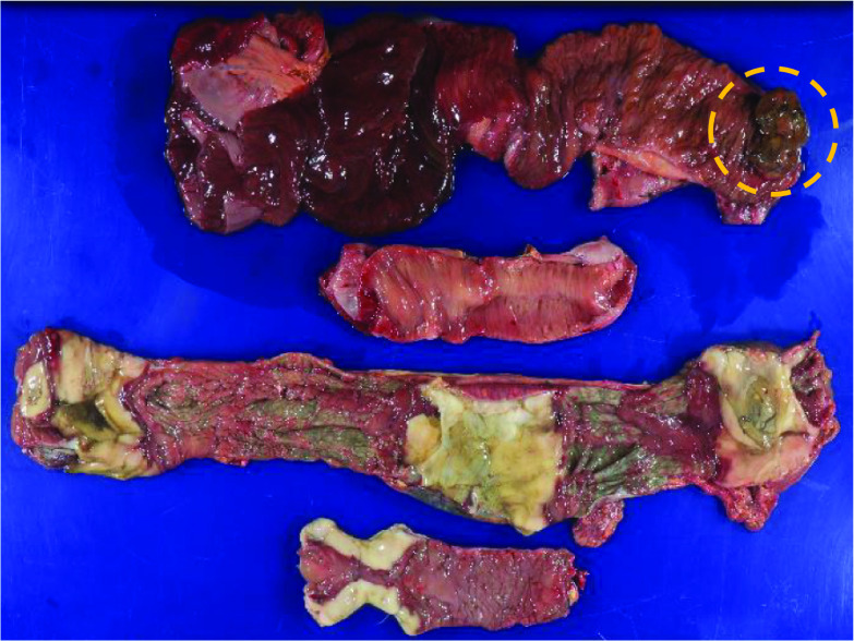
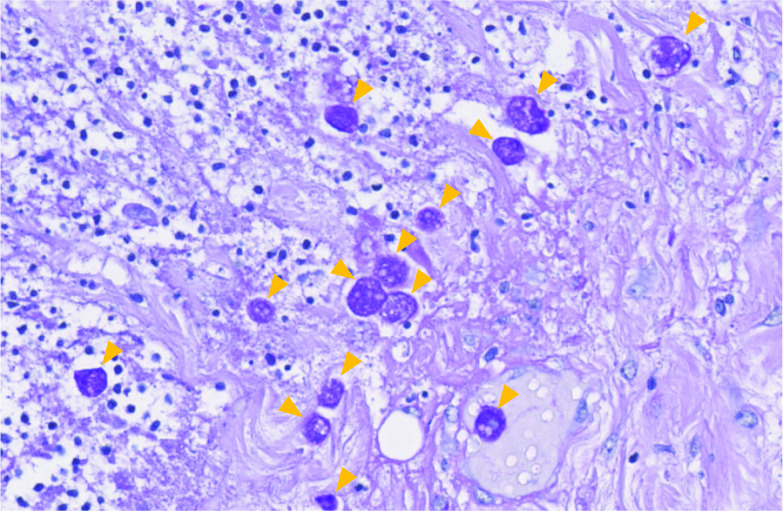
Case presentation: A 64-year-old man presented with anorexia, diarrhea, and altered consciousness on arrival to our hospital. Computed tomography revealed a large mass in the upper right lobe of his lung, and the patient was admitted for close investigation. Bloody diarrhea, lower abdominal pain, and hypotension were observed soon after admission. Urgent abdominal contrast-enhanced computed tomography scan revealed extensive intestinal ischemia, intestinal pneumatosis, and free intra-abdominal gas. The preoperative diagnosis was bowel necrosis and perforation with intussusception of the small intestinal tumor. Emergency subtotal colectomy and enterectomy were performed soon after the contrast-enhanced computed tomography. He was taken to an intensive care unit after surgery. However, he could not recover from sepsis and died with disseminated intravascular coagulation and multiple organ failure on the 10th-day post-surgery. A histopathological examination of the resected colon showed transmural necrosis and massive amoebae invasion. He was diagnosed with FAC. An autopsy revealed that he had developed pulmonary large cell carcinoma with small intestinal metastasis. The death was caused by intestinal ischemia, necrosis and the perforation of the residual bowel caused by amoebae invasion.
Conclusions: Since FAC is a lethal disease with a high mortality rate and antibiotic therapies except metronidazole are ineffective, preoperative serological testing and perioperative metronidazole therapy in FAC patients can dramatically improve their survival rates.
Keywords: Bowel perforation; Colectomy; Fulminant amoebic colitis; Intestinal necrosis; Metronidazole; Serological testing.
Conflict of interest statement
The authors declare that they have no competing interests.
Figures
References
LinkOut - more resources
Full Text Sources
Other Literature Sources
