. 2021 Jan 19;32(1):1.
doi: 10.1007/s10856-020-06475-6.

Effect of dexamethasone as osteogenic supplementation in in vitro osteogenic differentiation of stem cells from human exfoliated deciduous teeth

Affiliations

Effect of dexamethasone as osteogenic supplementation in in vitro osteogenic differentiation of stem cells from human exfoliated deciduous teeth

Mariane Beatriz Sordi et al. J Mater Sci Mater Med. .

Abstract

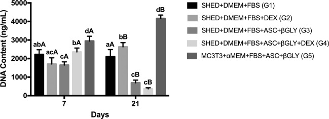
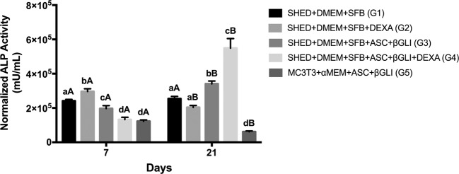
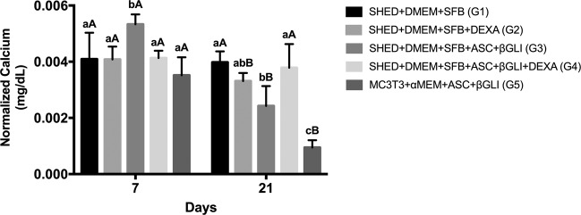
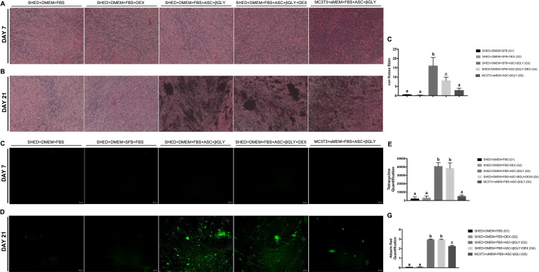
In in vitro culture systems, dexamethasone (DEX) has been applied with ascorbic acid (ASC) and β-glycerophosphate (βGLY) as culture media supplementation to induce osteogenic differentiation of mesenchymal stem cells. However, there are some inconsistencies regarding the role of DEX as osteogenic media supplementation. Therefore, this study verified the influence of DEX culture media supplementation on the osteogenic differentiation, especially the capacity to mineralize the extracellular matrix of stem cells from human exfoliated deciduous teeth (SHED). Five groups were established: G1-SHED + Dulbecco's Modified Eagles' Medium (DMEM) + fetal bovine serum (FBS); G2-SHED + DMEM + FBS + DEX; G3-SHED + DMEM + FBS + ASC + βGLY; G4-SHED + DMEM + FBS + ASC + βGLY + DEX; G5-MC3T3-E1 + α Minimal Essential Medium (MEM) + FBS + ASC + βGLY. DNA content, alkaline phosphatase (ALP) activity, free calcium quantification in the extracellular medium, and extracellular matrix mineralization quantification through staining with von Kossa, alizarin red, and tetracycline were performed on days 7 and 21. Osteogenic media supplemented with ASC and β-GLY demonstrated similar effects on SHED in the presence or absence of DEX for DNA content (day 21) and capacity to mineralize the extracellular matrix according to alizarin red and tetracycline quantifications (day 21). In addition, the presence of DEX in the osteogenic medium promoted less ALP activity (day 7) and extracellular matrix mineralization according to the von Kossa assay (day 21), and more free calcium quantification at extracellular medium (day 21). In summary, the presence of DEX in the osteogenic media supplementation did not interfere with SHED commitment into mineral matrix depositor cells. We suggest that DEX may be omitted from culture media supplementation for SHED osteogenic differentiation in vitro studies.


Conflict of interest statement

The authors declare that they have no conflict of interest. We affirm that we have no financial affiliation (e.g., employment, direct payment, stock holdings, retainers, consultant ships, patent-licensing arrangements or honoraria), or involvement with any commercial organization with a direct financial interest in the subject or materials discussed in this manuscript, nor have any such arrangements existed in the past years. Any other potential conflict of interest is disclosed.

Figures

Fig. 1
Fig. 1
DNA content assay by PicoGreen® reagent on days 7 (n = 12 for each group) and 21 (n = 12 for each group). Different lower-case letters refer to a significant difference (ANOVA/Tukey test, p < 0.05) among groups at the same experimental time. Different capital letters indicate significant differences (Student test, p < 0.05) between the experimental times for the same group
Fig. 2
Fig. 2
Alkaline phosphatase (ALP) activity by ALP Assay Kit Fluorimetric on days 7 (n = 12 for each group) and 21 (n = 12 for each group). ALP quantification results were normalized to the DNA content. Different lower-case letters refer to a significant difference (ANOVA/Tukey test, p < 0.05) among groups at the same experimental time. Different capital letters indicate significant differences (Student test, p < 0.05) between the experimental times for the same group
Fig. 3
Fig. 3
Free calcium quantification by the QuantiChromTM Calcium Assay Kit in the extracellular media on days 7 (n = 12 for each group) and 21 (n = 12 for each group). Calcium quantification results were normalized to the DNA content. Different lower-case letters refer to a significant difference (ANOVA/Tukey test, p < 0.05) among groups at the same experimental time. Different capital letters indicate significant differences (Student test, p < 0.05) between the experimental times for the same group
Fig. 4
Fig. 4
Extracellular matrix mineralization. A Von kossa staining on day 7 (n = 12 for each group) and B on day 21 (n = 12 for each group); C quantification of extracellular matrix mineralization by te Von kossa assay on day 21 using Image J software (NIH) (n = 12 for each group). Different lower-case letters refer to a significant difference (ANOVA/Tukey test, p < 0.05) among groups at the same experimental time; D tetracycline fluorescence on day 7 (n = 12 for each group) and E on day 21 (n = 12 for each group); F quantification of mineralization nodules by the tetracycline assay on day 21 (n = 12 for each group). Different lower-case letters refer to a significant difference (ANOVA/Tukey test, p < 0.05) among groups at the same experimental time; G red alizarin quantification on day 21 (n = 12 for each group). Different lower-case letters refer to a significant difference (ANOVA/Tukey test, p < 0.05) among groups at the same experimental time

Similar articles

Cited by

References

    1. Li Z, Jiang C, An S, Cheng Q, Huang Y, Wang Y, et al. Immunomodulatory properties of dental tissue-derived mesenchymal stem cells. Oral Dis. 2014;20:25–34. doi: 10.1111/odi.12086. - DOI - PubMed
    1. Bakopoulou A, About I. Stem cells of dental origin: current research trends and key milestones towards clinical application. Stem Cells Int. 2016;2016:1–20. doi: 10.1155/2016/4209891. - DOI - PMC - PubMed
    1. Wang H, Zhong QI, Yang T, Qi Y, Fu M, Yang XI, et al. Comparative characterization of SHED and DPSCs during extended cultivation in vitro. Mol Med Rep. 2018;17:6551–9. - PMC - PubMed
    1. Liu J, Yu F, Jiang B, Zhang W, Yang J, Xu G-T, et al. Characteristics and potential applications of human dental tissue-derived mesenchymal stem cells. Stem Cells. 2015;33:627–38. doi: 10.1002/stem.1909. - DOI - PubMed
    1. Miura M, Gronthos S, Zhao M, Lu B, Fisher LW, Robey PG, et al. SHED: stem cells from human exfoliated deciduous teeth. Proc Natl Acad Sci U S A. 2003;100:5807–12. doi: 10.1073/pnas.0937635100. - DOI - PMC - PubMed