. 2021 Feb;88(2):141-157.
doi: 10.1002/mrd.23449. Epub 2021 Jan 20.

Genome-wide chromatin occupancy of BRDT and gene expression analysis suggest transcriptional partners and specific epigenetic landscapes that regulate gene expression during spermatogenesis

Affiliations

Genome-wide chromatin occupancy of BRDT and gene expression analysis suggest transcriptional partners and specific epigenetic landscapes that regulate gene expression during spermatogenesis

Yoon Ra Her et al. Mol Reprod Dev. 2021 Feb.

Abstract

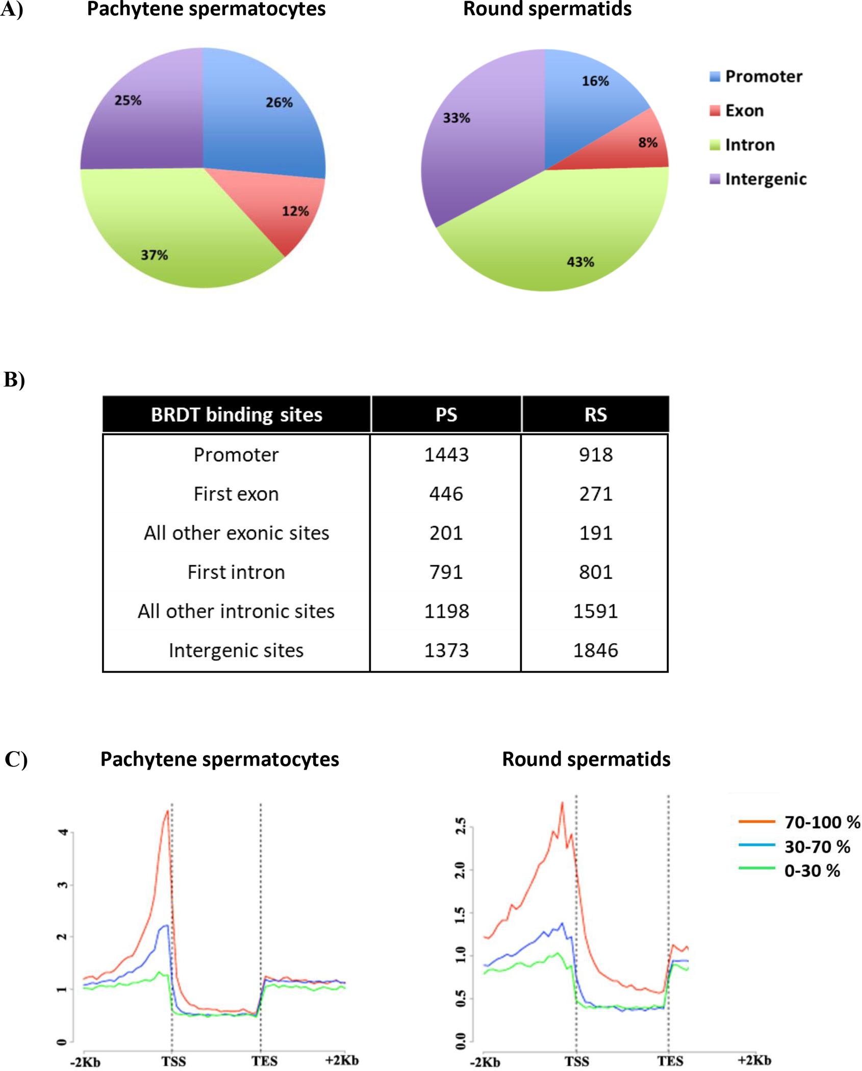
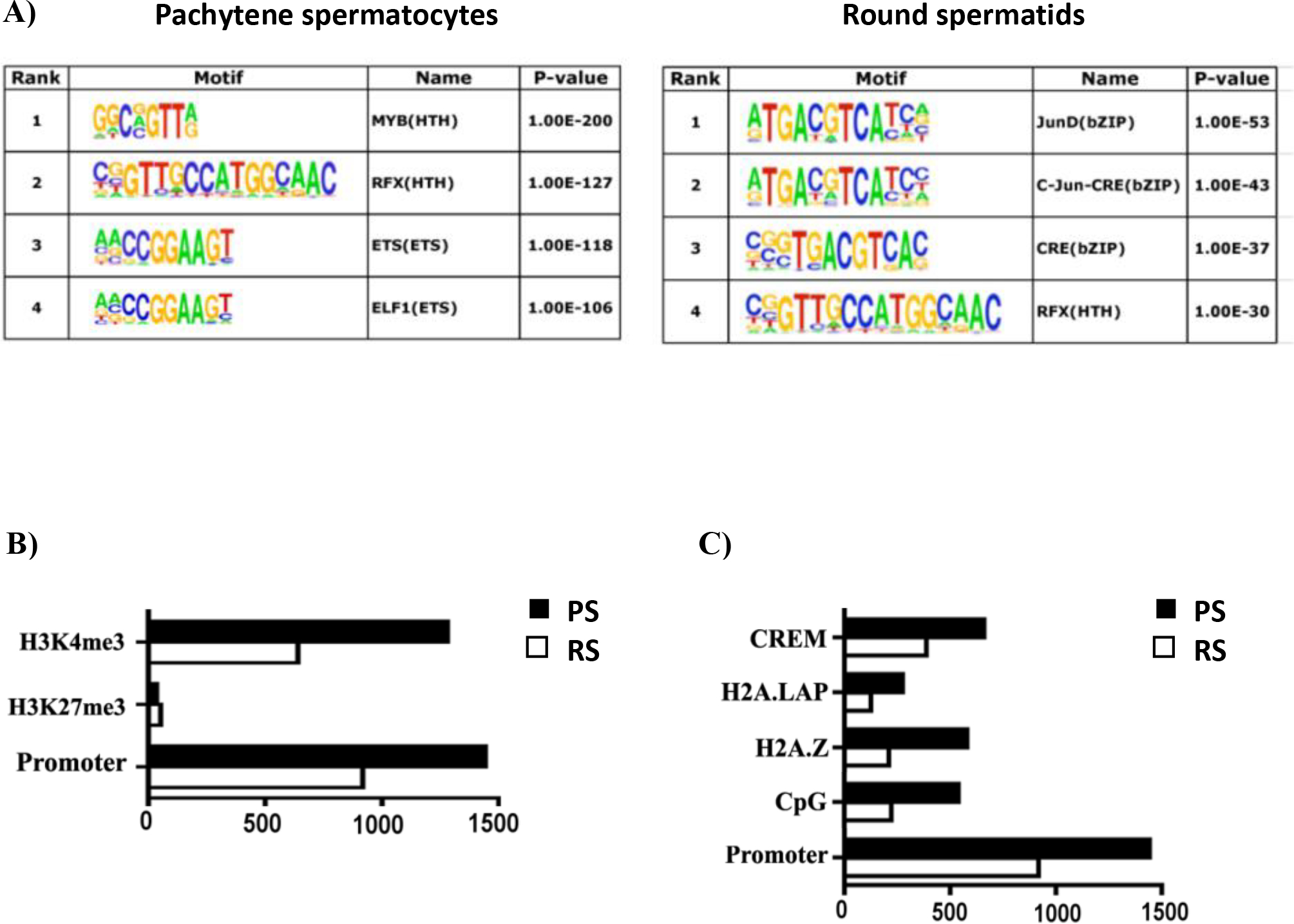
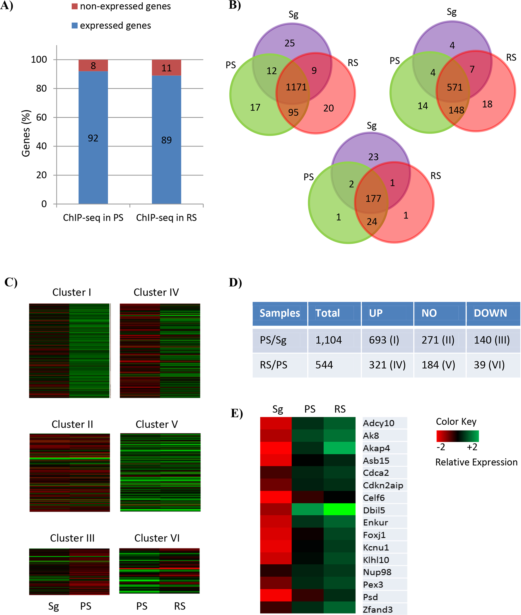
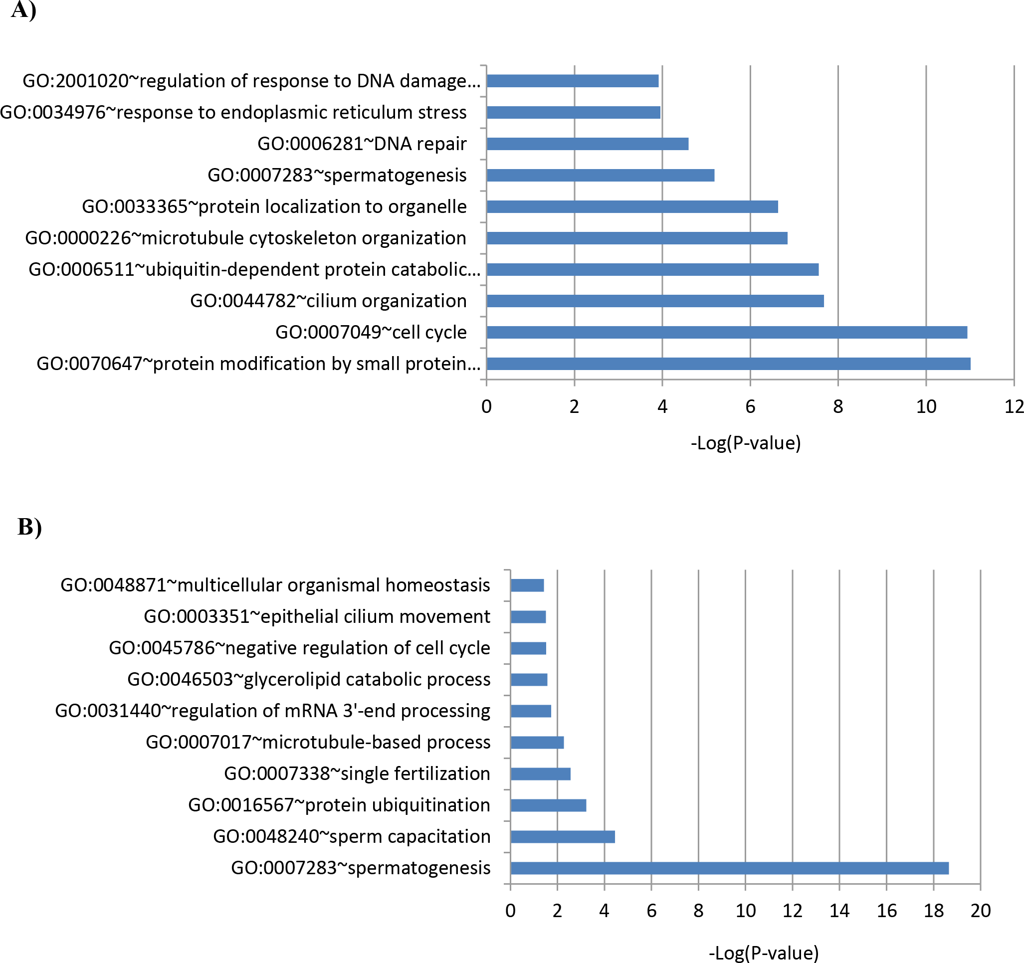
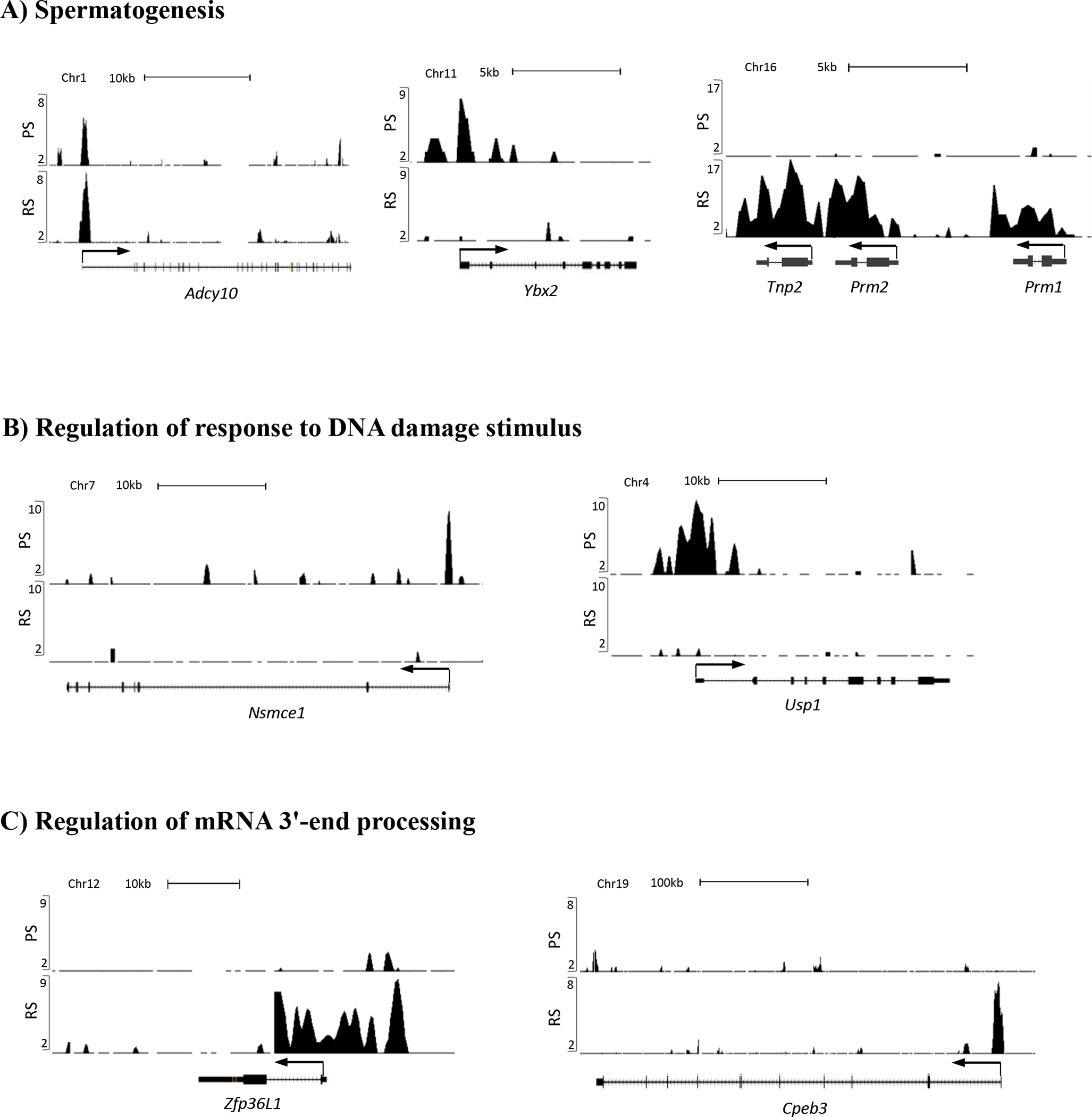
BRDT, a member of the BET family of double bromodomain-containing proteins, is essential for spermatogenesis in the mouse and has been postulated to be a key regulator of transcription in meiotic and post-meiotic cells. To understand the function of BRDT in these processes, we first characterized the genome-wide distribution of the BRDT binding sites, in particular within gene units, by ChIP-Seq analysis of enriched fractions of pachytene spermatocytes and round spermatids. In both cell types, BRDT binding sites were mainly located in promoters, first exons, and introns of genes. BRDT binding sites in promoters overlapped with several histone modifications and histone variants associated with active transcription, and were enriched for consensus sequences for specific transcription factors, including MYB, RFX, ETS, and ELF1 in pachytene spermatocytes, and JunD, c-Jun, CRE, and RFX in round spermatids. Subsequent integration of the ChIP-seq data with available transcriptome data revealed that stage-specific gene expression programs are associated with BRDT binding to their gene promoters, with most of the BDRT-bound genes being upregulated. Gene Ontology analysis further identified unique sets of genes enriched in diverse biological processes essential for meiosis and spermiogenesis between the two cell types, suggesting distinct developmentally stage-specific functions for BRDT. Taken together, our data suggest that BRDT cooperates with different transcription factors at distinctive chromatin regions within gene units to regulate diverse downstream target genes that function in male meiosis and spermiogenesis.

Keywords: BRDT; male meiosis; spermiogenesis; transcription.


Conflict of interest statement

Conflict of Interest Statement

The authors declare they have no competing interests.

Figures

Figure 1.
Figure 1.
High–resolution genome-wide analysis of BRDT-containing complexes occupancy in pachytene spermatocytes (PS) and round spermatids (RS) of wild type mice. (A-B) Pie chart shows genomic annotation of location and number of BRDT binding sites across the genome in wild type PS and RS. (C) High-resolution analysis of average tag density across a gene body in wild type mouse testicular cells. In each plot, genes were classified into 3 groups based on expression levels in PS and RS: highly expressed (the top 30% of genes indexed by expression levels, red lines), medium expression (the middle 40% of genes, blue lines) and lowly expressed (the lowest 40% of genes, green lines). Average tag density across the gene unit was plotted for each group. TSS, Transcription Start Sites; TES, Transcription End Sites
Figure 2.
Figure 2.
Characterization of BRDT binding site occupancy in pachytene spermatocytes (PS) and round spermatids (RS). (A) BRDT in vivo consensus binding site predicted by MEME and presented as a sequence LOGO. A total of 500 peak regions were analyzed for overrepresented motifs. Enriched transcription factor motifs lying within BRDT binding sites in PS and RS. (B) Correlative analysis of H3K4me3, H3K27me3 with BRDT binding site occupancy. (C) Correlative analysis of CpG, H2AZ, H2A.LAP, CREM with BRDT binding site occupancy. The x-axis in (B) and (C) refers to the number of associations with BRDT binding sites.
Figure 3.
Figure 3.
Comparative BRDT ChIP-seq and transcriptome analysis corresponding to the staged mouse spermatogenic cells. (A) Respective proportions of BRDT-bound genes in PS and RS, which are expressed and non-expressed in PS and RS, respectively. A total of 1,444 and 888 BRDT-bound genes in PS and RS, respectively were included. (B) Venn diagram of the genes expressed in three cell types, Sg, PS and RS, within the BRDT-bound genes either in PS (left) or RS (right) and both PS and RS (bottom). Distinct and overlapping expression between and among the cell types is shown. Purple, green and red cycles represent Sg, PS, and RS, respectively. (C) Heat maps showing the expression of 6 clusters of BRDT-bound genes that are expressed in PS and RS. The following comparisons were performed: PS vs. Sg; RS vs. PS (Log2 Fold Change (|FC|) ≥ 2; p < 0.01). Columns indicate specific cell types and rows indicate genes. The low versus high expression levels are represented on a red to green scale. (D) Total number of up-, down-, and non-differentially regulated genes belonging to each of the Clusters (|FC| ≥ 2; p < 0.01 (E) Heat map showing relative expression levels of the 16 genes overlapping between Cluster I and Cluster IV. The low versus high expression levels are represented on a red to green scale.
Figure 4.
Figure 4.
Enriched biological process GO terms of the genes belonging to (A) Cluster I (up-regulated genes in PS vs. Sg) and (B) Cluster IV (up-regulated genes in RS vs. PS).
Figure 5.
Figure 5.
UCSC Genome Browser screenshot localization of BRDT ChIP-seq signals at the gene promoters implicated in (A) spermatogenesis, (B) regulation of response to DNA damage stimulus, and (C) regulation of mRNA 3’-end processing. Graphic representation of ChIP-Seq reads derived from pachytene spermatocytes (PS) and round spermatids (RS) are shown on the vertical axis on all panels. ChIP-seq signals are displayed as normalized RPM (reads per million reads) values.

Similar articles

Cited by

References

    1. Agricola E, Randall RA, Gaarenstroom T, Dupont S, & Hill CS (2011). Recruitment of TIF1gamma to chromatin via its PHD finger-bromodomain activates its ubiquitin ligase and transcriptional repressor activities. Mol Cell, 43(1), 85–96. doi:10.1016/j.molcel.2011.05.020 - DOI - PubMed
    1. Al-Harthi L, & Roebuck KA (1998). Human immunodeficiency virus type-1 transcription: role of the 5’-untranslated leader region (review). Int J Mol Med, 1(5), 875–881. Retrieved from http://www.ncbi.nlm.nih.gov/pubmed/9852310 - PubMed
    1. Alcivar AA, Hake LE, Kwon YK, & Hecht NB (1991). junD mRNA expression differs from c-jun and junB mRNA expression during male germinal cell differentiation. Mol Reprod Dev, 30(3), 187–193. doi:10.1002/mrd.1080300304 - DOI - PubMed
    1. Ayoubi TAY, & VanDeVen WJM (1996). Regulation of gene expression by alternative promoters. Faseb Journal, 10(4), 453–460. Retrieved from <Go to ISI>://WOS:A1996UE55700009 - PubMed
    1. Bailey TL, Boden M, Buske FA, Frith M, Grant CE, Clementi L, . . . Noble WS (2009). MEME SUITE: tools for motif discovery and searching. Nucleic Acids Res, 37(Web Server issue), W202–208. doi:10.1093/nar/gkp335 - DOI - PMC - PubMed

Publication types