. 2021 Dec 17;117(14):2767-2780.
doi: 10.1093/cvr/cvab014.

Chitinase 3 like 1 is a regulator of smooth muscle cell physiology and atherosclerotic lesion stability

Affiliations

Chitinase 3 like 1 is a regulator of smooth muscle cell physiology and atherosclerotic lesion stability

Pavlos Tsantilas et al. Cardiovasc Res. .

Abstract

Aims: Atherosclerotic cerebrovascular disease underlies the majority of ischaemic strokes and is a major cause of death and disability. While plaque burden is a predictor of adverse outcomes, plaque vulnerability is increasingly recognized as a driver of lesion rupture and risk for clinical events. Defining the molecular regulators of carotid instability could inform the development of new biomarkers and/or translational targets for at-risk individuals.

Methods and results: Using two independent human endarterectomy biobanks, we found that the understudied glycoprotein, chitinase 3 like 1 (CHI3L1), is up-regulated in patients with carotid disease compared to healthy controls. Further, CHI3L1 levels were found to stratify individuals based on symptomatology and histopathological evidence of an unstable fibrous cap. Gain- and loss-of-function studies in cultured human carotid artery smooth muscle cells (SMCs) showed that CHI3L1 prevents a number of maladaptive changes in that cell type, including phenotype switching towards a synthetic and hyperproliferative state. Using two murine models of carotid remodelling and lesion vulnerability, we found that knockdown of Chil1 resulted in larger neointimal lesions comprised by de-differentiated SMCs that failed to invest within and stabilize the fibrous cap. Exploratory mechanistic studies identified alterations in potential downstream regulatory genes, including large tumour suppressor kinase 2 (LATS2), which mediates macrophage marker and inflammatory cytokine expression on SMCs, and may explain how CHI3L1 modulates cellular plasticity.

Conclusion: CHI3L1 is up-regulated in humans with carotid artery disease and appears to be a strong mediator of plaque vulnerability. Mechanistic studies suggest this change may be a context-dependent adaptive response meant to maintain vascular SMCs in a differentiated state and to prevent rupture of the fibrous cap. Part of this effect may be mediated through downstream suppression of LATS2. Future studies should determine how these changes occur at the molecular level, and whether this gene can be targeted as a novel translational therapy for subjects at risk of stroke.

Keywords: CHI3L1; Carotid stenosis; Dedifferentiation; Stroke; Vascular smooth muscle cells; Vulnerable plaque.


Figures

None
Graphical abstract
Figure 1
Figure 1
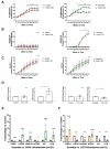
CHI3L1 mRNA and protein expression in human plaques. (A) BiKE mRNA expression analysis of CHI3L1 by microarrays. Normal arteries: n =10 (iliac and one aorta), carotid plaques: n=127, asympt: n=40 plaques from patients asymptomatic at surgery, sympt: n=87 plaques from patients symptomatic at surgery, event: indicates previous cardio- or cerebrovascular event n=27, no event n=98. CHI3L1 mRNA expression fold-change: plaques vs. normal =+33.2, P<0.0001, sympt vs. asympt =+1.6, P<0.0001, yes vs. no =+1.5, P=0.04. Statistical test: linear regression model and Student’s t-test. (B) BiKE protein quantification of CHI3L1 by LC-MS. Adjacent tissues: n=18 matched adjacent arterial tissues used as controls, plaques: n=18 carotid plaques, asympt: n=18 plaques from patients asymptomatic at surgery, sympt: n=18 plaques from patients symptomatic at surgery. CHI3L1 protein fold-change: plaques vs. adjacent =+1.23, P=0.0005, sympt vs. asympt =+1.38, P=0.0001. Statistical test: see (A). (C) CHI3L1 mRNA expression of laser capture micro-dissection of human fibrous cap of 10 stable plaques vs. 10 unstable plaques of the Munich Vascular Biobank. Scale bar: 3 mm. CHI3L1 expression fold-change: +2.1, P=0.0089. Shown as mean±SEM. Statistical test: Student’s t-test. (D) H&E and immunohistochemistry of human atherosclerotic plaques from the Munich Vascular Biobank. CHI3L1 and αSMA co-stain in advanced unstable plaques. Stable plaques show low CHI3L1 expression and minimal co-localization with αSMA. Scale bar: 2 mm (stable), 3 mm (unstable). *P<0.05; **P<0.01; ***P<0.001, ****P<0.0001; n.s., non-significant; TMT, tandem mass tag; M, media; NC, necrotic core; FC, fibrous cap; H&E, haematoxylin and eosin stain; CHI3L1, chitinase 3 like 1; αSMA, alpha smooth muscle actin; LGALS3, galectin-3.
Figure 2
Figure 2
Live-cell imaging analysis to determine the functional role of CHI3L1 and expression of vascular SMC and macrophage markers upon CHI3L1 and LATS2 modulation. Results are presented as the mean±standard deviation. Statistical test IncuCyte: multiple t-tests corrected for multiple comparisons (Holm–Sidak) for each individual time point and two-way repeated measures ANOVA for the whole time course. (A) Proliferation: occupied area (confluence %, n=3), CHI3L1-knockdown: P=0.0021; CHI3L1-overexpression: P=0.0059. (B) Apoptosis: IncuCyte apoptosis assay (n=3). CHI3L1-knockdown: P<0.0001; CHI3L1-overexpression: P=0.4210. (C) Migration: IncuCyte scratch wound assay (n=3). CHI3L1-knockdown: P=0.1978; CHI3L1-overexpression: P=0.2526. (D) Measurements of relative mRNA levels of the macrophage biomarker (CD68) and vascular SMC biomarker ACTA2 (αSMA). Differences between groups were presented as fold-change with error bars as standard deviation (n=3). Statistical test: Student’s t-test. Knockdown of CHI3L1: CD68 expression (fold-change 2.2, P=0.0335), ACTA2 expression (fold-change 0.7, P=0.0455). (E) Treatment of HCtASMC with anti-CHI3L1 siRNA before 12 h of oxLDL (100 µg/mL) exposure (n=5). Quantification of mRNA expression with qPCR: CHI3L1 fold-change 0.2, P<0.0001; LATS2 fold-change 4.9, P=0.0002; LGALS3 fold-change 2.9, P=0.006; ACTA2 fold-change 0.2, P=0.0007; IL6 fold-change 5.7, P <0.0001; IL1β, fold-change 4.8, P=0.0002. Statistical test: Multiple t-tests corrected for multiple comparisons using the Holm–Sidak method. (F) Treatment of HCtASMC with anti-LATS2 siRNA before 12 h of oxLDL (100 µg/mL) exposure (n=5). Quantification of mRNA expression with qPCR: CHI3L1 fold-change 1.2, P=0.1; LATS2 fold-change 0.2, P=0.001; LGALS3 fold-change 0.3, P=0.004; ACTA2 fold-change 1.4, P=0.4; IL6 fold-change 0.3, P=0.007; IL1β, fold-change 0.2, P=0.001. Statistical test: Multiple t-tests corrected for multiple comparisons using the Holm–Sidak method. *P<0.05; **P<0.01; ***P<0.001; ****P<0.0001. n.s., non-significant; OE, overexpression of CHI3L1; KD, knockdown of CHI3L1; Cas3/7, Caspase 3/7; CD68, cluster of differentiation 68; ACTA2, actin alpha 2; CHI3L1, chitinase 3 like 1, HCtASMC, human carotid artery SMCs; LATS2, large tumour suppressor kinase 2; LGALS3, galectin-3; IL6, interleukin 6; IL1β, interleukin 1 beta; oxLDL, oxidized low-density lipoprotein.
Figure 3
Figure 3
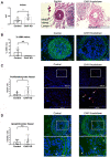
Functional role of Chil1 in vascular remodelling in the murine CLM. (A) Area intima was significantly increased in the knockdown group (P=0.0238). Scale bar: 200 µm. (B) Measurement of αSMA positive area. Threshold area is divided through the total ROI area and described as ‘% αSMA+ area’. Knockdown of Chil1 significantly decreased amount of αSMA staining in intima (P=0.0067). Scale bar: 100 µm. (C) Proliferation-index in the knockdown group was significantly increased (P=0.0419). Scale bar: 100 µm. (D) Apoptotic index: non-significant (P=0.244). Scale bar: 100 µm. Statistical test for (A–D): Student’s t-test. N=9 vs. 7 mice were used for all experiments (A–D). *P<0.05; **P<0.01; ***P<0.001; n.s., non-significant; αSMA, alpha smooth muscle actin; EdU, 5-ethynyl-2’-deoxyuridine; TUNEL, TdT-mediated dUTP-biotin nick end labelling; Chil1, chitinase 3 like 1.
Figure 4
Figure 4
Functional role of Chil1 on plaque vulnerability in the murine PRM. (A) Threshold area is divided through the total ROI area and described as ‘%+area’. Percentage of αSMA-positive area was significantly reduced in Chil1 knockdown group (P=0.015). No effect was detected when looking at percentage of MAC3-positive area (P=0.29). N=6 vs. 5 mice. Statistical test: Student’s t-test. Scale bar: 100 µm. (B) Representative picture of images stained with cross-linked fibrin to indicate atherothrombotic events and unstable/ruptured plaques in the inducible PRM in Apoe−/−. White arrows indicate increased fibrin signal mainly in the Anti-Chil1 group. (A and B): N=6 vs. 5 mice. Scale bar: 100 µm. (C) PRM of Tomato mice. Double immunofluorescent imaging (red fluorescence = initial MYH11 cells, green fluorescence = αSMA positive cells) supports results of reduced αSMA expression through down-regulation of Chil1 (red-dominant plaque in the knockdown group). Scale bar: 100 µm. N=2 vs. 2 mice. *P<0.05; **P<0.01; ***P<0.001; n.s., non-significant; αSMA, alpha smooth muscle actin; MAC3, macrophage antigen 3; MYH11, myosin heavy chain 11; Chil1, chitinase 3 like 1.
Figure 5
Figure 5
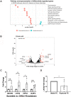
RNAseq to detect downstream effects of CHI3L1 in human carotid artery SMCs. (A) KEGG pathway analysis revealed 12 significantly differentially regulated pathways relevant for disease and tissue phenotype. This approach identifies genes belonging to a given pathway that are significantly up- or down-regulated after CHI3L1 overexpression. Numbers in circles represent the number of genes in the specific pathway that are down-regulated (orange circle) or up-regulated (turquoise circle). (B) A total of 33 genes (red dots) were identified as being significantly different between groups (fold-change >1.5 and Benjamini–Hochberg FDR<0.05). The six most significantly down-regulated genes upon CHI3L1 overexpression were chosen for further analysis. (C) All six genes were analysed in HCtASMCs in response to CHI3L1 knockdown using siRNA (n=4 vs. 4). Two genes (LATS2, P=0.005; HIPK2, P=0.022; ABLIM3, P= 0.190; ULBP2, P=0.190) were significantly increased upon CHI3L1 inhibition. Statistical test: multiple t-tests corrected for multiple comparisons using the Holm–Sidak method. (D) mRNA expression of the two genes being deregulated upon CHI3L1 modulation (inhibition and overexpression) in human fibrous caps from unstable vs. stable lesions (n=10 vs. 10). Here, only LATS2 appeared significantly regulated (P=0.0082). Statistical test: Student’s t-test. *P<0.05; **P<0.01; ***P<0.001; n.s., non-significant; ABLIM3, actin-binding LIM protein 3; LATS2, large tumour suppressor kinase 2; HIPK2, homeodomain-interacting protein kinase 2; ULBP2, UL16 binding protein; SCN3A, sodium channel, voltage-gated, type III, alpha subunit; CHI3L1, chitinase 3 like 1; TNFRSF10D, tumour necrosis factor receptor superfamily member 10D; FC, fibrous cap; SC, scramble; KD, knockdown; FDR, false discovery rate.

Comment in

References

    1. World Health Organisation - Healthinfo on Global Burden Disease. https://www.who.int/healthinfo/global_burden_disease/GHE2016_Deaths_Glob... (6 February 2020, date last accessed).
    1. Lozano R, Naghavi M, Foreman K, Lim S, Shibuya K, Aboyans V, Abraham J, Adair T, Aggarwal R, Ahn SY, Alvarado M, Anderson HR, Anderson LM, Andrews KG, Atkinson C, Baddour LM, Barker-Collo S, Bartels DH, Bell ML, Benjamin EJ, Bennett D, Bhalla K, Bikbov B, Bin Abdulhak A, Birbeck G, Blyth F, Bolliger I, Boufous S, Bucello C, Burch M, Burney P, Carapetis J, Chen H, Chou D, Chugh SS, Coffeng LE, Colan SD, Colquhoun S, Colson KE, Condon J, Connor MD, Cooper LT, Corriere M, Cortinovis M, de Vaccaro KC, Couser W, Cowie BC, Criqui MH, Cross M, Dabhadkar KC, Dahodwala N, De Leo D, Degenhardt L, Delossantos A, Denenberg J, Des Jarlais DC, Dharmaratne SD, Dorsey ER, Driscoll T, Duber H, Ebel B, Erwin PJ, Espindola P, Ezzati M, Feigin V, Flaxman AD, Forouzanfar MH, Fowkes FG, Franklin R, Fransen M, Freeman MK, Gabriel SE, Gakidou E, Gaspari F, Gillum RF, Gonzalez-Medina D, Halasa YA, Haring D, Harrison JE, Havmoeller R, Hay RJ, Hoen B, Hotez PJ, Hoy D, Jacobsen KH, James SL, Jasrasaria R, Jayaraman S, Johns N, Karthikeyan G, Kassebaum N, Keren A, Khoo JP, Knowlton LM, Kobusingye O, Koranteng A, Krishnamurthi R, Lipnick M, Lipshultz SE, Ohno SL, Mabweijano J, MacIntyre MF, Mallinger L, March L, Marks GB, Marks R, Matsumori A, Matzopoulos R, Mayosi BM, McAnulty JH, McDermott MM, McGrath J, Mensah GA, Merriman TR, Michaud C, Miller M, Miller TR, Mock C, Mocumbi AO, Mokdad AA, Moran A, Mulholland K, Nair MN, Naldi L, Narayan KM, Nasseri K, Norman P, O'Donnell M, Omer SB, Ortblad K, Osborne R, Ozgediz D, Pahari B, Pandian JD, Rivero AP, Padilla RP, Perez-Ruiz F, Perico N, Phillips D, Pierce K, Pope CA III, Porrini E, Pourmalek F, Raju M, Ranganathan D, Rehm JT, Rein DB, Remuzzi G, Rivara FP, Roberts T, De Leon FR, Rosenfeld LC, Rushton L, Sacco RL, Salomon JA, Sampson U, Sanman E, Schwebel DC, Segui-Gomez M, Shepard DS, Singh D, Singleton J, Sliwa K, Smith E, Steer A, Taylor JA, Thomas B, Tleyjeh IM, Towbin JA, Truelsen T, Undurraga EA, Venketasubramanian N, Vijayakumar L, Vos T, Wagner GR, Wang M, Wang W, Watt K, Weinstock MA, Weintraub R, Wilkinson JD, Woolf AD, Wulf S, Yeh PH, Yip P, Zabetian A, Zheng ZJ, Lopez AD, Murray CJ, AlMazroa MA, Memish ZA.. Global and regional mortality from 235 causes of death for 20 age groups in 1990 and 2010: a systematic analysis for the global burden of disease study 2010. Lancet 2012;380:2095–2128. - PMC - PubMed
    1. Furie KL, Kasner SE, Adams RJ, Albers GW, Bush RL, Fagan SC, Halperin JL, Johnston SC, Katzan I, Kernan WN, Mitchell PH, Ovbiagele B, Palesch YY, Sacco RL, Schwamm LH, Wassertheil-Smoller S, Turan TN, Wentworth D, American Heart Association Stroke Council CoCNCoCC, Interdisciplinary Council on Quality of C, Outcomes R. Guidelines for the prevention of stroke in patients with stroke or transient ischemic attack: a guideline for healthcare professionals from the American Heart Association/American Stroke Association. Stroke 2011;42:227–276. - PubMed
    1. Libby P, Ridker PM, Hansson GK.. Progress and challenges in translating the biology of atherosclerosis. Nature 2011;473:317–325. - PubMed
    1. Naylor AR, Ricco JB, de Borst GJ, Debus S, de Haro J, Halliday A, Hamilton G, Kakisis J, Kakkos S, Lepidi S, Markus HS, McCabe DJ, Roy J, Sillesen H, van den Berg JC, Vermassen F, Esvs Guidelines C, Kolh P, Chakfe N, Hinchliffe RJ, Koncar I, Lindholt JS, Vega de Ceniga M, Verzini F, Esvs Guideline R, Archie J, Bellmunt S, Chaudhuri A, Koelemay M, Lindahl AK, Padberg F, Venermo M.. Editor's choice - management of atherosclerotic carotid and vertebral artery disease: 2017 clinical practice guidelines of the European Society for Vascular Surgery (ESVS). Eur J Vasc Endovasc Surg 2018;55:3–81. - PubMed

Publication types

MeSH terms