. 2021 Jul;288(14):4311-4331.
doi: 10.1111/febs.15721. Epub 2021 Feb 18.

Pharmacological validation of TDO as a target for Parkinson's disease

Affiliations

Pharmacological validation of TDO as a target for Parkinson's disease

Paula Perez-Pardo et al. FEBS J. 2021 Jul.

Abstract

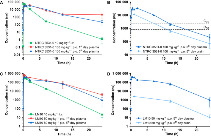
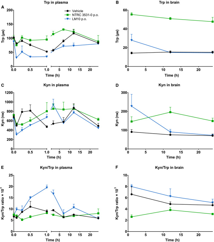
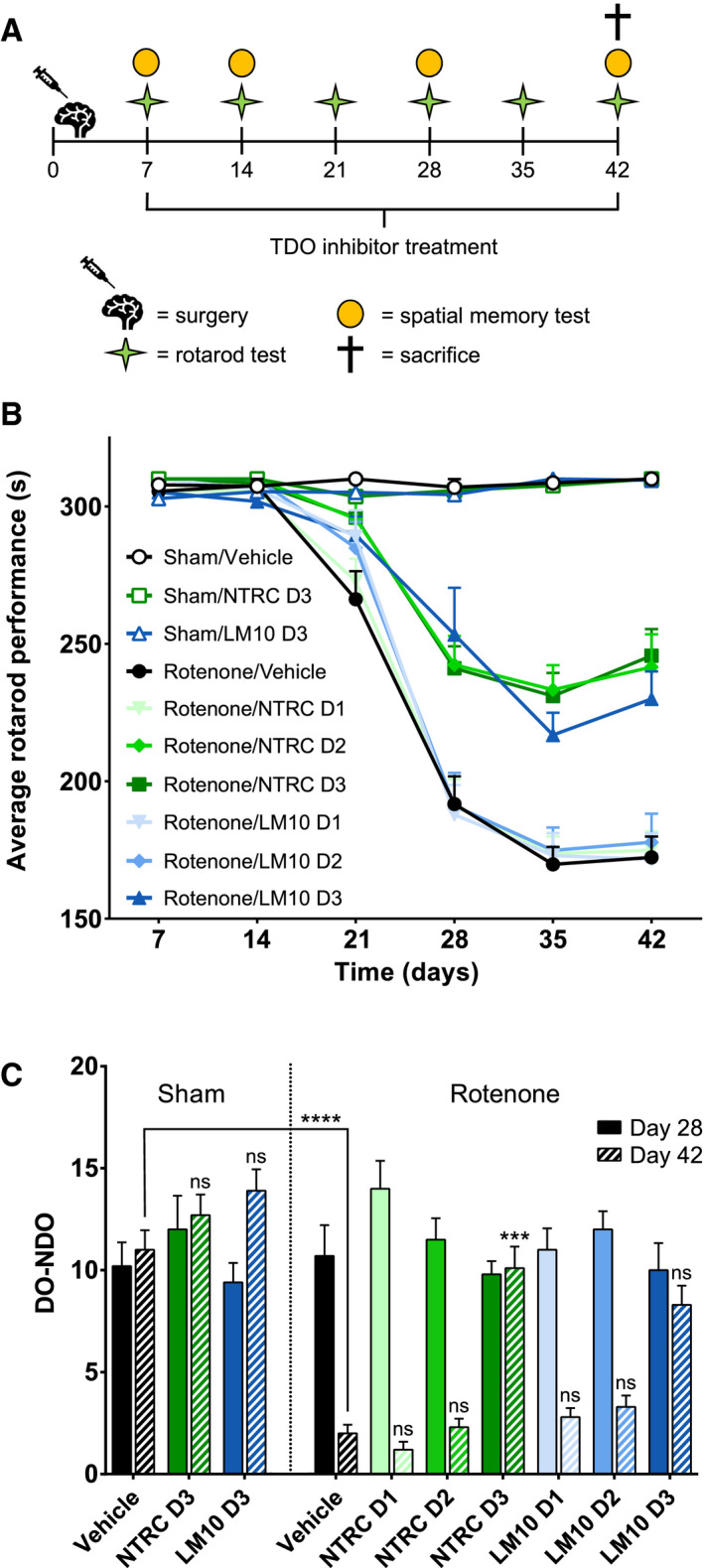
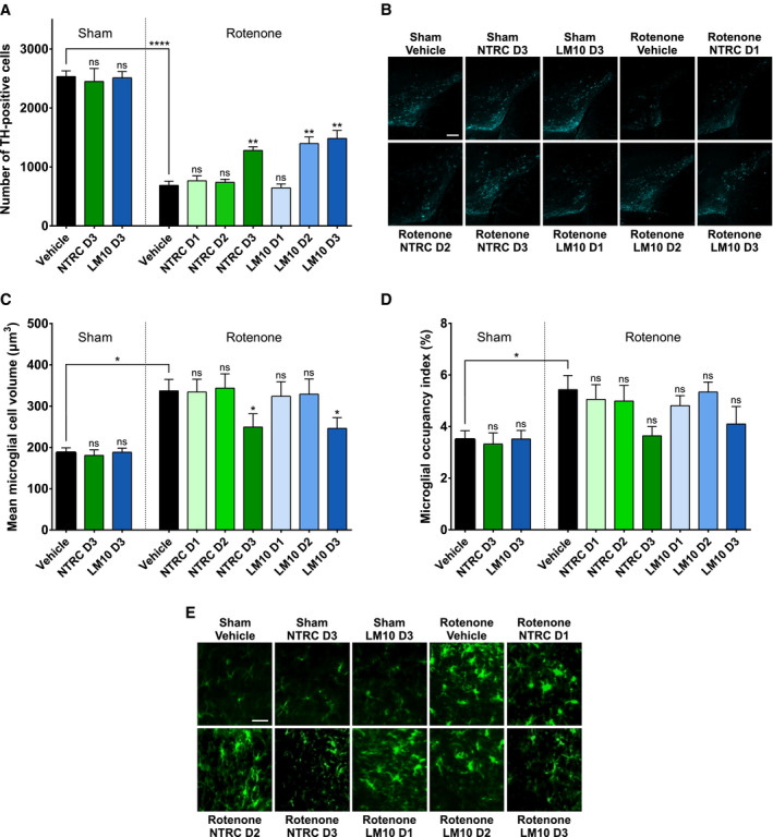
Parkinson's disease patients suffer from both motor and nonmotor impairments. There is currently no cure for Parkinson's disease, and the most commonly used treatment, levodopa, only functions as a temporary relief of motor symptoms. Inhibition of the expression of the L-tryptophan-catabolizing enzyme tryptophan 2,3-dioxygenase (TDO) has been shown to inhibit aging-related α-synuclein toxicity in Caenorhabditis elegans. To evaluate TDO inhibition as a potential therapeutic strategy for Parkinson's disease, a brain-penetrable, small molecule TDO inhibitor was developed, referred to as NTRC 3531-0. This compound potently inhibits human and mouse TDO in biochemical and cell-based assays and is selective over IDO1, an evolutionary unrelated enzyme that catalyzes the same reaction. In mice, NTRC 3531-0 increased plasma and brain L-tryptophan levels after oral administration, demonstrating inhibition of TDO activity in vivo. The effect on Parkinson's disease symptoms was evaluated in a rotenone-induced Parkinson's disease mouse model. A structurally dissimilar TDO inhibitor, LM10, was evaluated in parallel. Both inhibitors had beneficial effects on rotenone-induced motor and cognitive dysfunction as well as rotenone-induced dopaminergic cell loss and neuroinflammation in the substantia nigra. Moreover, both inhibitors improved intestinal transit and enhanced colon length, which indicates a reduction of the rotenone-induced intestinal dysfunction. Consistent with this, mice treated with TDO inhibitor showed decreased expression of rotenone-induced glial fibrillary acidic protein, which is a marker of enteric glial cells, and decreased α-synuclein accumulation in the enteric plexus. Our data support TDO inhibition as a potential therapeutic strategy to decrease motor, cognitive, and gastrointestinal symptoms in Parkinson's disease.

Keywords: L-tryptophan; blood-brain barrier; enzyme inhibitors; rotenone; tryptophan 2,3-dioxygenase.


Conflict of interest statement

R.C. Buijsman and G.J.R. Zaman are managing directors and shareholders of Netherlands Translational Research Center B.V. The other authors have no potential conflicts of interest.

Figures

Fig. 1
Fig. 1
Diagram of the enzymes and metabolites of the kynurenine pathway. ACMSD, aminocarboxymuconate semialdehyde decarboxylase; HAAO, 3‐hydroxyanthranilic acid oxygenase; IDO, indoleamine 2,3‐dioxygenase; KAT, kynurenine aminotransferase; KMO, kynurenine 3‐monooxygenase; KYNU, kynureninase; NAD, nicotinamide adenine dinucleotide; QPRT, quinolinate phosphoribosyltransferase; TDO, tryptophan 2,3‐dioxygenase.
Fig. 2
Fig. 2
Chemical structure of NTRC 3531‐0 (A) and LM10 (B).
Fig. 3
Fig. 3
Pharmacokinetics of NTRC 3531‐0 and LM10 in plasma and brain. (A) Plasma levels in time of NTRC 3531‐0 measured after single i.v. administration of 10 mg·kg−1, single p.o. administration of 100 mg·kg−1, or after the 5th dose of a 5‐day consecutive, once daily p.o. dosing of 100 mg·kg−1. (B) Levels of NTRC 3531‐0 in plasma and brain after the 5th dose. The horizontal dashed lines correspond to the IC50 and IC90 concentrations of NTRC 3531‐0 in the HEK‐hTDO assay. (C) Plasma levels in time of LM10 measured after single i.v. administration of 10 mg·kg−1, single p.o. administration of 50 mg·kg−1, or after the 5th dose of a of 5‐day consecutive, once daily p.o. dosing of 50 mg·kg−1. (D) Levels of LM10 in plasma and brain after the 5th dose. Plasma and brain levels are expressed as mean ± SEM of 3 mice per time point and dosing group.
Fig. 4
Fig. 4
In vivo effect of NTRC 3531‐0 on L‐tryptophan (Trp) and L‐kynurenine (Kyn) levels. (A,B) Plasma and brain levels in time of Trp after single p.o. administration of 100 mg·kg−1 NTRC 3531‐0 or 50 mg·kg−1 LM10. Two‐way ANOVA showed an overall effect of TDO inhibitor treatment on Trp levels in both plasma (P < 0.0001) and brain (P < 0.0001), an overall effect of time on Trp levels in plasma (P < 0.0001) and brain (P < 0.001), and an interaction effect between TDO inhibitor treatment and time in plasma (P < 0.0001). (C,D) Plasma and brain levels of Kyn. Two‐way ANOVA showed an overall effect of TDO inhibitor treatment on Kyn levels in brain (P < 0.01), an overall effect of time on Kyn levels in plasma (P < 0.0001) and brain (P < 0.01), and an interaction effect between TDO inhibitor treatment and time in plasma (P < 0.0001) and brain (P < 0.01). Basal levels of Trp and Kyn were determined after treatment with vehicle. (E,F) Kyn/Trp ratio in plasma and brain. Levels after single treatment were determined in naive mice, which regained access to feed 2 h after dosing. Plasma and brain levels are expressed as mean ± SEM of 3 mice per time point and dosing group.
Fig. 5
Fig. 5
Evaluation of motor and cognitive function of mice treated with rotenone and TDO inhibitors. (A) Experimental set‐up of the study with the numbers representing the days after surgery. PD was induced in mice by infusing rotenone in the striatum of the mice. Starting at day 7 after the operation, mice were treated once daily with an oral gavage of vehicle, NTRC 3531‐0 (D1: 25 mg·kg−1; D2: 50 mg·kg−1; D3: 100 mg·kg−1) or LM10 (D1: 12.5 mg·kg−1; D2: 25 mg·kg−1; D3: 50 mg·kg−1). (B) Rotarod performance of mice treated with rotenone and TDO inhibitors. Two‐way ANOVA showed an overall effect of rotenone injection on rotarod performance starting from day 21 (P < 0.0001). Repeated measures demonstrated that rotenone‐treated mice developed motor problems with time when compared to sham‐operated mice (interaction effect rotenone and time) (P < 0.0001). (C) Effect of TDO inhibitors on spatial memory at day 28 and day 42. Spatial discrimination is determined by comparing the time mice spent to explore a nondisplaced object (NDO) or a displaced object (DO), upon returning into a cage. Two‐way ANOVA showed an overall effect of rotenone injection on day 42 (P < 0.0001). N = 10 mice per group for panels B and C. Results are expressed as mean ± SEM. Indications of significance above individual bars represent the comparison to the respective vehicle control of the sham‐operated or rotenone‐injected groups. ns, not significant (P > 0.05); ***P < 0.001; ****P < 0.0001.
Fig. 6
Fig. 6
Evaluation of dopaminergic cell loss and neuroinflammation in mice treated with rotenone and TDO inhibitors. (A) Effect of rotenone and TDO inhibitors on the number of dopaminergic cells (tyrosine hydroxylase‐positive cells) in the substantia nigra. Two‐way ANOVA showed an overall effect of rotenone injection on the number of dopaminergic neurons in the substantia nigra (P < 0.0001). (B) Representative 2D images of anti‐TH‐labeled dopaminergic cells in the cleared brains of mice treated with rotenone and TDO inhibitors (scale bar: 250 μm). (C) Effect of rotenone and TDO inhibitors on the volume of microglia in the substantia nigra. Two‐way ANOVA showed an overall effect of rotenone injection on the volume of the microglia (P < 0.0001). (D) Effect of rotenone and TDO inhibitors on the space occupied by microglia in the substantia nigra. Rotenone injection decreased the space occupied by microglia compared to sham‐operated mice. The labeling of the groups is the same as listed for Fig. 5A. N = 4 mice per group for all groups in panels A, C and D, except for the sham/NTRC D3 and rotenone/LM10 D3 groups with N = 3, and the sham/LM10 D3 group with N = 5. Results are expressed as mean ± SEM. Indications of significance above individual bars represent the comparison to the respective vehicle control of the sham‐operated or rotenone‐injected groups. ns, not significant (P > 0.05); *P < 0.05; **P < 0.01; ****P < 0.0001. (E) Representative 2D images of anti‐Iba1‐labeled microglial cells in the cleared brains of mice treated with rotenone and TDO inhibitors (scale bar: 30 μm).
Fig. 7
Fig. 7
Gastrointestinal phenotype of mice treated with rotenone and TDO inhibitor. (A) Transit time. Two‐way ANOVA showed an overall effect of rotenone injection on intestinal transit (P < 0.0001) and an interaction effect between rotenone and TDO inhibitor treatment (P < 0.05). (B) Colon length. Two‐way ANOVA showed an overall effect of rotenone injection on colon length (P < 0.0001). (C) Expression of glial fibrillary acidic protein (GFAP) in enteric glial cells. Two‐way ANOVA showed an overall effect of rotenone injection on GFAP expression (P < 0.001). (D) α‐Synuclein expression in colonic tissue. The labeling of the groups is the same as listed for Fig. 5A. N = 10 mice per group for all groups in panels A to D, except for the sham/LM10 D3 and rotenone/vehicle groups in panel A with N = 9, the rotenone/LM10 D1 and D2 groups in panel A with N = 8, and the rotenone/NTRC D3 group in panel C with N = 9. Results are expressed as mean ± SEM. Indications of significance above individual bars represent the comparison to the respective vehicle control of the sham‐operated or rotenone‐injected groups. ns, not significant (P > 0.05); *P < 0.05; **P < 0.01; ***P < 0.001; ****P < 0.0001. (E) Representative images of GFAP expression (red) as a marker for enteric glial cells, α‐synuclein expression (green) and DAPI staining (blue) in the colonic tissue of mice treated with rotenone and TDO inhibitors (scale bar: 100 μm).

References

    1. Reich SG & Savitt JM (2019) Parkinson’s disease. Med Clin North Am 103, 337–350. - PubMed
    1. Beach TG, Adler CH, Sue LI, Vedders L, Lue L, White III CL, Akiyama H, Caviness JN, Shill HA, Sabbagh MN & Walker DG & Arizona Parkinson’s Disease Consortium (2010) Multi‐organ distribution of phosphorylated alpha‐synuclein histopathology in subjects with Lewy body disorders. Acta Neuropathol 119, 689–702. - PMC - PubMed
    1. Poewe W (2008) Non‐motor symptoms in Parkinson’s disease. Eur J Neurol 15 (Suppl 1), 14–20. - PubMed
    1. Tansey MG & Goldberg MS (2010) Neuroinflammation in Parkinson’s disease: its role in neuronal death and implications for therapeutic intervention. Neurobiol Dis 37, 510–508. - PMC - PubMed
    1. Goodarzi Z, Mrklas KJ, Roberts DJ, Jette N, Pringsheim T & Holroyd‐Leduc J (2016) Detecting depression in Parkinson disease: a systematic review and meta‐analysis. Neurology 87, 426–437. - PMC - PubMed

Publication types