

Characterization of the intestinal mucosal proteome in cats with inflammatory bowel disease and alimentary small cell lymphoma
- PMID: 33471936
- PMCID: PMC7848303
- DOI: 10.1111/jvim.16003
Characterization of the intestinal mucosal proteome in cats with inflammatory bowel disease and alimentary small cell lymphoma
Erratum in
-
Erratum for Characterization of the intestinal mucosal proteome in cats with inflammatory bowel disease and alimentary small cell lymphoma.J Vet Intern Med. 2021 Mar;35(2):1204. doi: 10.1111/jvim.16099. Epub 2021 Mar 6. J Vet Intern Med. 2021. PMID: 33769588 Free PMC article. No abstract available.
Abstract
Background: Current tests for diagnosis and differentiation of lymphoplasmacytic enteritis (LPE) and small cell lymphoma (SCL) in cats are expensive, invasive, and lack specificity. The identification of less invasive, more reliable biomarkers would facilitate diagnosis.
Objectives: To characterize the mucosal proteome in endoscopically obtained, small intestinal tissue biopsy specimens. We hypothesized that differentially expressed proteins could be identified and serve as biomarker candidates for the differentiation of LPE and SCL in cats.
Animals: Six healthy control cats, 6 cats with LPE, and 8 cats with SCL.
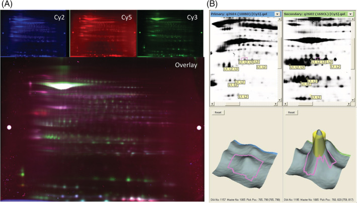
Methods: The mucosal proteome was analyzed using 2-dimensional fluorescence difference gel electrophoresis (2D DIGE) and nanoflow liquid chromatography tandem mass spectrometry. For 5 proteins, results were verified by Western blot analysis.
Results: A total of 2349 spots were identified, of which 9 were differentially expressed with a ≥2-fold change between healthy cats and cats with LPE and SCL (.01 < P < .001). Eight of these 9 spots were also differentially expressed between cats with LPE and cats with SCL (P .001 < P < .04). However, Western blot analysis for malate dehydrogenase-1, malate dehydrogenase-2, apolipoprotein, annexin IV, and annexin V did not confirm significant differential protein expression for any of the 5 proteins assessed.
Conclusions and clinical importance: Two-D DIGE did not identify potential biomarker candidates in the intestinal mucosa of cats with LPE and SCL. Future studies should focus on different techniques to identify biomarker candidates for cats with chronic enteropathies (CE).
Keywords: EATL; enteropathy-associated T-cell lymphoma; feline chronic enteropathy.
© 2021 The Authors. Journal of Veterinary Internal Medicine published by Wiley Periodicals LLC. on behalf of the American College of Veterinary Internal Medicine.
Conflict of interest statement
At the time of the study, authors Marsilio, Dröes, Lidbury, Suchodolski, and Steiner are or were employed by the Gastrointestinal Laboratory at Texas A&M University, which offers laboratory testing, including histopathology services, on a fee‐for‐service basis. The author Dangott is an employee of the Protein Chemistry Laboratory at Texas A&M, which offers laboratory testing, including 2D DIGE, on a fee‐for‐service basis. The author Ackermann is affiliated with the Gastrointestinal Laboratory at Texas A&M University. The author Estep is employed by Texas Veterinary Pathology, LLC, which offers histopathology for animals on a fee‐for‐service basis. The authors Chow and Hill have nothing to disclose.
Figures
References
-
- Jergens AE, Schreiner CA, Frank DE, et al. A scoring index for disease activity in canine inflammatory bowel disease. J Vet Intern Med. 2003;17:291‐297. - PubMed
-
- Simpson KW, Jergens AE. Pitfalls and progress in the diagnosis and management of canine inflammatory bowel disease. Vet Clin North Am Small Anim Pract. 2011;41:381‐398. - PubMed
-
- Sabattini S, Bottero E, Turba ME, Vicchi F, Bo S, Bettini G. Differentiating feline inflammatory bowel disease from alimentary lymphoma in duodenal endoscopic biopsies. J Small Anim Pract. 2016;57:396‐401. - PubMed
-
- Moore PF, Rodriguez‐Bertos A, Kass PH. Feline gastrointestinal lymphoma: mucosal architecture, immunophenotype, and molecular clonality. Vet Pathol. 2012;49:658‐668. - PubMed
MeSH terms
Substances
LinkOut - more resources
Full Text Sources
Other Literature Sources
Research Materials
Miscellaneous
