Case Reports
. 2021 Jan 20;21(1):89.
doi: 10.1186/s12879-021-05793-6.

Clinical course and challenging management of early COVID-19 infection after heart transplantation: case report of two patients

Affiliations
Case Reports

Clinical course and challenging management of early COVID-19 infection after heart transplantation: case report of two patients

Vincent Tchana-Sato et al. BMC Infect Dis. .

Abstract

Background: There are limited data on Coronavirus disease 2019 (COVID-19) in solid organ transplant patients, especially in heart transplant recipients, with only a few case reports and case series described so far. Heart transplant recipients may be at particular high risk due to their comorbidities and immunosuppressed state.

Case presentation: This report describes the clinical course and the challenging management of early COVID-19 infection in two heart transplant recipients who tested positive for the SARS-CoV-2 virus in the perioperative period of the transplant procedure. The two patients developed a severe form of the disease and ultimately died despite the initiation of an antiviral monotherapy with hydroxychloroquine coupled with the interruption of mycophenolate mofetil.

Conclusions: These two cases illustrate the severity and poor prognosis of COVID-19 in the perioperative period of a heart transplant. Thorough screening of donors and recipients is mandatory, and the issue of asymptomatic carriers needs to be addressed.

Keywords: Asymptomatic carrier; Case report; Coronavirus disease 2019 (COVID-19); Heart transplantation; Severe acute respiratory syndrome coronavirus 2 (SARS-Cov-2).


Conflict of interest statement

The authors declare that they have no competing interests.

Figures
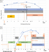
Fig. 1
Fig. 1
Clinical course of Recipient 1 (a) and Recipient 2 (b). From admission and day of surgery (HTx) to death highlighting the SARS-CoV-2 RNA load (Log10 copies/ml), the ward and ICU length of stay, diagnostic and therapeutic interventions such as chest CT, myocardial biopsy, BAL, initiation of antibiotics, and HCQ. a MM was withheld on POD 16. The clinical deterioration observed on POD 14 was close to the peak of the viral load observed on POD 16. b MM was withheld on POD 20. We can see that the deterioration of the patient’s clinical status on POD 19 coincides with a peak of the viral load. The SARS-CoV-2 RNA load is expressed by log10 copies per milliliter. Two RT-PCR assays were used due to a change in the testing method of the laboratory. The first target is the E and RdRP gene (Corman V.) The second test is the automated Cobas© SARS-CoV-2 molecular test (Cobas 6800 Roche) targeting the E and ORF1ab genes. Only the E gene is illustrated in the figure. ABt, antibiotics; BAL, bronchoalveolar lavage; Chest CT, chest computed tomography; HTx, heart transplantation; HCQ, hydroxychloroquine; ICU, intensive care unit; MM, mycophenolate mofetil; POD, postoperative day; RT-PCR, reverse transcriptase polymerase chain reaction
Fig. 2
Fig. 2
Chest X-ray and CT of recipient 1 (a) and recipient 2 (b. a) Progressive occurrence of pulmonary lesions during the postoperative course. The black arrows illustrate the ground-glass opacity lesions compatible with COVID-19 infection on POD 14. The blue arrows illustrate bilateral parenchymal patchy consolidations at POD 25. b Progressive occurrence of pulmonary lesions during the postoperative course until readmission in the intensive care unit on POD 20, where we can clearly see the ground-glass opacities lesions compatible with COVID-19 infection at POD 19 (black arrow), and bilateral patchy consolidations at POD 20 (blue arrow). POD, postoperative day
Fig. 3
Fig. 3
Electron microscopy (a-b) and immunohistology (c-d) of heart tissue from recipient 2. a-b Transmission electron microscopy representative examples demonstrating particles whose morphology is compatible with coronavirus particles (arrows). c-d Absence of immunoreactivity for the viral nucleocapsid protein (c) which contrasts with the intense staining in the positive control (lung tissue specimen of a SARS-CoV-2 infected hamster) (d). Transmission electron microscopy. Tissues were fixed at 4 °C in 4% glutaraldehyde (Laborimpex, Brussels, Belgium) in phosphate buffer at pH 7.4 and postfixed in 1% osmium tetroxide (Laborimpex, Brussels, Belgium) for 1 H at 4 °C. They were then dehydrated in graded (70, 90, 100%) ethanol solutions (VWR International, Leuven, Belgium) and propylene oxide (Laborimpex, Brussels, Belgium), embedded in epon (SERVA, Zandhoven, Belgium) and hardened at 60 °C. Semi-thin sections were stained with 0.5% toluidine blue and examined by light microscopy. Ultra-thin sections (80 nm) were stained with uranyl acetate (Fluka, Bornem, Belgium) and lead citrate (Leica, Aartselaar, Belgium). These sections were examined using an EM 910 transmission electron microscope (60 kV) (Zeiss, Belgium). Immunohistology Sections were deparaffinized and rehydrated in graded alcohols. After endogenous peroxydase inhibition with H2O2 (3%), nonspecific binding sites were blocked with the Protein Block Serum-Free solution (Dako). Slides were incubated for 1H at room temperature with a rabbit polyclonal antibody against the SARS-CoV-2 recombinant fusion nucleoprotein (ABclonal). Immunodetection was performed with the Polyview plus AP-Rabbit and alkaline phosphatase (Enzo Life Sciences)
Fig. 4
Fig. 4
Laboratory findings (Blood count and inflammatory markers) of Recipients 1 (A) and 2 (B). CRP, C-reactive protein; LDH, Lactate dehydrogenase; WBC, White cell blood count

References

    1. Li F, Cai J, Dong N. First cases of COVID-19 in heart transplantation from China. J Heart Lung Transplant. 2020;0(0) [cited 2020 Apr 25]. Available from: https://www.jhltonline.org/article/S1053-2498(20)31467-4/abstract. - PMC - PubMed
    1. Pereira MR, Mohan S, Cohen DJ, Husain SA, Dube GK, Ratner LE, et al. COVID-19 in solid organ transplant recipients: initial report from the US epicenter. Am J Transplant. [cited 2020 Apr 27];n/a(n/a). Available from: https://onlinelibrary.wiley.com/doi/abs/10.1111/ajt.15941. - DOI - PMC - PubMed
    1. Fernández-Ruiz M, Andrés A, Loinaz C, Delgado JF, López-Medrano F, San Juan R, et al. COVID-19 in solid organ transplant recipients: a single-center case series from Spain. Am J Transplant. 2020;20:1849–58. - PMC - PubMed
    1. Kates OS, Fisher CE, Stankiewicz-Karita HC, Shepherd AK, Church EC, Kapnadak SG, et al. Earliest cases of coronavirus disease 2019 (COVID-19) identified in solid organ transplant recipients in the United States. Am J Transplant. 2020;20:1885–90. - PMC - PubMed
    1. Mathies D, Rauschning D, Wagner U, Mueller F, Maibaum M, Binnemann C, et al. A case of SARS-CoV-2-pneumonia with successful antiviral therapy in a 77-year-old male with heart transplant. Am J Transplant. 2020;20:1925–9. - PMC - PubMed

Publication types