

Topical co-administration of zoledronate with recombinant human bone morphogenetic protein-2 can induce and maintain bone formation in the bone marrow environment
- PMID: 33472600
- PMCID: PMC7819170
- DOI: 10.1186/s12891-021-03971-w
Topical co-administration of zoledronate with recombinant human bone morphogenetic protein-2 can induce and maintain bone formation in the bone marrow environment
Abstract
Background: Bone morphogenetic proteins (BMPs) induce osteogenesis in various environments. However, when BMPs are used alone in the bone marrow environment, the maintenance of new bone formation is difficult owing to vigorous bone resorption. This is because BMPs stimulate the differentiation of not only osteoblast precursor cells but also osteoclast precursor cells. The present study aimed to induce and maintain new bone formation using the topical co-administration of recombinant human BMP-2 (rh-BMP-2) and zoledronate (ZOL) on beta-tricalcium phosphate (β-TCP) composite.
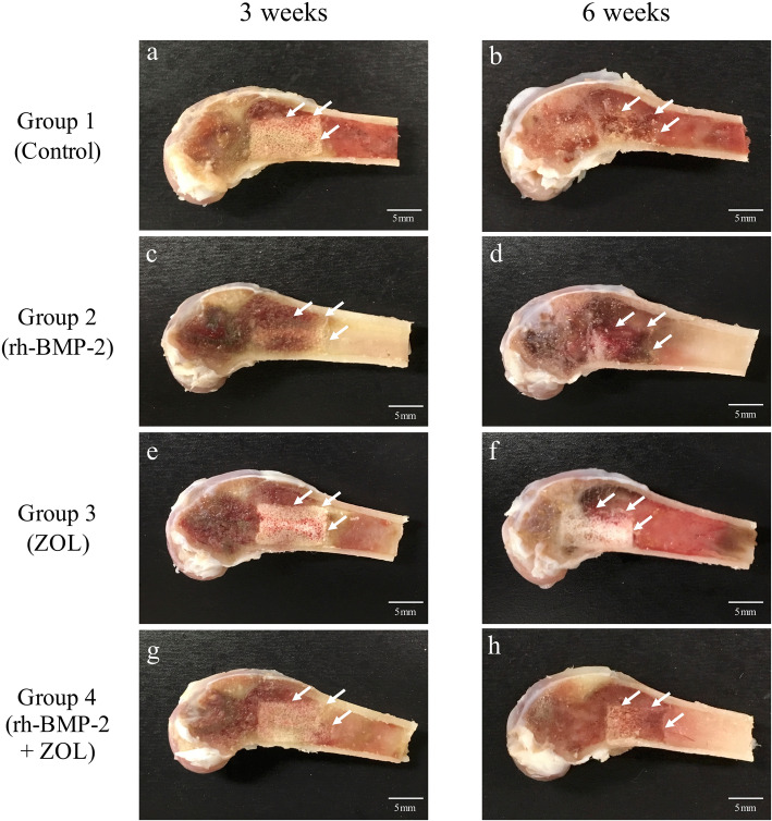
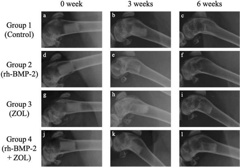
Methods: β-TCP columns were impregnated with both rh-BMP-2 (30 µg) and ZOL (5 µg), rh-BMP-2 alone, or ZOL alone, and implanted into the left femur canal of New Zealand white rabbits (n = 56). The implanted β-TCP columns were harvested and evaluated at 3 and 6 weeks after implantation. These harvested β-TCP columns were evaluated radiologically using plane radiograph, and histologically using haematoxylin/eosin (H&E) and Masson's trichrome (MT) staining. In addition, micro-computed tomography (CT) was performed for qualitative analysis of bone formation in each group (n = 7).
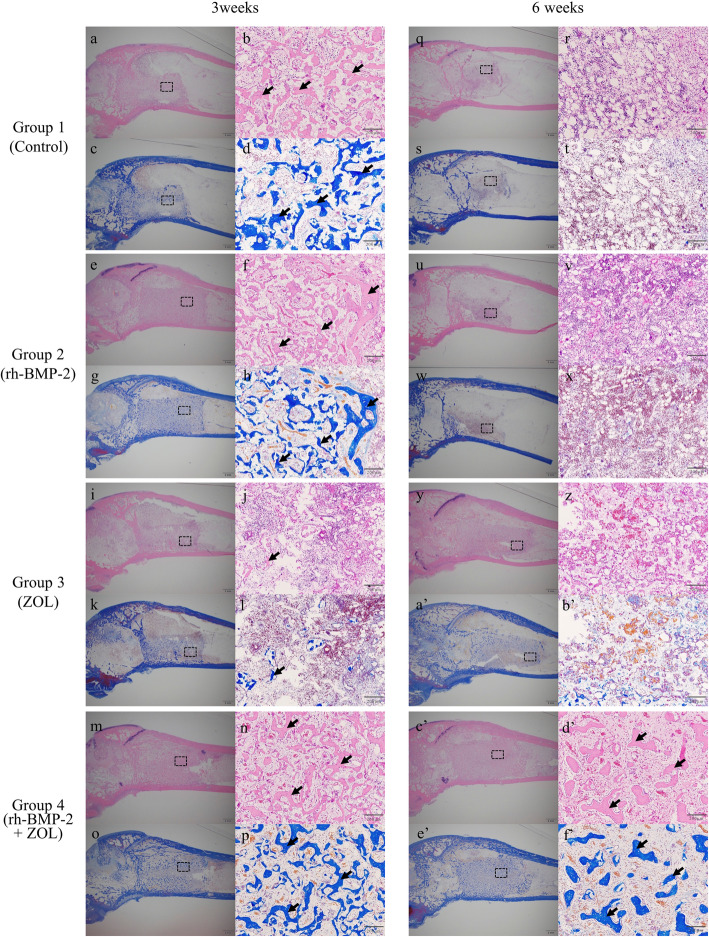
Results: Tissue sections stained with H&E and MT dyes revealed that new bone formation inside the β-TCP composite was significantly greater in those impregnated with both rh-BMP-2 and ZOL than in those from the other experimental groups at 3 and 6 weeks after implantations (p < 0.05). Micro-CT data also demonstrated that the bone volume and the bone mineral density inside the β-TCP columns were significantly greater in those impregnated with both rh-BMP-2 and ZOL than in those from the other experimental groups at 3 and 6 weeks after implantations (p < 0.05).
Conclusions: The topical co-administration of both rh-BMP-2 and ZOL on β-TCP composite promoted and maintained newly formed bone structure in the bone marrow environment.
Keywords: Bone morphogenetic proteins; Histology; Rabbit; micro computed tomography; β-tricalcium phosphate.
Conflict of interest statement
The authors declare that they have no competing interests.
Figures


Similar articles
-
Local co-application of zoledronate promotes long-term maintenance of newly formed bone induced by recombinant human bone morphogenetic protein 2.Biochem Biophys Res Commun. 2016 Nov 18;480(3):314-320. doi: 10.1016/j.bbrc.2016.10.034. Epub 2016 Oct 14. Biochem Biophys Res Commun. 2016. PMID: 27746180
-
The effect of bone morphogenic protein-2-coated tri-calcium phosphate/hydroxyapatite on new bone formation in a rat model of femoral distraction osteogenesis.Cytotherapy. 2012 Mar;14(3):315-26. doi: 10.3109/14653249.2011.630728. Epub 2011 Nov 28. Cytotherapy. 2012. PMID: 22122301
-
Escherichia coli-derived BMP-2-absorbed β-TCP granules induce bone regeneration in rabbit critical-sized femoral segmental defects.Int Orthop. 2019 May;43(5):1247-1253. doi: 10.1007/s00264-018-4079-4. Epub 2018 Aug 10. Int Orthop. 2019. PMID: 30097727
-
The importance of drug delivery to optimize the effects of bone morphogenetic proteins during periodontal regeneration.Curr Pharm Biotechnol. 2001 Jun;2(2):131-42. doi: 10.2174/1389201013378716. Curr Pharm Biotechnol. 2001. PMID: 11480418 Review.
-
Bone morphogenetic proteins.Growth Factors. 2004 Dec;22(4):233-41. doi: 10.1080/08977190412331279890. Growth Factors. 2004. PMID: 15621726 Review.
Cited by
-
Computed tomography provides a "one-stop-shop" targeted analysis for coronary artery calcification and osteoporosis: a review.Front Endocrinol (Lausanne). 2025 Feb 28;16:1356831. doi: 10.3389/fendo.2025.1356831. eCollection 2025. Front Endocrinol (Lausanne). 2025. PMID: 40093749 Free PMC article. Review.
-
The Role of BMP Signaling in Osteoclast Regulation.J Dev Biol. 2021 Jun 28;9(3):24. doi: 10.3390/jdb9030024. J Dev Biol. 2021. PMID: 34203252 Free PMC article. Review.
References
-
- Fujioka-Kobayashi M, Kobayashi E, Schaller B, Mottini M, Miron RJ, Saulacic N. Effect of recombinant human bone morphogenic protein 9 (rhBMP9) loaded onto bone grafts versus barrier membranes on new bone formation in a rabbit calvarial defect model. J Biomed Mater Res - Part A. 2017;105(10):2655–61. doi: 10.1002/jbm.a.36125. - DOI - PubMed
MeSH terms
Substances
LinkOut - more resources
Full Text Sources
Other Literature Sources
