. 2021 Feb 9;118(6):e2022310118.
doi: 10.1073/pnas.2022310118.

Coronavirus replication-transcription complex: Vital and selective NMPylation of a conserved site in nsp9 by the NiRAN-RdRp subunit

Affiliations

Coronavirus replication-transcription complex: Vital and selective NMPylation of a conserved site in nsp9 by the NiRAN-RdRp subunit

Heiko Slanina et al. Proc Natl Acad Sci U S A. .

Abstract

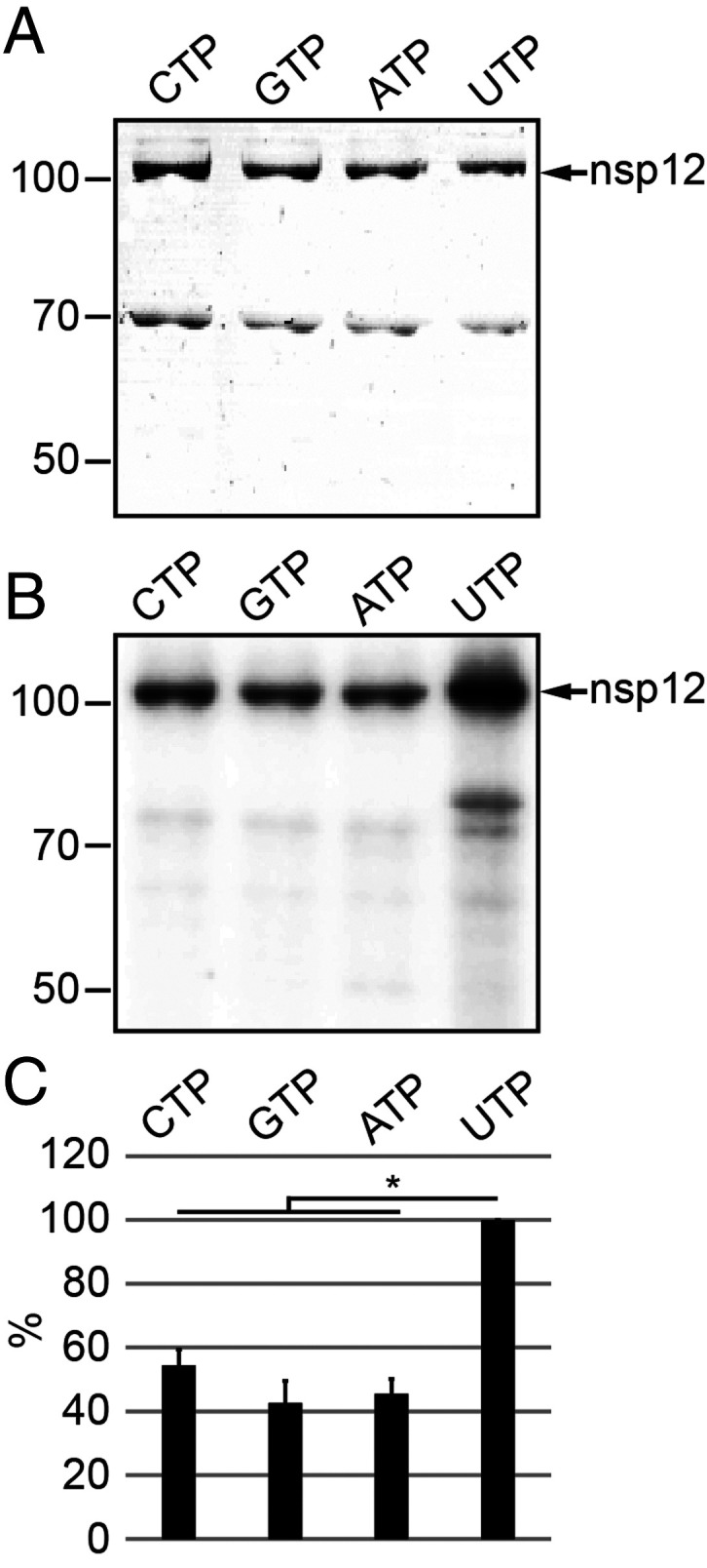
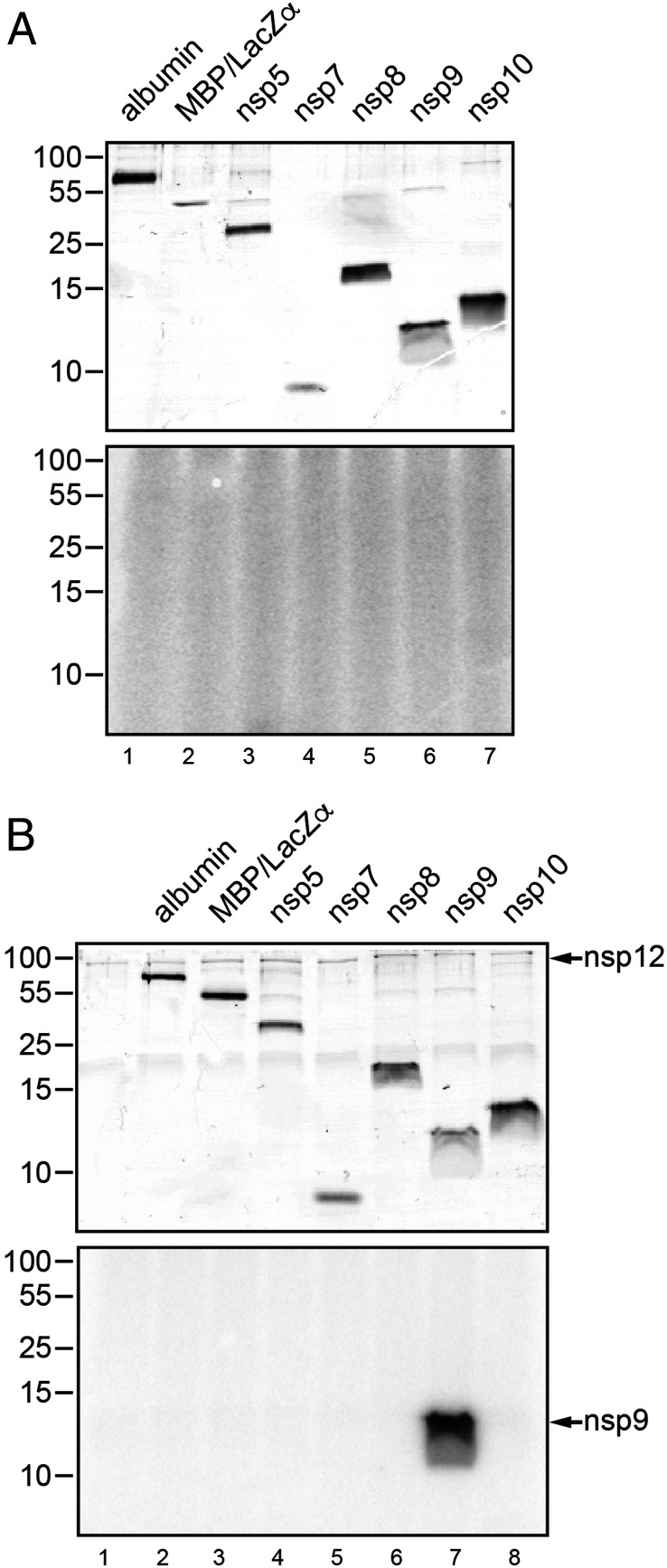
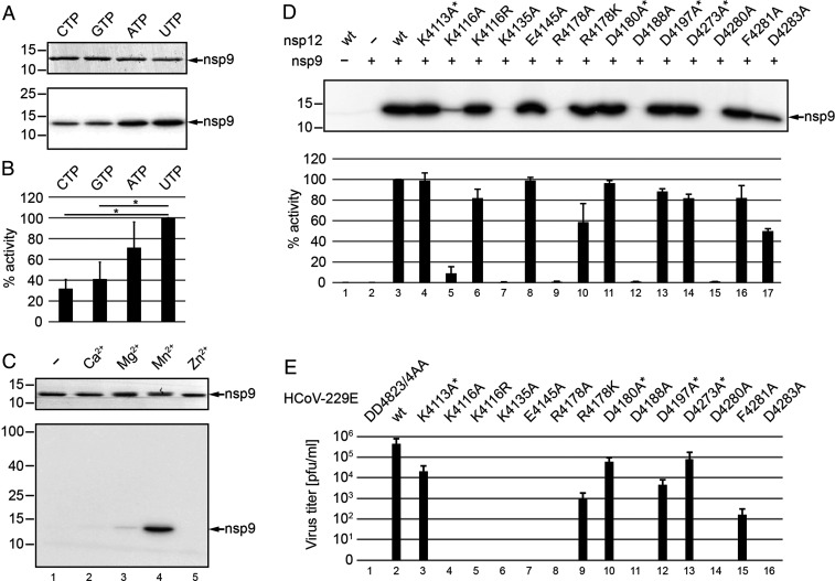
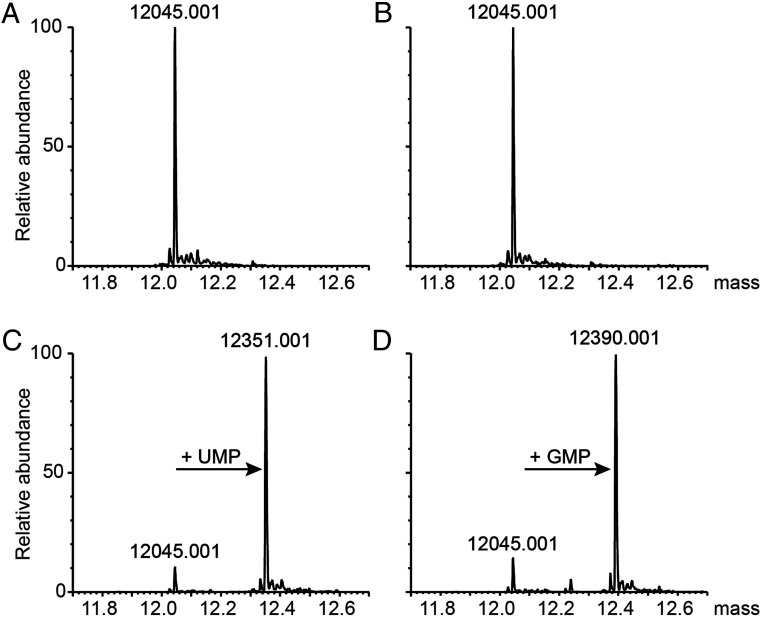
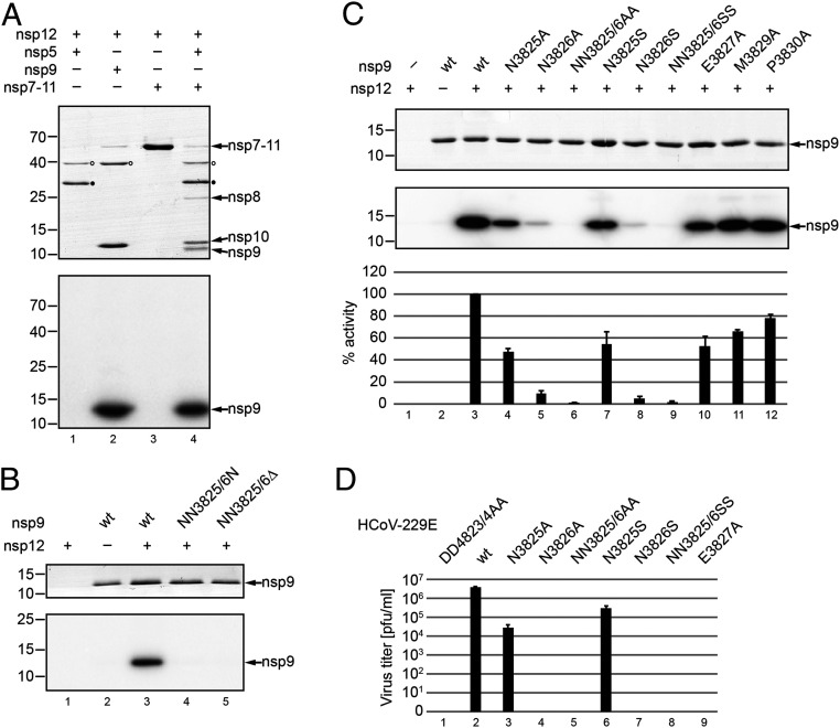
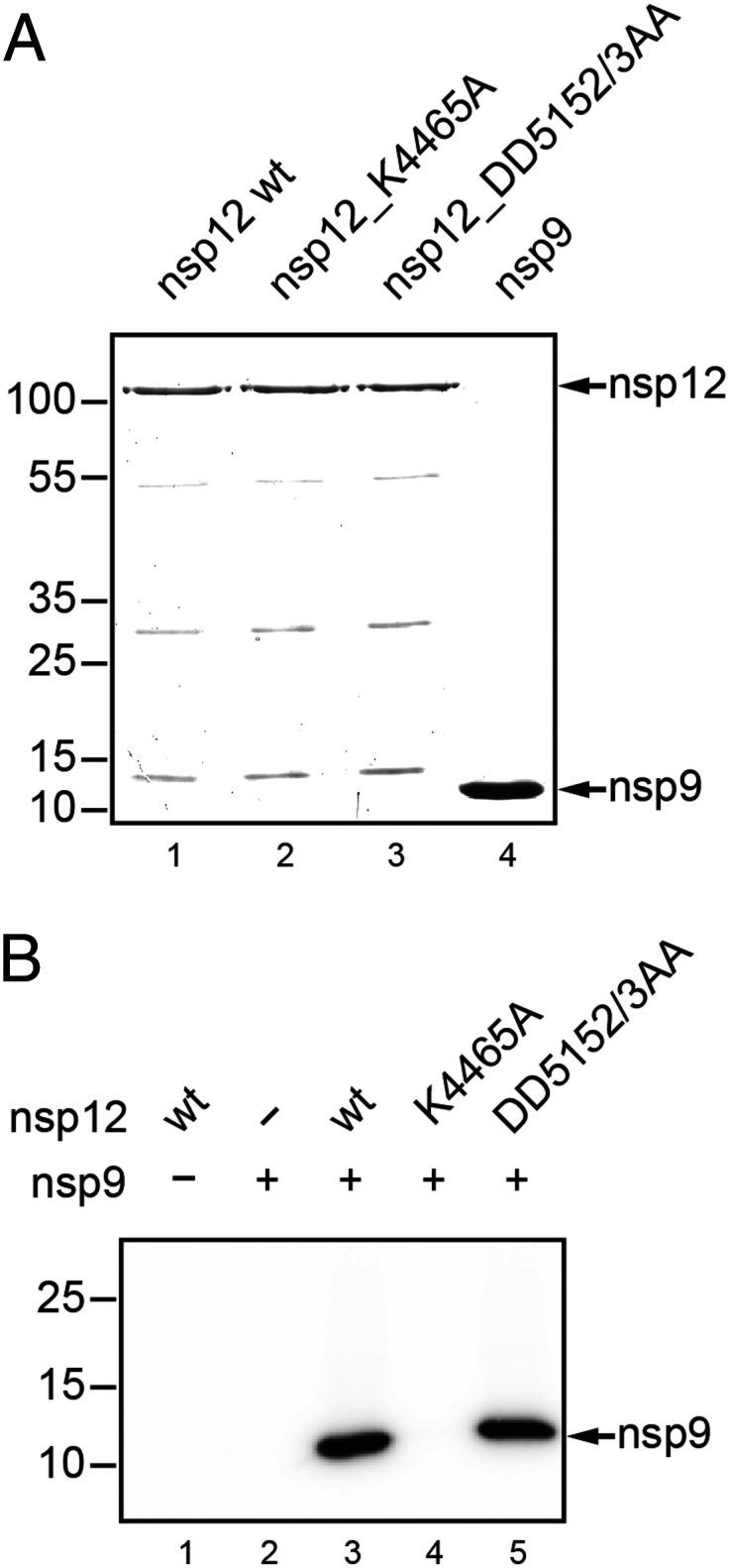
RNA-dependent RNA polymerases (RdRps) of the Nidovirales (Coronaviridae, Arteriviridae, and 12 other families) are linked to an amino-terminal (N-terminal) domain, called NiRAN, in a nonstructural protein (nsp) that is released from polyprotein 1ab by the viral main protease (Mpro). Previously, self-GMPylation/UMPylation activities were reported for an arterivirus NiRAN-RdRp nsp and suggested to generate a transient state primed for transferring nucleoside monophosphate (NMP) to (currently unknown) viral and/or cellular biopolymers. Here, we show that the coronavirus (human coronavirus [HCoV]-229E and severe acute respiratory syndrome coronavirus 2) nsp12 (NiRAN-RdRp) has Mn2+-dependent NMPylation activity that catalyzes the transfer of a single NMP to the cognate nsp9 by forming a phosphoramidate bond with the primary amine at the nsp9 N terminus (N3825) following Mpro-mediated proteolytic release of nsp9 from N-terminally flanking nsps. Uridine triphosphate was the preferred nucleotide in this reaction, but also adenosine triphosphate, guanosine triphosphate, and cytidine triphosphate were suitable cosubstrates. Mutational studies using recombinant coronavirus nsp9 and nsp12 proteins and genetically engineered HCoV-229E mutants identified residues essential for NiRAN-mediated nsp9 NMPylation and virus replication in cell culture. The data corroborate predictions on NiRAN active-site residues and establish an essential role for the nsp9 N3826 residue in both nsp9 NMPylation in vitro and virus replication. This residue is part of a conserved N-terminal NNE tripeptide sequence and shown to be the only invariant residue in nsp9 and its homologs in viruses of the family Coronaviridae The study provides a solid basis for functional studies of other nidovirus NMPylation activities and suggests a possible target for antiviral drug development.

Keywords: NiRAN; RNA polymerase; coronavirus; nidovirus; nucleotidyltransferase.


Conflict of interest statement

The authors declare no competing interest.

Figures

Fig. 1.
Fig. 1.
Self-NMPylation activity of HCoV-229E nsp12. (A) HCoV-229E nsp12-His6 (106 kDa) was incubated for 30 min with the indicated [α-32P]NTPs in the presence of 6 mM MnCl2 (for details, see Materials and Methods). Reaction products were separated by SDS-PAGE and stained with Coomassie brilliant blue. (B) Radiolabeled proteins were visualized by phosphorimaging. Positions of nsp12-His6 and protein molecular mass markers (in kilodaltons) are indicated in A and B. (C) Radioactive signal intensities (mean values ± SEM) were determined from three independent experiments. *P ≤ 0.05. Signal intensities (in percentages) are given in relation to UTP.
Fig. 2.
Fig. 2.
HCoV-229E nsp12-mediated UMPylation of nsp9. HCoV-229E nsp12-His6–mediated protein UMPylation activity was assessed using a range of protein substrates, including bovine serum albumin, MBP-lacZα, and a range of C-terminally His6-tagged HCoV-229E nsps encoded by ORF1a. The proteins were incubated for 10 min with [α-32P]UTP in the absence (A) or presence (B) of nsp12, as described in Materials and Methods. In the Top of A and B, the Coomassie brilliant blue-stained SDS-polyacrylamide gels are shown, and, in the Bottom of A and B, the corresponding autoradiograms are shown. Positions of protein molecular mass markers (in kilodaltons) are given to the left. Also indicated is the position of nsp12-His6 (B, Top) and the radioactive signal observed upon incubation of nsp12-His6 with nsp9-His6 (B, lane 7), the latter indicating nsp12-His6–mediated transfer of [α-32P]UMP to nsp9-His6 (12.9 kDa), which was not observed for other proteins tested.
Fig. 3.
Fig. 3.
Biochemical and virological characterization of HCoV-229E NiRAN-mediated nsp9 NMPylation. (A and B) Role of nucleotide cosubstrate used in the reaction. Nsp12-His6 and nsp9-His6 were mixed and incubated in the presence of different [α-32P]NTPs in the standard NMPylation assay. (A, Top) The Coomassie-stained nsp9-His6 separated by SDS-PAGE. (A, Bottom) An autoradiogram of the same area of the gel. (B) Relative activities (mean values ± SEM) in the presence of the indicated nucleotide cofactors were determined from three independent experiments. *P ≤ 0.05. (C) Role of metal ions. Shown is a standard NMPylation assay in the presence of [α-32P]UTP and different metal ions, each at 1 mM. In C, Top, the Coomassie-stained nsp9-His6 is shown, and, in C, Bottom, the corresponding autoradiogram is shown. Sizes of marker proteins (in kilodaltons) are indicated to the left in A and C. (D) Mutant forms of HCoV-229E nsp12-His6 carrying the indicated amino acid substitutions were incubated with nsp9-His6 in the presence of [α-32P]UTP, as described in Materials and Methods. Radiolabeled nsp9-His6 produced in the NMPylation reaction was detected by phosphorimaging (D, Top), and relative activities compared to the wild-type (wt) protein are presented in D, Bottom as mean values (±SEM) determined from three independent experiments. Asterisks indicate substitutions of nonconserved residues. (E) Virus titers in p1 cell culture supernatants obtained at 24 h p.i. were determined by plaque assay. Codon substitutions in the NiRAN domain of the engineered HCoV-229E mutants are indicated (residue numbering is according to their position in pp1ab). The replication-deficient RdRp active-site mutant nsp12_DD4823/4AA was used as control.
Fig. 4.
Fig. 4.
Mono-NMPylation of nsp9 in the presence of nsp12 and UTP or GTP. Shown are deconvoluted intact protein mass spectra of HCoV-229E nsp9 (SI Appendix, Table S1) (AD). (A) nsp9 alone, (B) nsp9 + nsp12-His6, (C) nsp9 + nsp12-His6 in the presence of UTP, (D) nsp9 + nsp12-His6 in the presence of GTP.
Fig. 5.
Fig. 5.
Roles of proteolytic processing and N-terminal residues of nsp9 in nsp12-mediated UMPylation. (A) Requirement of a free nsp9 N terminus for nsp9 UMPylation. Nsp7-11-His6 was preincubated at 30 °C in the presence or absence of recombinant Mpro (nsp5-His6) in NMPylation assay buffer containing UTP. After 3 h, the NMPylation assay was started by the addition of nsp12-His6, as described in Materials and Methods. Reactions containing nsp5-His6 (lane 1) and nsp9-His6 (lane 2) were used as controls. After 10 min, the reactions were terminated and the reaction mixtures separated by SDS-PAGE. Proteins were stained with Coomassie brilliant blue (A, Top). Nsp7-11-His6 precursor and processing products resulting from nsp5-His6–mediated cleavage are indicated to the right. Note that (due to their small size) nsp7 and nsp11-His6 are not detectable in this gel and reactions supplemented with nsp5-His6 (lanes 1 and 4; position of nsp5-His6 is indicated by a closed circle) or nsp9-His6 (lane 2) contain small amounts of MBP (indicated by open circles) as a residual impurity resulting from their expression as MBP fusion proteins (SI Appendix, Table S1). (B) Nsp9-His6 variants lacking one or two N-terminal Asn residues (residue numbering is according to the position in pp1a/pp1ab) were purified and incubated with nsp12-His6 and [α-32P]UTP. B, Top shows the Coomassie-stained SDS-PAGE and B, Bottom shows the corresponding autoradiogram. Positions of molecular weight markers (in kilodaltons) are indicated to the left. (C) Conserved residues at the N terminus of HCoV-229E nsp9-His6 were replaced with Ala or Ser, and equal amounts of protein were used in nsp12-His6–mediated UMPylation reactions. Reaction products were separated by SDS-PAGE and stained with Coomassie brilliant blue (C, Top), and radiolabeled nsp9-His6 was detected by phosphorimaging (C, Middle). Relative NMPylation activities (mean values ± SEM) were calculated from three independent experiments using the wild-type (wt) protein as a reference (set to 100%). (D) Virus titers in p1 cell culture supernatants of Huh-7 cells infected with HCoV-229E wild type and mutants carrying the indicated amino acid replacements in nsp9 were determined by plaque assay. The replication-deficient RdRp motif C double mutant, DD4823/4AA, was used as negative control.
Fig. 6.
Fig. 6.
SARS-CoV-2 nsp12-mediated NMPylation of nsp9. (A) Coomassie-stained SDS-polyacrylamide gel showing the recombinant proteins used in the NMPylation assay. As controls, mutant proteins with active-site replacements in the NiRAN domain (K4465A) and the RdRp domain (DD5152/3AA) of SARS-CoV-2 nsp12 were used. Residue numbering is according to the position in pp1ab. (B) Autoradiogram of an UMPylation assay using nsp9-His6 and [α-32P]UTP as substrates for nsp12-His6 (wild type [wt] and mutants). Molecular masses (in kilodaltons) of marker proteins are indicated to the left.

References

    1. de Groot R. J., et al. ., “Order Nidovirales” in Virus Taxonomy, King A. M. Q., Adams M. J., Carstens E. B., Lefkowitz E. J., Eds. (Elsevier, Amsterdam, The Netherlands, 2012), pp. 785–795.
    1. Gorbalenya A. E., Enjuanes L., Ziebuhr J., Snijder E. J., Nidovirales: Evolving the largest RNA virus genome. Virus Res. 117, 17–37 (2006). - PMC - PubMed
    1. Walker P. J., et al. ., Changes to virus taxonomy and the statutes ratified by the International Committee on Taxonomy of Viruses (2020). Arch. Virol. 165, 2737–2748 (2020). - PubMed
    1. Wu F., et al. ., A new coronavirus associated with human respiratory disease in China. Nature 579, 265–269 (2020). - PMC - PubMed
    1. Zhou P., et al. ., A pneumonia outbreak associated with a new coronavirus of probable bat origin. Nature 579, 270–273 (2020). - PMC - PubMed

Publication types

MeSH terms

LinkOut - more resources