. 2021 Mar 10;95(7):e02434-20.
doi: 10.1128/JVI.02434-20. Epub 2021 Jan 20.

Cellular and Viral Determinants of HSV-1 Entry and Intracellular Transport towards Nucleus of Infected Cells

Affiliations

Cellular and Viral Determinants of HSV-1 Entry and Intracellular Transport towards Nucleus of Infected Cells

Farhana Musarrat et al. J Virol. .

Abstract

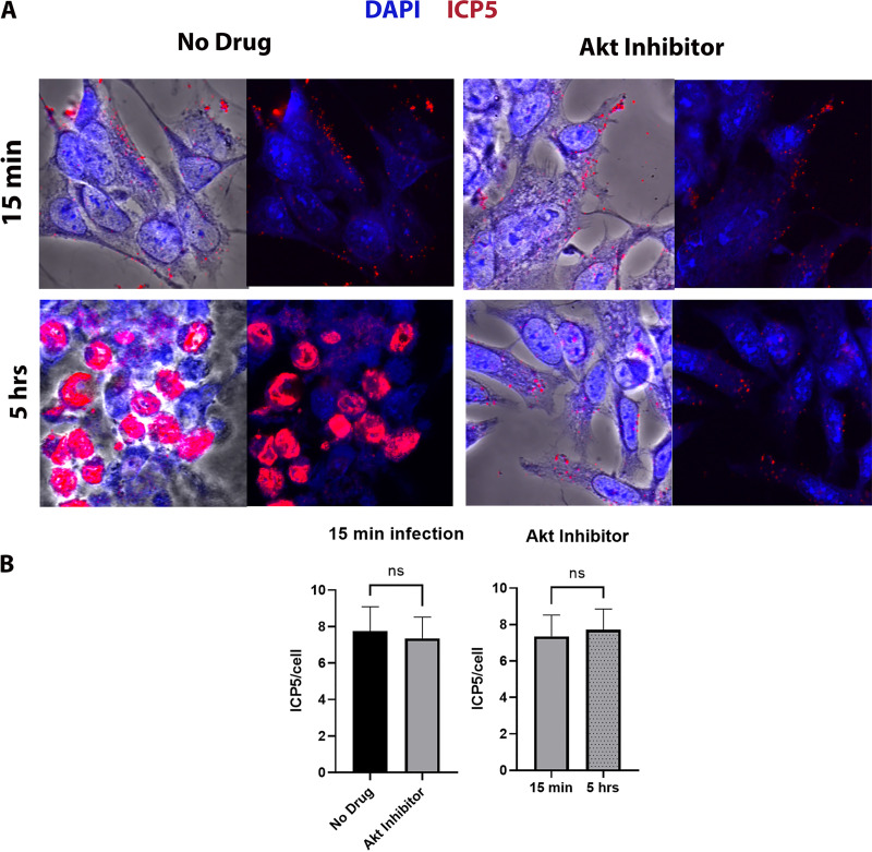
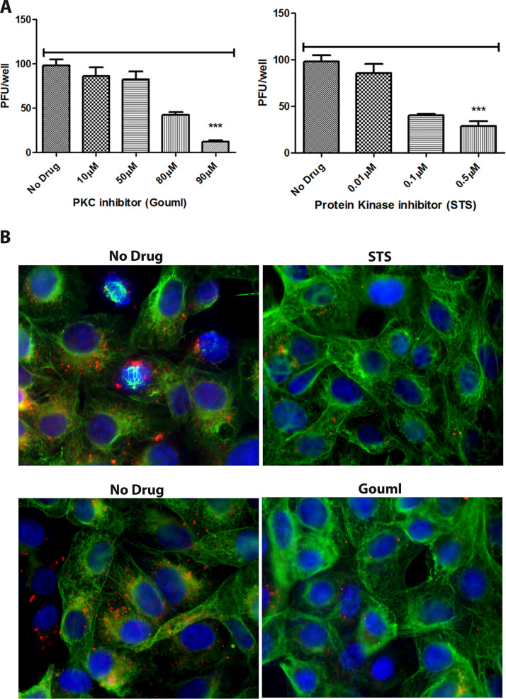
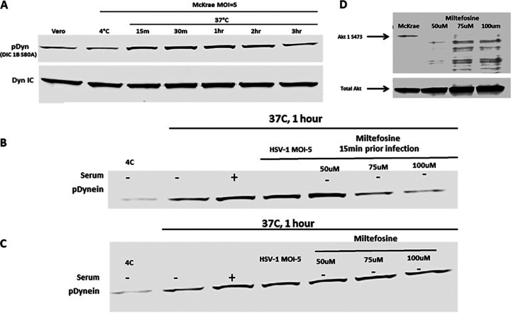
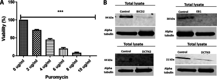
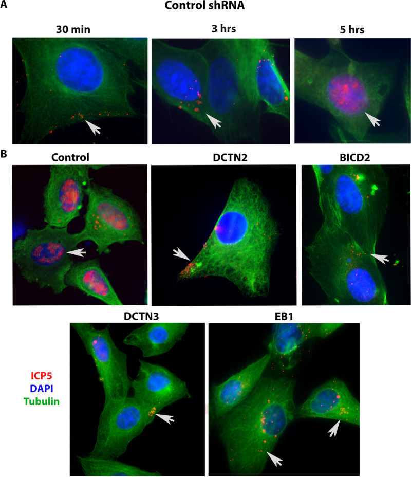
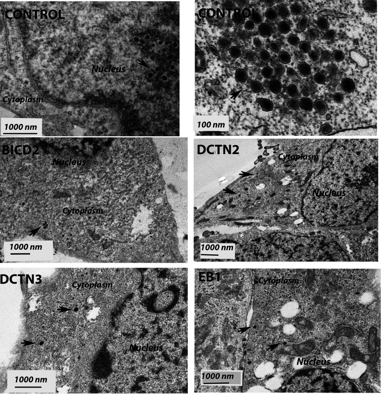
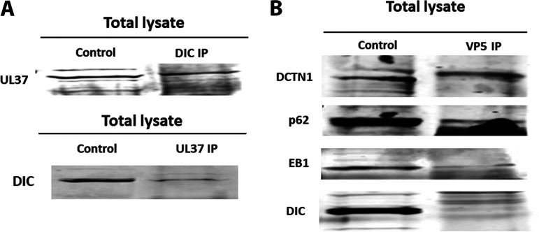
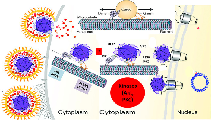
HSV-1 employs cellular motor proteins and modulates kinase pathways to facilitate intracellular virion capsid transport. Previously, we and others have shown that the Akt inhibitor miltefosine inhibited virus entry. Herein, we show that the protein kinase C inhibitors staurosporine (STS) and gouml inhibited HSV-1 entry into Vero cells, and that miltefosine prevents HSV-1 capsid transport toward the nucleus. We have reported that the HSV-1 UL37 tegument protein interacts with the dynein motor complex during virus entry and virion egress, while others have shown that the UL37/UL36 protein complex binds dynein and kinesin causing a saltatory movement of capsids in neuronal axons. Co-immoprecipitation experiments confirmed previous findings from our laboratory that the UL37 protein interacted with the dynein intermediate chain (DIC) at early times post infection. This UL37-DIC interaction was concurrent with DIC phosphorylation in infected, but not mock-infected cells. Miltefosine inhibited dynein phosphorylation when added before, but not after virus entry. Inhibition of motor accessory protein dynactins (DCTN2, DCTN3), the adaptor proteins EB1 and the Bicaudal D homolog 2 (BICD2) expression using lentiviruses expressing specific shRNAs, inhibited intracellular transport of virion capsids toward the nucleus of human neuroblastoma (SK-N-SH) cells. Co-immunoprecipitation experiments revealed that the major capsid protein Vp5 interacted with dynactins (DCTN1/p150 and DCTN4/p62) and the end-binding protein (EB1) at early times post infection. These results show that Akt and kinase C are involved in virus entry and intracellular transport of virion capsids, but not in dynein activation via phosphorylation. Importantly, both the UL37 and Vp5 viral proteins are involved in dynein-dependent transport of virion capsids to the nuclei of infected cells.Importance. Herpes simplex virus type-1 enter either via fusion at the plasma membranes or endocytosis depositing the virion capsids into the cytoplasm of infected cells. The viral capsids utilize the dynein motor complex to move toward the nuclei of infected cells using the microtubular network. This work shows that inhibitors of the Akt kinase and kinase C inhibit not only viral entry into cells but also virion capsid transport toward the nucleus. In addition, the work reveals that the virion protein ICP5 (VP5) interacts with the dynein cofactor dynactin, while the UL37 protein interacts with the dynein intermediate chain (DIC). Importantly, neither Akt nor Kinase C was found to be responsible for phosphorylation/activation of dynein indicating that other cellular or viral kinases may be involved.


Figures

FIG 1
FIG 1
Effect of Akt HSV-1 transport. (A) SK-N-SH cells were adsorbed at 4°C with HSV-1 (McKrae) at an MOI of 20 for 1 h, shifted to 37°C for 15 min, and then washed with low-pH buffer. The Akt inhibitor miltefosine was added at a concentration of 30 μM, and the mixture was incubated for 5 h at 37°C. The 8-well chamber slide was then fixed with formalin, permeabilized with methanol, and prepared for fluorescence microscopy. Antibody against VP5 (ICP5) (red) was used, and nuclei were stained with DAPI (blue). Capsids colocalized in the nuclei appear purple. Magnification is 63× under oil immersion. (B) The amount of fluorescence detected with the anti-ICP5 antibody (virion capsids) was quantified by fluorescence imaging of 10 different random sections of slides, using ImageJ software. The fluorescence signals per cell were counted, and their average value was used to plot the graph. Statistical comparison was conducted by Graph Pad Prism using Student's t test. Error bars represent the 95% confidence interval about the mean. Differences were determined significant at P < 0.05.
FIG 2
FIG 2
Effect of PKC inhibitors on HSV-1 entry. (A) Vero cells were treated with a series of dilutions of the PKC inhibitor Gouml or the protein kinase inhibitor staurosporine (STS) for 4 h and then infected with HSV-1 (McKrae) (100 PFU) for 1 h at 4°C. Cells were incubated at 37°C for 48 h for plaque assay. Viral plaques were counted after crystal violet staining. ***, P < 0.001 versus no-drug-treated control. Statistical comparison was conducted by Graph Pad prism using ANOVA with a post hoc t test with Bonferroni adjustment. Error bars represent the 95% confidence interval about the mean. (B) Vero cells were treated with a series of dilutions of PKC inhibitor (Gouml) or protein kinase inhibitor (staurosporine) for 4 h and then infected with McKrae (MOI of 20) for 1 h at 4°C. Cells were incubated at 37°C for 3 h and then fixed with formalin and permeabilized with methanol. Antibodies against VP5 (ICP5) (red), and α-tubulin (green) were used, and nuclei were stained with DAPI (blue). Magnification, 40×.
FIG 3
FIG 3
Dynein activation by HSV-1. (A) Serum-starved Vero cells were infected with HSV-1 (McKrae) at 4°C for 1 h at an MOI of 5 and then shifted to 37°C for 15 min, 30 min, 1 h, 2 h, and 3 h. Cells were lysed with the NP-40 lysis buffer containing a protease inhibitor cocktail and analyzed for phosphorylated dynein intermediate chain (DIC 1B S80A) by SDS-PAGE. The total dynein intermediate chain (DIC) protein was used as the loading control. (B) Vero cells were pretreated with miltefosine at 37°C. Cells were infected with HSV-1 (McKrae) at 4°C for 1 h at an MOI of 5 and then shifted to 37°C for 1 h. Cells were lysed with NP-40 lysis buffer containing a protease inhibitor cocktail and analyzed for the presence of the phosphorylated dynein intermediate chain (DIC 1B S80A) by SDS-PAGE. (C) Following infection with HSV-1 (McKrae) at 4°C for 1 h at an MOI of 5, cells were incubated at 37°C for 15 min, and then miltefosine was added and the mixture was incubated for 1 h. Cell lysates were prepared the same way for SDS-PAGE analysis. + and − indicate whether serum was added (+) or not (−). (D) Effect of miltefosine on Akt phosphorylation. Total Akt was used as loading control.
FIG 4
FIG 4
shRNA-mediated silencing of dynein cofactors in the SK-N-SH cell line. (A) Puromycin toxicity assay on SK-N-SH cells. A range of different concentrations (0 to 10 μg/ml) of puromycin was used to determine the appropriate amount of puromycin that is toxic to SK-N-SH cells. ***, P < 0.001 versus the no-drug (0 μg/ml)-treated control. Statistical comparison was conducted by GraphPad Prism software using ANOVA with a post hoc t test with Bonferroni adjustment. Error bars represent 95% confidence intervals about the means. (B) Lentivirus shRNA (human)-mediated silencing in SK-N-SH cells. The left lanes in each panel show SK-N-SH lysate treated with control shRNA, and the right lanes show lysates treated with shRNA targeted against motor accessory proteins BICD2, EB1, and the dynactin complex (DCTN2 and DCTN3), respectively.
FIG 5
FIG 5
Role of motor accessory proteins in HSV-1 intracellular transport. shRNA-treated SK-N-SH cells were synchronously infected with HSV-1 (McKrae) at an MOI of 20 for 30 min, 3 h, and 5 h at 37°C. Control-shRNA-treated SK-N-SH shows the presence of VP5 (ICP5) in the nucleus at 5 hpi (A). (B) Side-by-side comparison between control shRNA and shRNA against motor accessory protein-treated SK-N-SH cells infected with HSV-1 (McKrae) at an MOI of 20 for 5 h. The 8-well chamber slide was fixed with formalin, permeabilized with methanol, and prepared for fluorescence microscopy. Antibody against VP5 (ICP5) (red) was used, and nuclei were stained with DAPI (blue). Capsids colocalized in the nuclei are visible as purple (red plus blue). Magnification of 60× with oil immersion.
FIG 6
FIG 6
Transmission electron micrograph (TEM) of HSV-1 (McKrae)-infected SK-N-SH cells treated with control, EB1, BICD2, DCTN2, and DCTN3 shRNAs. The knockout SK-N-SH cells were synchronously infected with HSV-1 (McKrae) at an MOI of 20 for 5 h at 37°C. Following infection, cells were fixed and prepared for electron microscopy. SK-N-SH cells treated with control shRNA shows the presence of HSV-1 (McKrae) capsid-like structure in the nucleus. A higher magnification (100 nm) is used for better visibility. Other than the control, HSV-1 (McKrae) capsids were visible only in the cytoplasm, but not in the nucleus of the motor accessory protein knockout SK-N-SH cells. Black arrowheads specify the location of HSV-1 McKrae capsids.
FIG 7
FIG 7
Interactions between HSV-1 capsid/tegument proteins and cellular motor and accessory proteins. (A) Two-way immunoprecipitations (IP) showing UL37 and dynein intermediate-chain (DIC) interaction in HSV-1 (McKrae)-infected SK-N-SH lysates at 2 h postinfection at an MOI of 10. (B) Immunoprecipitation assay showing potential interactions between VP5 and DCTN1, VP5 and p62, and VP5 and EB1 and no interaction between VP5 and DIC proteins. SK-N-SH cells were infected with HSV-1 (McKrae) at an MOI of 10 for an hour, and lysates were prepared with NP-40 lysis buffer with protease inhibitor cocktail. The left lane represents SK-N-SH lysates (control), and the right lane represents lysates immunoprecipitated for HSV-1 VP5 and subsequently probed for dynactin complex (DCTN1 [p150], p62 [DCTN4]), EB1, and DIC.
FIG 8
FIG 8
Model of HSV-1 transport into host cells. The schematic shows the molecules and intracellular signaling, which may be involved in intracellular capsid transport. Following entry into host cells, HSV-1 (McKrae) activates/phosphorylates the dynein motor and the Akt and MAPK pathways (data not shown). The major capsid protein VP5 (ICP5) and the inner tegument protein UL37 recruit the dynein motor and other accessory proteins, such as BICD2, EB1, and the dynactin complex (DCTN) to facilitate retrograde transport of virion capsids. Akt and PKC activities are required for efficient entry and transport along the microtubules toward the nucleus, where the virus replicates its DNA. (Adapted from reference .)

Similar articles

Cited by

References

    1. Evans RD, Robinson C, Briggs DA, Tooth DJ, Ramalho JS, Cantero M, Montoliu L, Patel S, Sviderskaya EV, Hume AN. 2014. Myosin-Va and dynamic actin oppose microtubules to drive long-range organelle transport. Curr Biol 24:1743–1750. doi:10.1016/j.cub.2014.06.019. - DOI - PMC - PubMed
    1. Cramer L. 2008. Organelle transport: dynamic actin tracks for myosin motors. Curr Biol 18:R1066–R1068. doi:10.1016/j.cub.2008.09.048. - DOI - PubMed
    1. Schuh M. 2011. An actin-dependent mechanism for long-range vesicle transport. Nat Cell Biol 13:1431–1436. doi:10.1038/ncb2353. - DOI - PMC - PubMed
    1. Barlan K, Gelfand VI. 2017. Microtubule-based transport and the distribution, tethering, and organization of organelles. Cold Spring Harb Perspect Biol 9:a025817. doi:10.1101/cshperspect.a025817. - DOI - PMC - PubMed
    1. Kapitein LC, Hoogenraad CC. 2011. Which way to go? Cytoskeletal organization and polarized transport in neurons. Mol Cell Neurosci 46:9–20. doi:10.1016/j.mcn.2010.08.015. - DOI - PubMed

LinkOut - more resources