. 2021 Mar 25;95(8):e02370-20.
doi: 10.1128/JVI.02370-20. Epub 2021 Jan 20.

Comparison of Subgenomic and Total RNA in SARS-CoV-2 Challenged Rhesus Macaques

Affiliations

Comparison of Subgenomic and Total RNA in SARS-CoV-2 Challenged Rhesus Macaques

Gabriel Dagotto et al. J Virol. .

Abstract

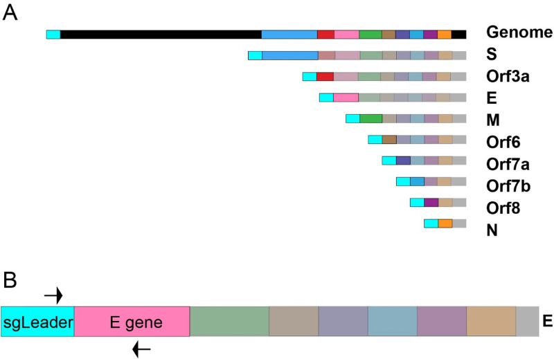
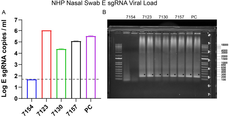
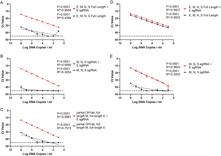
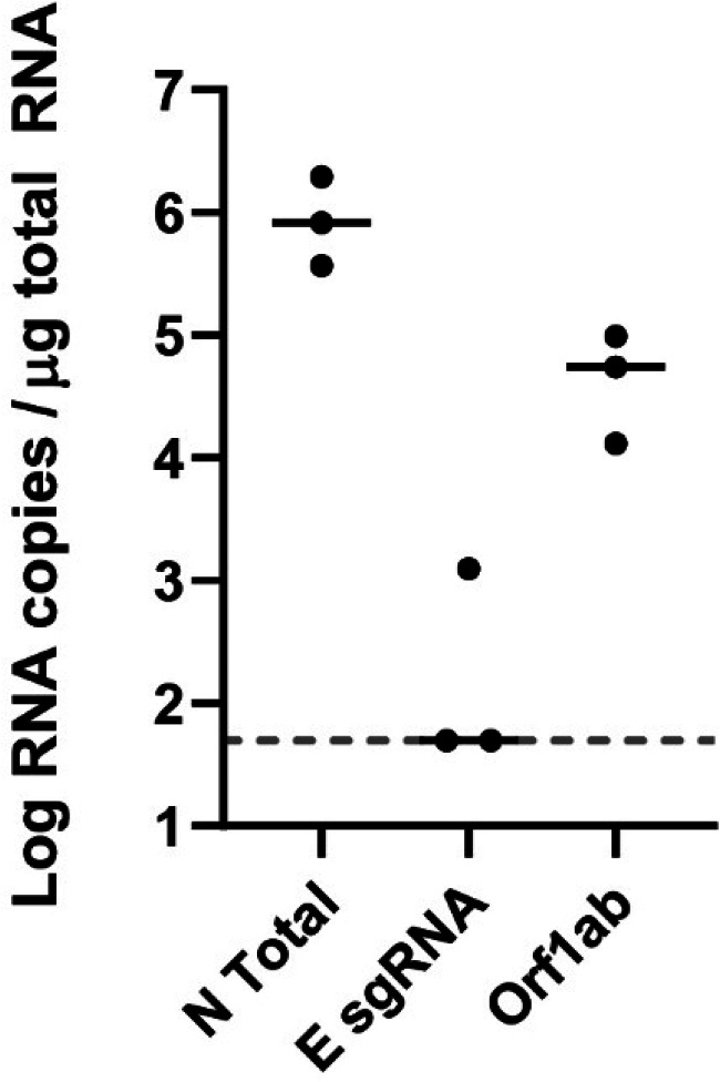
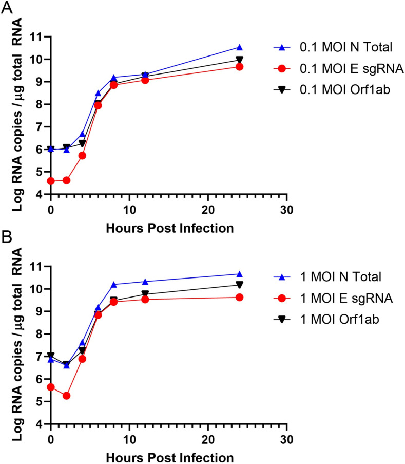
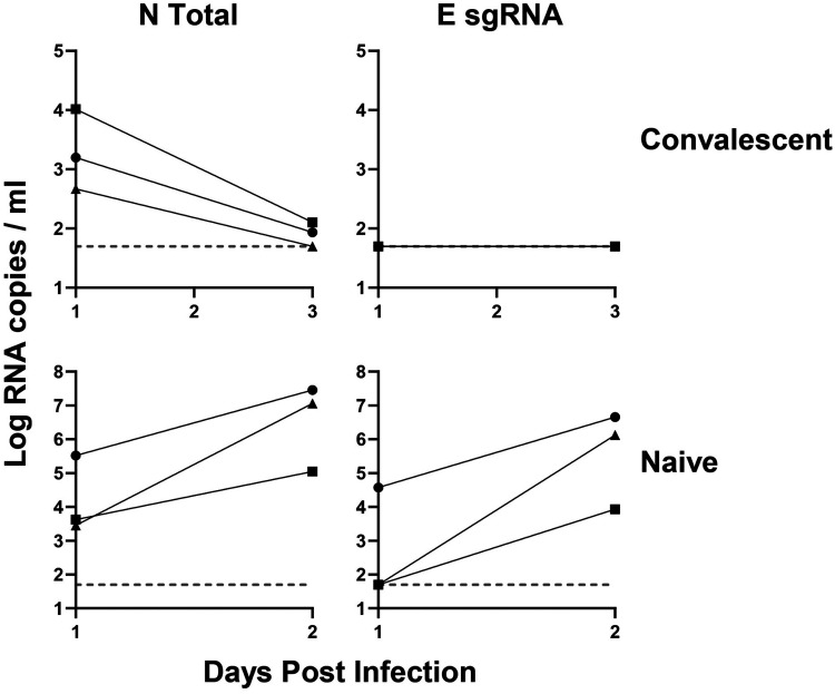
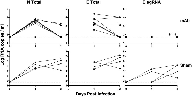
Respiratory virus challenge studies involve administration of the challenge virus and sampling to assess for protection from the same anatomical locations. It can therefore be difficult to differentiate actively replicating virus from input challenge virus. For SARS-CoV-2, specific monitoring of actively replicating virus is critical to investigate the protective and therapeutic efficacy of vaccines, monoclonal antibodies, and antiviral drugs. We developed a SARS-CoV-2 subgenomic RNA (sgRNA) RT-PCR assay to differentiate productive infection from inactivated or neutralized virus. Subgenomic RNAs are generated after cell entry and are poorly incorporate into mature virions, and thus may provide a marker for actively replicating virus. We show envelope (E) sgRNA was degraded by RNase in infected cell lysates, while genomic RNA (gRNA) was protected, presumably due to packaging into virions. To investigate the capacity of the sgRNA assay to distinguish input challenge virus from actively replicating virus in vivo, we compared the E sgRNA assay to a standard nucleoprotein (N) or E total RNA assay in convalescent rhesus macaques and in antibody-treated rhesus macaques after experimental SARS-CoV-2 challenge. In both studies, the E sgRNA assay was negative, suggesting protective efficacy, whereas the N and E total RNA assays remained positive. These data suggest the potential utility of sgRNA to monitor actively replicating virus in prophylactic and therapeutic SARS-CoV-2 studies.ImportanceDeveloping therapeutic and prophylactic countermeasures for the SARS-CoV-2 virus is a public health priority. During challenge studies, respiratory viruses are delivered and sampled from the same anatomical location. It is therefore important to distinguish actively replicating virus from input challenge virus. The most common assay for detecting SARS-CoV-2 virus, reverse transcription polymerase chain reaction (RT-PCR) targeting nucleocapsid total RNA, cannot distinguish neutralized input virus from replicating virus. In this study, we assess SARS-CoV-2 subgenomic RNA as a potential measure of replicating virus in rhesus macaques.


Figures

FIG 1
FIG 1
Graphical representation of sgRNAs and the E sgRNA assay. (A) Graphical representation of SARS-CoV-2 virus and sgRNA. Upon cellular entry SARS-CoV-2 generates sgRNAs for structural genes and accessory proteins before they are produced. The subgenomic leader sequence is colored cyan to highlight its position in the genomic and subgenomic RNAs. (B) Graphical representation of the primer binding sites for the E sgRNA assay on subgenomic E RNA. The forward primer binds to the subgenomic leader sequence present on all subgenomic RNAs as well as the genomic RNA. The reverse primer binds to the E gene (pink).
FIG 2
FIG 2
SARS-CoV-2-infected NHPs were sampled through nasal swabs on day 4 postinfection. (A) RNA was extracted from the nasal swabs and an E sgRNA RT-PCR assay was performed. (B) The assay RT-PCR results were then run in duplicate on a 0.8% agarose gel to confirm a single amplicon. Error bars define the standard deviation of the mean of two technical replicates for each macaque. PC indicates positive control. Asterisk indicates expected band.
FIG 3
FIG 3
Assay specificity with linear DNA mixtures. RT-PCR was performed on DNA fragment mixtures with and without the addition of E sgRNA linear DNA fragments. These mixtures were serially diluted 10-fold from 108 to 10 copies per ml. (A) Mixture of E, M, N, and S full-length DNA fragments. (B) Mixture of M, N, and S subgenomic partial DNA fragments. (C) Mixture of E and M full-length DNA fragments and the 5′ end of Orf1a containing the subgenomic leader sequence. In all mixtures, linearity was only present after the addition of E sgRNA. RT-PCR targeting E gRNA was performed on DNA fragment mixtures with and without the addition of an E sgRNA DNA fragment. (D) Mixture of E, M, N, and S full-length DNA fragments. (E) Mixture of M, N, and S subgenomic DNA fragments. Error bars denote the 95% confidence intervals of the mean of eight technical replicates. Lines represent simple linear regressions.
FIG 4
FIG 4
Infectious cell lysate treated with RNase A. Infectious cell lysate was treated with RNase A for 1 h and then RNA was extracted and RT-PCR for the N gene (N total), subgenomic E (E sgRNA), and genomic RNA (Orf1ab) was performed. Black bars represent median responses.
FIG 5
FIG 5
Longitudinal SARS-CoV-2 infection. Vero-E6 cells were infected at 0.1 MOI (A) or 1.0 MOI (B) in 12-well plates. Wells were harvested in triplicate at the following time points: 0, 2, 4, 6, 8, 12, and 24 h postinfection.
FIG 6
FIG 6
Convalescent NHP SARS-CoV-2 RT-PCR. NHPs were challenged with SARS-CoV-2 and rechallenged 35 days later. RNA extracted from nasal swabs from the rechallenge macaques was run for N total and E sgRNA in naive and the same convalescent animals.
FIG 7
FIG 7
RT-PCR of monoclonal antibody-protected NHPs challenged with SARS-CoV-2. NHPs were given 50 mg/kg of a monoclonal SARS-CoV-2 antibody and then challenged 3 days later with SARS-CoV-2. RNA extracted by BAL fluid was measured for N total, E total, and E sgRNA. Protected macaques (MAb) were compared to unprotected macaques (sham) to demonstrate assay success.

References

    1. Fehr AR, Perlman S. 2015. Coronaviruses: an overview of their replication and pathogenesis, p 1–23. In Maier HJ, Bickerton E, Britton P (ed), Coronaviruses: methods and protocols. Springer New York, New York, NY. doi:10.1007/978-1-4939-2438-7_1 - DOI - PMC - PubMed
    1. Hou YJ, Okuda K, Edwards CE, Martinez DR, Asakura T, Dinnon KH, Kato T, Lee RE, Yount BL, Mascenik TM, Chen G, Olivier KN, Ghio A, Tse LV, Leist SR, Gralinski LE, Schäfer A, Dang H, Gilmore R, Nakano S, Sun L, Fulcher ML, Livraghi-Butrico A, Nicely NI, Cameron M, Cameron C, Kelvin DJ, de Silva A, Margolis DM, Markmann A, Bartelt L, Zumwalt R, Martinez FJ, Salvatore SP, Borczuk A, Tata PR, Sontake V, Kimple A, Jaspers I, O'Neal WK, Randell SH, Boucher RC, Baric RS. 2020. SARS-CoV-2 reverse genetics reveals a variable infection gradient in the respiratory tract. Cell 182:429–446. doi:10.1016/j.cell.2020.05.042. - DOI - PMC - PubMed
    1. Song Z, Xu Y, Bao L, Zhang L, Yu P, Qu Y, Zhu H, Zhao W, Han Y, Qin C. 2019. From SARS to MERS, thrusting coronaviruses into the spotlight. Viruses 11:59. doi:10.3390/v11010059. - DOI - PMC - PubMed
    1. Sawicki SG, Sawicki DL, Siddell SG. 2007. A contemporary view of coronavirus transcription. J Virol 81:20–29. doi:10.1128/JVI.01358-06. - DOI - PMC - PubMed
    1. Sola I, Almazán F, Zúñiga S, Enjuanes L. 2015. Continuous and discontinuous RNA synthesis in coronaviruses. Annu Rev Virol 2:265–288. doi:10.1146/annurev-virology-100114-055218. - DOI - PMC - PubMed