. 2021 Jan 20;12(1):477.
doi: 10.1038/s41467-020-20735-7.

Structure of a microtubule-bound axonemal dynein

Affiliations

Structure of a microtubule-bound axonemal dynein

Travis Walton et al. Nat Commun. .

Abstract

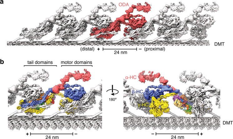
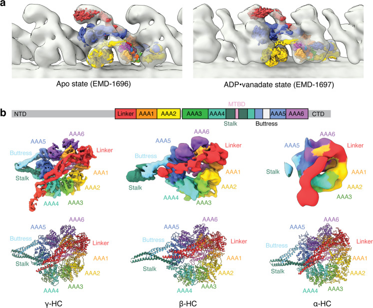
Axonemal dyneins are tethered to doublet microtubules inside cilia to drive ciliary beating, a process critical for cellular motility and extracellular fluid flow. Axonemal dyneins are evolutionarily and biochemically distinct from cytoplasmic dyneins that transport cargo, and the mechanisms regulating their localization and function are poorly understood. Here, we report a single-particle cryo-EM reconstruction of a three-headed axonemal dynein natively bound to doublet microtubules isolated from cilia. The slanted conformation of the axonemal dynein causes interaction of its motor domains with the neighboring dynein complex. Our structure shows how a heterotrimeric docking complex specifically localizes the linear array of axonemal dyneins to the doublet microtubule by directly interacting with the heavy chains. Our structural analysis establishes the arrangement of conserved heavy, intermediate and light chain subunits, and provides a framework to understand the roles of individual subunits and the interactions between dyneins during ciliary waveform generation.


Conflict of interest statement

The authors declare no competing interests.

Figures

Fig. 1
Fig. 1. Structure of the ODA axonemal dynein bound to a doublet microtubule.
a Composite map showing the 24-nm repeat of the ODA bound to protofilaments A07 and A08 of the doublet microtubule (DMT). A single ODA complex (colored red) forms an elongated and inclined structure that interacts with both its neighbors. b Two views showing the ODA complex colored by subunit. The motor domains form a triple stack in which the motors of both β- and γ-HC interact with the tail domains of the proximal ODA. The minus (−) and plus (+) ends of the microtubule are indicated on the scale bar.
Fig. 2
Fig. 2. The ODA complexes are in an apo state.
a The composite map of the ODA generated from our single-particle cryo-EM approach was docked into the subtomogram volumes of the C. reinhardtii ODA in apo and ADP•Vanadate states (representing the post- and pre-powerstroke states, respectively). The map fits well into the apo state. b General domain organization of an axonemal dynein heavy chain. Density maps and models of the motor domains of the three heavy chains colored by subdomain. The linker of β-HC adopts a different angle relative to the stalk compared to the linkers of α- and γ-HC.
Fig. 3
Fig. 3. Arrangement of the HC tails and IC–LC block and comparison with cytoplasmic dyneins.
a Map of the IC–LC block of the native C. reinhardtii ODA complex colored by subunit. b Atomic model of the IC–LC block of the C. reinhardtii ODA complex colored by subunit. c Atomic model of the IC–LC block of recombinant human dynein-1 (PDB 5NVU). d Atomic model of the IC–LC block of recombinant human dynein-2 (PDB 6RLB).
Fig. 4
Fig. 4. Structural basis for the docking of axonemal dyneins to the doublet microtubule surface.
a The ODA docking complex (ODA-DC) is a heterotrimer consisting of a DC1/DC2 coiled-coil and a globular DC3 subunit that repeats every 24 nm. Each ODA complex interacts with one copy of DC1/DC2 coiled-coil, and the DC3 subunit from the proximal ODA-DC. Thus, each ODA-DC contributes to the binding of two ODA complexes. b Atomic model of the interactions between the ODA and the ODA-DC. The C terminus of the DC1/DC2 coiled-coil interacts with the tail domains of β- and γ-HC. DC3 interacts with helical bundle 3 of the γ-HC tail. NDD N-terminal dimerization domain.

References

    1. Gibbons IR, Rowe AJ. Dynein: a protein with adenosine triphosphatase activity from cilia. Science. 1965;149:424–426. doi: 10.1126/science.149.3682.424. - DOI - PubMed
    1. Roberts AJ. Emerging mechanisms of dynein transport in the cytoplasm versus the cilium. Biochem. Soc. Trans. 2018;46:967–982. doi: 10.1042/BST20170568. - DOI - PMC - PubMed
    1. King SM. Axonemal dynein arms. Cold Spring Harbor Perspect. Biol. 2016;8:a028100. doi: 10.1101/cshperspect.a028100. - DOI - PMC - PubMed
    1. Nonaka S, et al. Randomization of left-right asymmetry due to loss of nodal cilia generating leftward flow of extraembryonic fluid in mice lacking KIF3B motor protein. Cell. 1998;95:829–837. doi: 10.1016/S0092-8674(00)81705-5. - DOI - PubMed
    1. Faubel R, Westendorf C, Bodenschatz E, Eichele G. Cilia-based flow network in the brain ventricles. Science. 2016;353:176–178. doi: 10.1126/science.aae0450. - DOI - PubMed

Publication types

Substances