. 2021 Jan 20;12(1):485.
doi: 10.1038/s41467-020-20794-w.

Single-cell analysis of Schistosoma mansoni identifies a conserved genetic program controlling germline stem cell fate

Affiliations

Single-cell analysis of Schistosoma mansoni identifies a conserved genetic program controlling germline stem cell fate

Pengyang Li et al. Nat Commun. .

Abstract

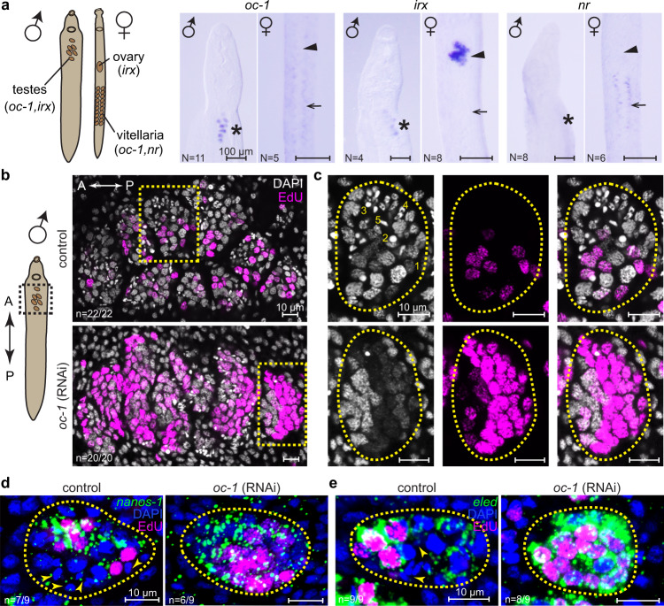
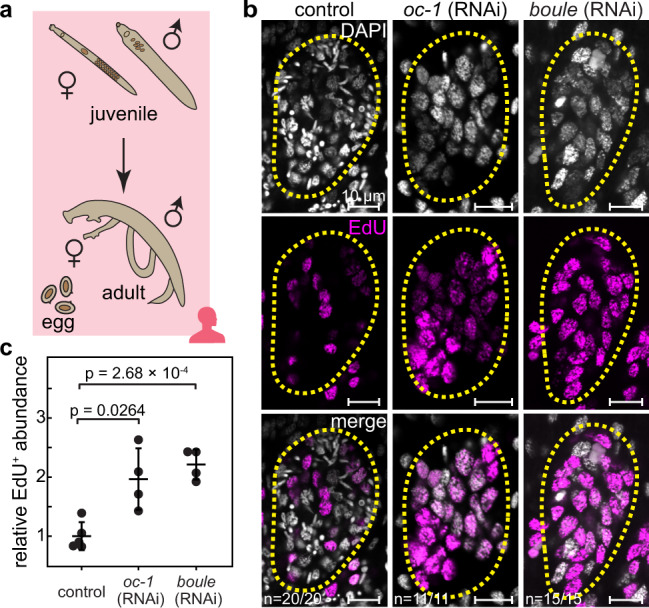
Schistosomes are parasitic flatworms causing one of the most prevalent infectious diseases from which millions of people are currently suffering. These parasites have high fecundity and their eggs are both the transmissible agents and the cause of the infection-associated pathology. Given its biomedical significance, the schistosome germline has been a research focus for more than a century. Nonetheless, molecular mechanisms that regulate its development are only now being understood. In particular, it is unknown what balances the fate of germline stem cells (GSCs) in producing daughter stem cells through mitotic divisions versus gametes through meiosis. Here, we perform single-cell RNA sequencing on juvenile schistosomes and capture GSCs during de novo gonadal development. We identify a genetic program that controls the proliferation and differentiation of GSCs. This program centers around onecut, a homeobox transcription factor, and boule, an mRNA binding protein. Their expressions are mutually dependent in the schistosome male germline, and knocking down either of them causes over-proliferation of GSCs and blocks germ cell differentiation. We further show that this germline-specific regulatory program is conserved in the planarian, schistosome's free-living evolutionary cousin, but the function of onecut has changed during evolution to support GSC maintenance.


Conflict of interest statement

The authors declare no competing interests.

Figures

Fig. 1
Fig. 1. scRNA-seq identifies schistosome stem cell subpopulations and GSC transcriptional signatures.
UMAP projections showing scRNA-seq data of a all cells and b ago2-1+ stem cells (boxed in a) only. The manifolds are reconstructed by SAM. In a, ago2-1 expression is overlaid. In b, cells are color-coded by Leiden clusters and annotated based on marker gene expression. c Heatmap showing the expression of genes enriched in specific stem cell populations, with the mean expression of each gene shifted to 0. Top: the populations are grouped through hierarchical clustering in the PC space, with the height of the branches representing correlation distance between populations. Left: annotated transcription factors (GO: 0003677 DNA binding and GO: 0003700 DNA-binding transcription factor activity) are indicated by black lines. Yellow boxes: genes that lack expression in the presumptive multipotent group compared to the progenitor group. Green box: genes enriched in nanos-1+ population. For the sake of visualization, we only show the top 50 markers per cluster. The complete list of markers is provided in Supplementary Data 1. d UMAP projections showing the expression of nanos-1 in stem cells (left) and the expression of eled (a previously characterized GSC marker), oc-1, nr, and boule in nanos-1+ cells (right). nr and boule expression separate nanos-1+ cells into S1 stem cells and GSCs.
Fig. 2
Fig. 2. oc-1 RNAi leads to over-proliferation of male GSCs.
a WISH images of oc-1, irx, and nr expression in male and female juvenile parasites. Left: schematic showing the locations of major reproductive organs in schistosome juveniles. Asterisks: primordial testes; arrowheads: ovary primordia; arrows: vitellaria primordia. N: number of samples showing similar results over two independent experiments, each using parasites collected from a separate infection. b Confocal images of DAPI/EdU-stained testes in control and oc-1 RNAi male juvenile parasites. The imaged areas correspond to the dashed box in the schematic on the left. A: anterior; P: posterior. c Magnified views of individual testis lobules corresponding to yellow boxes in b. Dashed circles: testis lobule boundary. Nuclear morphologies are characteristic of each cell type and labeled in the image as (1) GSC, (2) spermatocyte, (3) round spermatid, (4) elongating spermatid, and (5) sperm. Stage 2-5 are considered as differentiated germ cells. Note the increased fraction of EdU+ nuclei and reduced number of nuclei corresponding to differentiated germ cells after RNAi compared to controls. Quantifications of EdU+ nuclei abundance, GSC number density, and the fraction of differentiated germ cells are provided in Fig. 3c-e along with other RNAi experiments for direct comparison. d, e FISH images showing broader expression of nanos-1 (d) and eled (e) in testes after oc-1 RNAi. Arrowheads: differentiated germ cells that do not express nanos-1 or eled. n: number of samples exhibiting the reported phenotype out of the total number of samples analyzed. RNAi experiments were repeated on at least three biological replicates.
Fig. 3
Fig. 3. An RNAi screen identifies that boule phenocopies oc-1 after RNAi in the juvenile male germline.
a WISH images showing the expression of boule, akkn, lsm14, zc3h31, ccnb1ip1, and uaf in male juvenile parasites. Asterisks: testes. The imaged areas correspond to the dashed box in the schematic on the left. N: number of samples showing similar results over two independent experiments. b Confocal images showing representative individual testis lobules stained by DAPI and EdU in control and boule, akkn, lsm14, zc3h31, ccnb1ip1, and uaf RNAi juvenile parasites. Insets: magnified boxed areas. Dashed circles: testis lobule boundary. Arrowheads in ccnb1ip1 RNAi image: incomplete separation of meiotic nuclei. Nuclear morphologies are labeled in the images as in Fig. 2c. c Fraction of EdU+ nuclei, d number density of GSCs and e fraction of differentiated germ cells in testes in control and RNAi parasites. Each data point represents the mean across all testis lobules in a single parasite. N = 12 (control RNAi); N = 12 (oc-1 RNAi), N = 12 (boule RNAi), N = 4 (akkn RNAi); N = 13 (lsm1 RNAi); N = 6 (zc3h31 RNAi); N = 6 (ccnb1ip1 RNAi). Data are plotted as average ± standard deviation and p-values are calculated using two-sided Welch’s t-test. f, g FISH images showing broader expression of nanos-1 (f) and eled (g) in testes after boule RNAi. Arrowheads: differentiated germ cells that do not express nanos-1 or eled. n: number of samples exhibiting the reported phenotype out of the total number of samples analyzed. RNAi experiments were repeated on at least three biological replicates.
Fig. 4
Fig. 4. Knockdowns of boule and oc-1 cause male GSC over-proliferation in adult parasites.
a Schematic of the sexual maturation process from juveniles to adults, which mate and lay eggs. b Confocal images showing representative individual testis lobules stained by DAPI and EdU in adult parasites after control, oc-1, and boule RNAi. Dashed circles: testis lobule boundary. Note a significant increase of EdU+ nuclei and a marked reduction of differentiated germ cells. n: number of samples exhibiting the reported phenotype out of the total number of samples analyzed. c Since accurate counting of germ cells, which are densely packed in adult testes, is challenging, the number density of EdU+ nuclei per unit image area in testes normalized against the abundance in control parasites is quantified instead. N = 5 (control RNAi); N = 4 (oc-1 RNAi); N = 4 (boule RNAi). Data are plotted as average ± standard deviation and p-values are calculated using two-sided Welch’s t-test. RNAi experiments were repeated on at least three biological replicates.
Fig. 5
Fig. 5. Epistatic interactions of oc-1, boule, nanos-1, and eled.
a WISH images showing that the expression of oc-1 and boule are mutually dependent. Knockdown of either one causes the elimination of both. Asterisks: testes. b Confocal images showing representative individual testis lobules stained by DAPI and EdU in juvenile parasites after RNAi treatments. Dashed circles: testis lobule boundary. n: number of samples exhibiting the reported phenotype out of the total number of samples analyzed. c Fraction of EdU+ nuclei in testes in control and RNAi parasites. Each data point represents the average across all testis lobules in a single parasite. N = 10 (control RNAi); N = 10 (eled RNAi); N = 10 (oc-1 RNAi); N = 11 (eled;oc-1 RNAi); N = 10 (boule RNAi); N = 10 (eled;boule RNAi). nanos-1 RNAi experiments were not quantified because only a few GSC nuclei are present per testis lobule, which is insufficient for reliable statistics. Data are plotted as average ± standard deviation and p-values are calculated using two-sided Welch’s t-test. RNAi experiments were repeated on at least three biological replicates. d Proposed model of genetic interactions between oc-1, boule, nanos-1, and eled in regulating male GSC identity, proliferation, and differentiation.
Fig. 6
Fig. 6. Smed-oc-1 is required for the maintenance of spermatogonial cells.
a Schematic of planarian testes lobules containing spermatogonial cells (GSCs and mitotic spermatogonia) at the periphery and differentiated germ cells (spermatocytes, round spermatids, elongating spermatids, and sperm) distributed in the internal layers. b WISH images showing the expression of Smed-oc-1 in sexually immature (left) and mature (right) planarians. The punctuated signal in the immature planarian corresponds to presumptive testis primordia. In the mature planarian, the signal is specific to testes. c FISH images of Smed-oc-1 showing its expression in testis primordia within sexually immature planarians (top) and in spermatogonial cells (including GSCs and mitotic spermatogonia) in sexually mature planarians (bottom). N: number of samples showing similar results over three independent experiments. d Dorsal view of sexually mature planarians after control and Smed-oc-1 RNAi. Note that testes, which are distributed beneath the dorsal epithelium and visualized by strong DAPI signal, are completely eliminated after Smed-oc-1 RNAi. Animals were fixed after eight feedings of dsRNA. e Testes of control (left) and Smed-oc-1 (right) RNAi planarians after three feedings of dsRNA. In control animals, Smed-germinal histone H4 (gH4) labels the spermatogonial cells at the periphery of testis lobules, which are mostly absent after Smed-oc-1 RNAi, while differentiated germ cells remain at the center of the testis lobules. gH4 expression in individually isolated somatic stem cells maintains after Smed-oc-1 RNAi. f WISH images showing that the expression of Smed-oc-1, Smed-nanos, and Smed-boule-2 are disrupted after three feedings of Smed-oc-1 RNAi. The imaged areas correspond to the dashed box in the schematic on the left. n: number of samples exhibiting the reported phenotype out of the total number of samples analyzed. RNAi experiments were repeated on at least three biological replicates, each containing a group of planarians fed with dsRNA separately.

References

    1. Hoffmann KF, Brindley PJ, Berriman M. Halting harmful helminths. Science. 2014;346:168–169. doi: 10.1126/science.1261139. - DOI - PubMed
    1. King CH, Dangerfield-Cha M. The unacknowledged impact of chronic schistosomiasis. Chronic Illn. 2008;4:65–79. doi: 10.1177/1742395307084407. - DOI - PubMed
    1. Hotez PJ, Fenwick A. Schistosomiasis in Africa: an emerging tragedy in our new global health decade. PLoS Negl. Trop. Dis. 2009;3:e485. doi: 10.1371/journal.pntd.0000485. - DOI - PMC - PubMed
    1. Wang B, Collins JJ, III, Newmark PA. Functional genomic characterization of neoblast-like stem cells in larval Schistosoma mansoni. eLife. 2013;2:e00768. doi: 10.7554/eLife.00768. - DOI - PMC - PubMed
    1. Wang B, et al. Stem cell heterogeneity drives the parasitic life cycle of Schistosoma mansoni. eLife. 2018;7:e35449. doi: 10.7554/eLife.35449. - DOI - PMC - PubMed

Publication types