. 2021 Jan 20;12(1):469.
doi: 10.1038/s41467-020-20789-7.

Molecular determinants and mechanism for antibody cocktail preventing SARS-CoV-2 escape

Affiliations

Molecular determinants and mechanism for antibody cocktail preventing SARS-CoV-2 escape

Zhiqiang Ku et al. Nat Commun. .

Erratum in

Abstract

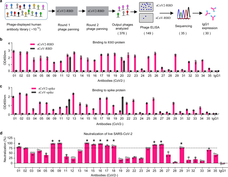
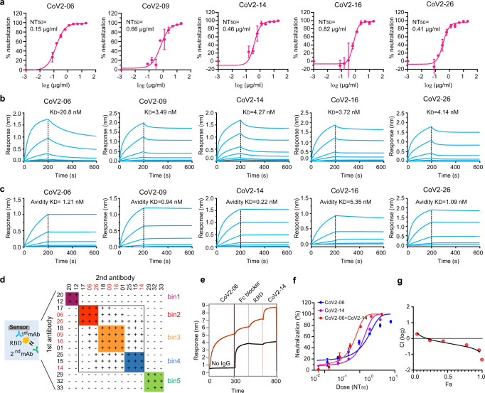
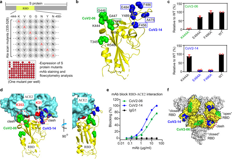
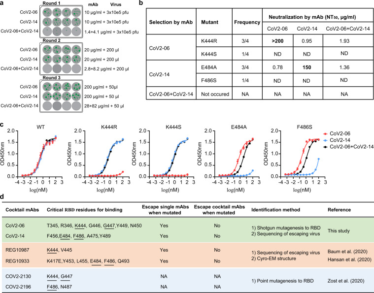
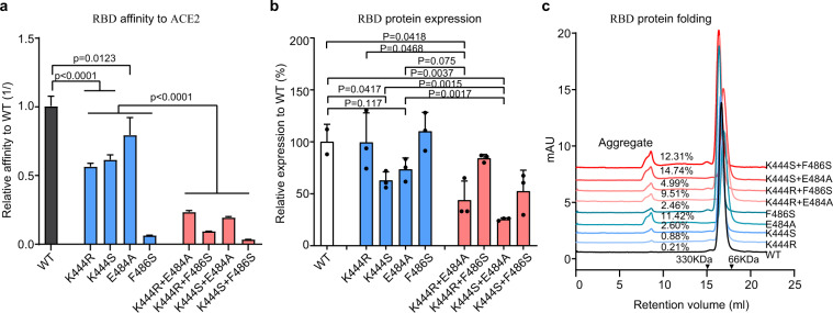
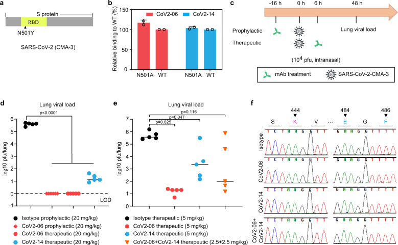
Antibody cocktails represent a promising approach to prevent SARS-CoV-2 escape. The determinants for selecting antibody combinations and the mechanism that antibody cocktails prevent viral escape remain unclear. We compared the critical residues in the receptor-binding domain (RBD) used by multiple neutralizing antibodies and cocktails and identified a combination of two antibodies CoV2-06 and CoV2-14 for preventing viral escape. The two antibodies simultaneously bind to non-overlapping epitopes and independently compete for receptor binding. SARS-CoV-2 rapidly escapes from individual antibodies by generating resistant mutations in vitro, but it doesn't escape from the cocktail due to stronger mutational constraints on RBD-ACE2 interaction and RBD protein folding requirements. We also identified a conserved neutralizing epitope shared between SARS-CoV-2 and SARS-CoV for antibody CoV2-12. Treatments with CoV2-06 and CoV2-14 individually and in combination confer protection in mice. These findings provide insights for rational selection and mechanistic understanding of antibody cocktails as candidates for treating COVID-19.


Conflict of interest statement

Z.K., N.Z., Z.A., X.X., and P.-Y.S. have filed a patent on the reverse genetic system and reporter SARS-CoV-2. Other authors declare no competing interests.

Figures

Fig. 1
Fig. 1. Isolation of RBD-directed human mAbs with neutralizing activates against SARS-CoV-2.
a Flowcharts of the scFv phage library panning and mAb selection process. b, c ELISA binding of purified mAbs to the RBD proteins (b) and the S proteins (c) of SARS-CoV-2 and SARS-CoV. The dashed line is 2× the OD450nm of a control IgG1 and as a cut-off for binders. d Neutralization of live SARS-CoV-2 by the antibodies at 10 µg/ml. The dashed line indicates a 75% neutralization. The stars indicate the 11 mAbs with neutralization above 75%. Error bars indicate SD of triplicates.
Fig. 2
Fig. 2. Identification of CoV2-06 and CoV2-14 as two neutralizing mAbs suitable for cocktail.
a Neutralization titration curves of the top five mAbs with 50% neutralization titer (NT50) below 1 µg/ml. Each data point is the mean ± SD of two replicates. b, c Kinetic binding curves of the top five mAbs to the RBD protein (b) and the prefusion S protein (c) of SARS-CoV-2. The vertical dashed lines indicate the separation of association and dissociation phases. d Epitope binning of 15 mAbs by a BLI-based cross-competition assay. Antibodies grouped into different bins shown in different colors. The top five neutralizing mAbs are shown in red; “+” denotes that the first antibody competes with the second antibody and “−” denotes that the first antibody does not compete with the second antibody. e Simultaneous binding of CoV2-06 and CoV2-14 on the sCoV2-RBD protein. f Dose-dependent percent neutralization of SARS-CoV-2 by individual CoV2-06, CoV2-14 mAbs, and a cocktail of the two mAbs; n = 3 biologically independent cells. g Plot of calculated log-scale CI values (y-axis) versus fractional effects (x-axis). CI value =1 indicates additive effect, <1 means synergism, and >1 indicates antagonism. Error bars indicate SD of triplicates.
Fig. 3
Fig. 3. Molecular determinants on the RBD for CoV2-06 and CoV2-14 binding and the mechanism of neutralization.
a Schematic diagram of the shotgun and high-throughput epitope mapping strategy. Representative alanine scan mutations in the RBD region of S and the critical procedures for mapping are shown. b The residues critical for CoV2-06 and CoV2-14 are shown as green and blue spheres, respectively, on a structure of RBD (PDB: 6M0J). The residues that make direct contact with ACE2 are boxed. c CoV2-06 or Cov2-14 binding to the sCoV2-RBD proteins with indicated mutations. Error bars indicate SD of duplicates wells. d The critical residues for CoV2-06 and CoV2-14 at the interface of RBD-ACE2 complex (PDB: 6M0J). The arrows indicate the K353 and K31 residues in ACE2, which are two virus-binding hotspots. The dashed circles indicate the steric clash of the two mAbs and ACE2 in binding to the RBD. e Dose-dependent blocking of RBD binding to ACE2 by CoV2-06 and Cov2-14. f The landscape of CoV2-06 and CoV2-14 epitopes on the trimeric S structure (PDB: 6VSB). The RBD in each monomer is outlined and colored in yellow. The CoV2-06 epitope is colored in green and the CoV2-14 epitope in blue. The dashed circle indicates a steric clash of CoV2-14 and an adjacent “open” RBD in binding to a “closed” RBD.
Fig. 4
Fig. 4. Molecular determinants on the RBD for binding by CoV2-26, CoV2-09, and VH3-53-like antibodies.
a, b The residues critical for CoV2-26 (a) and CoV2-09 (b) binding are shown as magenta spheres on the RBD–ACE2 complex (PDB: 6M0J). The arrows indicate the K353 and K31 residues in ACE2, which are two virus-binding hotspots. The dashed circles indicate the clash of mAb and ACE2 in binding to the RBD. c Dose-dependent blocking of RBD binding to ACE2 by the mAbs. d The residues critical for the VH3-53 antibody CC12.1 are shown as blue spheres on the RBD–ACE2 complex (PDB: 6M0J). e Comparison of the critical residues for the CoV2-09 and the CC12.1 antibody. f The RBD residues critical for binding of the indicated mAbs.
Fig. 5
Fig. 5. CoV2-06 and CoV2-14 cocktail prevents escape mutation of live SARS-CoV-2.
a Schematic diagram for the procedures of evaluating SARS-CoV-2 escape mutation under individual or cocktail mAbs. Green dots represent cell clusters expressing the mNeonGreen due to viral infection. b The mutated RBD residue, occurring frequency, and mAb neutralization of the mutant viruses. ND not determined, NA not available. c ELISA binding curves of indicated mAb to wild-type (WT) or mutant sCoV2-RBD proteins. Data points are mean ± SD of two replicates. d Summary of the key residues, the ability to inhibit mutant virus, and the methods of identifying the critical residues for cocktail mAbs in this study and published studies.
Fig. 6
Fig. 6. Effects of single-site or double-site mutations on the RBD affinity to ACE2, the expression level, and the folding stability of RBD.
a The relative binding affinities of the sCoV2-RBD mutant proteins ACE2. The y-axis indicates the reversed value of KD of mutants/WT. Data are mean ± SD of the KD values from fitting of five kinetic curves. Two-tailed Student’s t-test. The distribution of data points is not available from the Octet Data Analysis software. b The relative expressing levels of the sCoV2-RBD mutant proteins to wild-type (WT) protein. The y-axis indicates the value of protein concentration of mutants/WT. Data are mean ± SD of triplicate wells of transfection. Two-tailed Student’s t-test. c The size-exclusion chromatography (SEC) analysis of purified sCoV2-RBD mutant or wild-type proteins. The retention volume of proteins with indicated molecular weight are shown by arrowheads. The percentages of protein aggregates are shown.
Fig. 7
Fig. 7. Sequence analysis of SARS-CoV-2 isolates with natural mutations at the K444, E484, or F486 sites of the RBD.
a Summary of total numbers, accession ID, collection date, and geographic locations for the clinical SARS-CoV-2 isolates with indicated mutations. A total of 70,943 viral genome sequences were queried from GISAID and analyzed. b Alignment of the RBD sequences of the mutant viruses with the reference Wuhan-Hu-1 strain. c Frequency of the virus variants with single mutations of the K444, E484, and F486 residues, or simultaneous mutations of K444+E484 or K444+F486 residues in the total analyzed viral sequences.
Fig. 8
Fig. 8. Antibody protection of SARS-CoV-2 infection in mice.
a A diagram showing the N501Y adapted mutation in the S protein RBD of the SARS-CoV-2 mouse-adapted strain (CMA-3). b ELISA binding of CoV2-06 and CoV2-14 to the WT sCoV2-RBD or the N501A mutant. Error bars indicate SD of duplicates wells. c Schematic diagram of prophylactic or therapeutic evaluations of the antibodies. d The infectious viral load in the lung of CoV-06- or CoV2-14-treated mice compared to that of isotype IgG1-treated mice. The dashed line indicates the limit of detection (LOD) of the assay. e The infectious viral load in the lung of mice with indicated treatment. The median levels of the lung viral load were shown as solid lines. N = 5 mice. Ordinary one-way ANOVA with Sidak’s multiple comparison test. f Representative sequencing results of the RBD regions of the viruses harvested from each treatment groups. The amino acid residues critical for antibody binding were indicated by triangles.

References

    1. Sanders, J. M., Monogue, M. L., Jodlowski, T. Z. & Cutrell, J. B. Pharmacologic treatments for coronavirus disease 2019 (COVID-19): a review. JAMA323, 1824–1836 (2020). - PubMed
    1. Thanh Le,T, et al. The COVID-19 vaccine development landscape. Nat. Rev. Drug Discov. 2020;19:305–306. doi: 10.1038/d41573-020-00073-5. - DOI - PubMed
    1. Hoffmann M, et al. SARS-CoV-2 cell entry depends on ACE2 and TMPRSS2 and is blocked by a clinically proven protease inhibitor. Cell. 2020;181:271–280.e278. doi: 10.1016/j.cell.2020.02.052. - DOI - PMC - PubMed
    1. Li F. Structure, function, and evolution of coronavirus spike proteins. Annu. Rev. Virol. 2016;3:237–261. doi: 10.1146/annurev-virology-110615-042301. - DOI - PMC - PubMed
    1. Wrapp D, et al. Cryo-EM structure of the 2019-nCoV spike in the prefusion conformation. Science. 2020;367:1260–1263. doi: 10.1126/science.abb2507. - DOI - PMC - PubMed

Publication types

MeSH terms