. 2021 Jan 20;11(1):1848.
doi: 10.1038/s41598-020-80406-x.

Research on Golay-coded excitation in real-time imaging of high frequency ultrasound biomicroscopy

Affiliations

Research on Golay-coded excitation in real-time imaging of high frequency ultrasound biomicroscopy

Xiaochun Wang et al. Sci Rep. .

Abstract

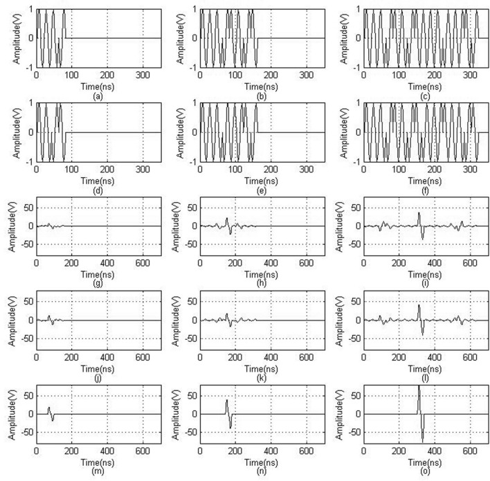
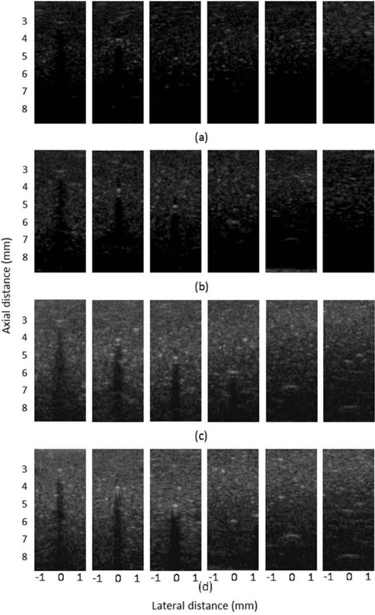
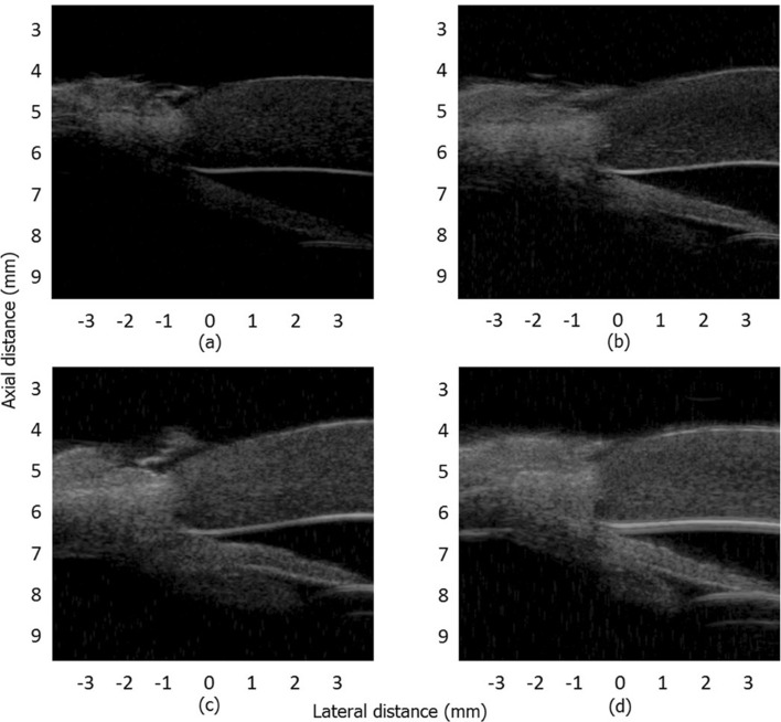
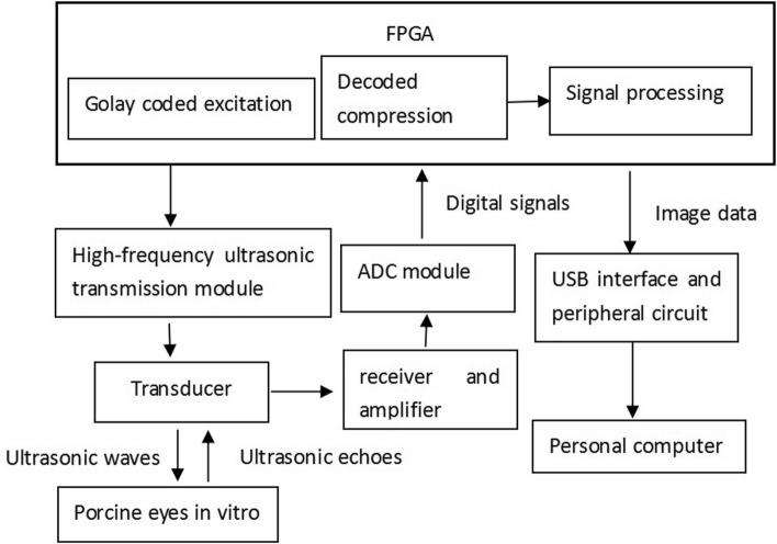
High frequency ultrasonic imaging provides clinicians with high-resolution diagnostic images and more accurate measurement results. The technique is now widely used in ophthalmology, dermatology, and small animal imaging. However, since ultrasonic attenuation in tissue increases rapidly with increasing frequency, the depth of detection of high frequency ultrasound in tissue is limited to a few millimeters. In this paper, a novel method of using Golay-coded excitation as a replacement for conventional single-pulse excitation in high frequency ultrasound biomicroscopy was proposed, and real-time imaging was realized. While maintaining the transmission voltage and image resolution unchanged, the detection depth can be effectively improved. The ultrasonic transmission frequency is 30 MHz and the transmission voltage is ± 60 V p-p. In this study, 4-bit, 8-bit, and 16-bit coding sequences and decoding compression were used. To verify the effectiveness of the coding sequence in real-time imaging of ultrasound biomicroscopy, we designed a 10-μm diameter line target echo experiment, an ultrasound phantom experiment, and an in vitro porcine eye experiment. The experimental results show that the code/decode method of signal processing can not only maintain a resolution consistent with that of single-pulse transmission, but can also improve the detection depth and signal-to-noise ratio.


Conflict of interest statement

The authors declare no competing interests.

Figures

Figure 1
Figure 1
Computer simulation results for Golay coding and decoding. (ac) are respectively the 4-bit, 8-bit, and 16-bit Golay sequence A codes for encoding. (df) are respectively the 4-bit, 8-bit, and 16-bit Golay sequence B codes for encoding. (gi) are respectively the 4-bit, 8-bit, and 16-bit Golay sequence A codes for decoding. (jl) are respectively the 4-bit, 8-bit, and 16-bit Golay sequence B codes for decoding. (mo) are respectively the 4-bit, 8-bit, and 16-bit Golay decoding results.
Figure 2
Figure 2
Experimental results of wire target echoes. (a) Single-pulse transmission waveforms. (bd) are respectively the 4-bit, 8-bit, and 16-bit Golay sequence A coded transmission waveforms. (eg) are respectively the 4-bit, 8-bit, and 16-bit Golay sequence B coded transmission waveforms. (hj) are respectively the 4-bit, 8-bit, and 16-bit Golay sequence A coded return echoes. (km) are respectively the 4-bit, 8-bit, and 16-bit Golay sequence B coded return echoes. (n) is the return echo of a single pulse. (oq) are respectively the 4-bit, 8-bit, and 16-bit Golay sequence decoded return echoes. (ru) are respectively the frequency spectra of the single-pulse echo and the 4-bit, 8-bit, and 16-bit Golay sequence echoes.
Figure 3
Figure 3
SNR improvement of the echo signal under Golay code excitations.
Figure 4
Figure 4
Envelope of decoded echo signals.
Figure 5
Figure 5
The results of resolution test imaging: (a) Single-pulse transmission. (b) 4-bit Golay-coded transmission. (c) 8-bit Golay-coded transmission. (d) 16-bit Golay-coded transmission.
Figure 6
Figure 6
Tissue-mimicking ultrasound phantom imaging results: (a) Single-pulse real-time scan image result. (bd) are respectively the real-time imaging results for 4-bit, 8-bit, and 16-bit coded excitation after decoding compression.
Figure 7
Figure 7
Imaging results of a corner segment of an in vitro porcine eyeball. (a) Real-time scan image using a single pulse. (bd) Real-time imaging results using 4-bit, 8-bit, and 16-bit coded excitation and decoding compression, respectively.
Figure 8
Figure 8
Pulse compression of the Golay code.
Figure 9
Figure 9
Block diagram of the echo experimental system.
Figure 10
Figure 10
Block diagram of the real-time imaging system.
Figure 11
Figure 11
Diagram of the line target for the resolution test. (a) The wire target for resolution test. (b) Diagram of ultrasonic scanning system.

References

    1. Lockwood GR, et al. Beyond 30 MHz—applications of high-frequency ultrasound imaging. IEEE Eng. Med. Biol. Mag. 1996;15:60–71. doi: 10.1109/51.544513. - DOI
    1. Kuerten D, et al. Evaluation of long-term anatomic changes following canaloplasty with anterior segment spectral-domain optical coherence tomography and ultrasound biomicroscopy. J. Glaucoma. 2018;27:87–93. doi: 10.1097/IJG.0000000000000827. - DOI - PubMed
    1. Farhan HMA, AlMutairi RN. Anterior segment biometry using ultrasound biomicroscopy and the Artemis-2 very high frequency ultrasound scanner. Clin. Ophthalmol. 2013;7:141–147. doi: 10.2147/OPTH.S39463. - DOI - PMC - PubMed
    1. Farhan HMA. Agreement between Orbscan II, VuMAX UBM and Artemis-2 very-high frequency ultrasound scanner for measurement of anterior chamber depth. BMC Ophthalmol. 2014;14:1–7. doi: 10.1186/1471-2415-14-20. - DOI - PMC - PubMed
    1. Silverman RH, et al. Clinical applications of very high frequency ultrasound in ophthalmology. J. Acoust. Soc. Am. 2004;115:2376–2376. doi: 10.1121/1.4780096. - DOI

Publication types

LinkOut - more resources