. 2021 May;23(5):900-908.
doi: 10.1038/s41436-020-01071-z. Epub 2021 Jan 20.

A pathogenic UFSP2 variant in an autosomal recessive form of pediatric neurodevelopmental anomalies and epilepsy

Affiliations

A pathogenic UFSP2 variant in an autosomal recessive form of pediatric neurodevelopmental anomalies and epilepsy

Min Ni et al. Genet Med. 2021 May.

Abstract

Purpose: Neurodevelopmental disabilities are common and genetically heterogeneous. We identified a homozygous variant in the gene encoding UFM1-specific peptidase 2 (UFSP2), which participates in the UFMylation pathway of protein modification. UFSP2 variants are implicated in autosomal dominant skeletal dysplasias, but not neurodevelopmental disorders. Homozygosity for the variant occurred in eight children from four South Asian families with neurodevelopmental delay and epilepsy. We describe the clinical consequences of this variant and its effect on UFMylation.

Methods: Exome sequencing was used to detect potentially pathogenic variants and identify shared regions of homozygosity. Immunoblotting assessed protein expression and post-translational modifications in patient-derived fibroblasts.

Results: The variant (c.344T>A; p.V115E) is rare and alters a conserved residue in UFSP2. Immunoblotting in patient-derived fibroblasts revealed reduced UFSP2 abundance and increased abundance of UFMylated targets, indicating the variant may impair de-UFMylation rather than UFMylation. Reconstituting patient-derived fibroblasts with wild-type UFSP2 reduced UFMylation marks. Analysis of UFSP2's structure indicated that variants observed in skeletal disorders localize to the catalytic domain, whereas V115 resides in an N-terminal domain possibly involved in substrate binding.

Conclusion: Different UFSP2 variants cause markedly different diseases, with homozygosity for V115E causing a severe syndrome of neurodevelopmental disability and epilepsy.


Conflict of interest statement

R.J.D. is a member of the Scientific Advisory Board for Agios Pharmaceuticals and Vida Ventures.

Figures
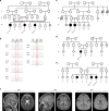
Fig. 1
Fig. 1. Pedigrees of four unrelated families with affected children carrying the homozygous UFSP2 p.V115E variant.
(a) Pedigree showing the relationship of the three affected patients indicated as P1, P2, and P3 from a consanguineous Pakistani kindred (family 1). Genotype annotations show the nucleotide as T for wild-type and A for variant. (b) Chromatogram of UFSP2 sequences confirming parental heterozygosity (III.3, III.4, III.5, and III.8) for c.344T>A, homozygosity for the variant in the three patients (IV.1, IV.4, and IV.6) and homozygous wild-type in the unaffected sister (IV.2). (c) Family of patients P4 and P5 (family 2). Three individuals (II.6, II.7, and III.1) who died with unknown neurological disorders are shaded in light gray. (d) Family of patient P6 (family 3). AW alive and well. (e) Family of patients P7 and P8 (family 4). The female sibling III.4 had a clinically similar disorder and died at 8 years of age. DNA was not available for UFSP2 sequencing. (f) Representative magnetic resonance images (MRIs), including P5, cerebellar volume loss (left) and cortical volume loss (right); P6, mild cerebellar volume loss (left) and mild hypomyelination (right); P7, moderate prominence of cortical cerebrospinal fluid (CSF) space. High-resolution electronic images were not available for P1–P3.
Fig. 2
Fig. 2. Runs of homozygosity analysis in families 1 and 3.
(a) Chromosomal distribution of homozygous regions in patients P1, P2, P3, and P6. The displayed regions are larger than 1 Mb and are homozygous in the patients but not the parents. The sole homozygous region shared by all four patients is indicated by the red frame on chromosome 4. (b) Schematic of homozygous segments on chromosome 4q in families 1 and 3. For each individual, the top line displays markers with homozygous genotypes and the bottom line displays markers with heterozygous genotypes. The homozygous regions are highlighted in color blocks: red for regions common to more than one individual, gray for regions unique to one individual. The UFSP2 locus is indicated.
Fig. 3
Fig. 3. UFSP2 expression and UFMylation marks in patient and control fibroblasts.
(a) Immunoblot analysis of primary human fibroblasts from four control subjects and patients P1, P2 and P3. (b) Quantitative analysis of the immunoblots for UFMylated proteins in Fig. 3a. The intensities for total anti-UFM1 signal were normalized to GAPDH. **p < 0.01. (c) Quantitative real-time polymerase chain reaction (RT-PCR) of UFSP2 messenger RNA (mRNA) in fibroblasts from three control subjects and patients P1, P2, and P3. (d) Immunoblot analysis of UFMylated proteins in P1 and P3 fibroblasts ectopically expressing wild-type (WT) or V115E variant of UFSP2.
Fig. 4
Fig. 4. Structural analysis of human UFSP2 variants and expression of UFSP2 and its targets in human and mouse tissues.
(a) Schematic of human UFSP2 functional domains. The patient-derived variants are indicated, with those causing dominantly inherited disease in blue and the new variant causing recessively inherited disease in red. (b) Conservation of the V115 residue (red frame) across multiple species. (c) Three-dimensional structure of mouse Ufsp2. The homologous residues mutated in patients are indicated. Val107, Tyr282, Asp418, and His420 correspond to Val115, Tyr290, Asp426, and His428 respectively, in human UFSP2. (d) Gene expression of UFSP2 and DDRGK1 in 24 human tissues from the Genotype–Tissue Expression (GTEx) database. (e) Immunoblots showing expression of Ufsp2 and its targets, Ddrgk1, Trip4, and Rpl26, in mouse (M) tissues and human (H) brain. Calnexin is used as a loading control.

References

    1. Kjeldsen MJ, Corey LA, Christensen K, Friis ML. Epileptic seizures and syndromes in twins: the importance of genetic factors. Epilepsy Res. 2003;55:137–146. doi: 10.1016/S0920-1211(03)00117-7. - DOI - PubMed
    1. Hildebrand MS, et al. Recent advances in the molecular genetics of epilepsy. J. Med. Genet. 2013;50:271–279. doi: 10.1136/jmedgenet-2012-101448. - DOI - PubMed
    1. Yang Y, et al. Clinical whole-exome sequencing for the diagnosis of mendelian disorders. N. Engl. J. Med. 2013;369:1502–1511. doi: 10.1056/NEJMoa1306555. - DOI - PMC - PubMed
    1. Liu P, et al. Reanalysis of clinical exome sequencing data. N. Engl. J. Med. 2019;380:2478–2480. doi: 10.1056/NEJMc1812033. - DOI - PMC - PubMed
    1. Gerakis Y, Quintero M, Li H, Hetz C. The UFMylation system in proteostasis and beyond. Trends Cell. Biol. 2019;29:974–986. doi: 10.1016/j.tcb.2019.09.005. - DOI - PMC - PubMed

Publication types