Review
. 2021 Jan 5;6(1):1-11.
doi: 10.1016/j.synbio.2020.12.001. eCollection 2021 Mar.

Complex natural product production methods and options

Affiliations
Review

Complex natural product production methods and options

Dongwon Park et al. Synth Syst Biotechnol. .

Abstract

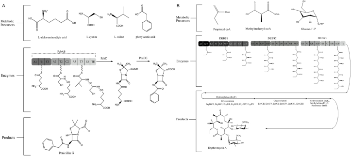
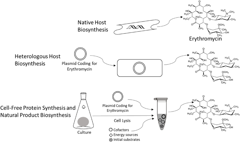
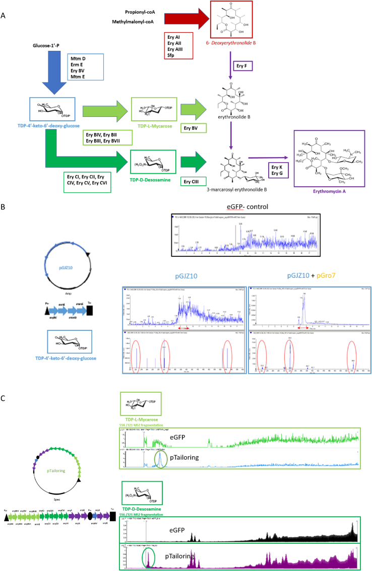
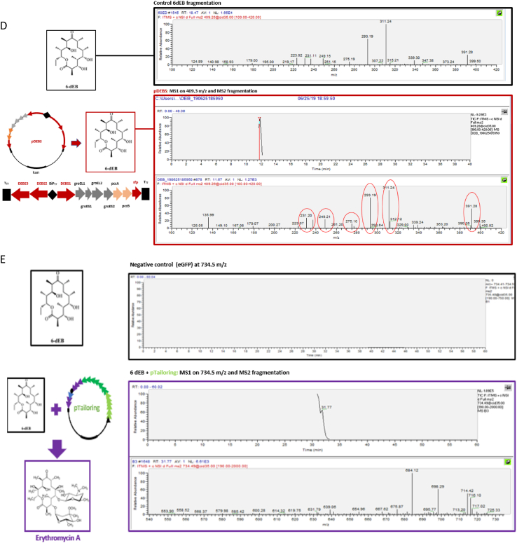
Natural products have had a major impact upon quality of life, with antibiotics as a classic example of having a transformative impact upon human health. In this contribution, we will highlight both historic and emerging methods of natural product bio-manufacturing. Traditional methods of natural product production relied upon native cellular host systems. In this context, pragmatic and effective methodologies were established to enable widespread access to natural products. In reviewing such strategies, we will also highlight the development of heterologous natural product biosynthesis, which relies instead on a surrogate host system theoretically capable of advanced production potential. In comparing native and heterologous systems, we will comment on the base organisms used for natural product biosynthesis and how the properties of such cellular hosts dictate scaled engineering practices to facilitate compound distribution. In concluding the article, we will examine novel efforts in production practices that entirely eliminate the constraints of cellular production hosts. That is, cell free production efforts will be introduced and reviewed for the purpose of complex natural product biosynthesis. Included in this final analysis will be research efforts made on our part to test the cell free biosynthesis of the complex polyketide antibiotic natural product erythromycin.

Keywords: Bio-manufacturing; Biosynthesis; Heterologous host; Native host; Natural product.


Conflict of interest statement

The Authors Declare No Conflicts.

Figures

Fig. 1
Fig. 1
Biosynthetic pathways for the natural product antibiotics penicillin (a) and erythromycin (b). Highlighted are the multi-domain biosynthetic enzymes for nonribosomal peptide (penicillin) and polyketide (erythromycin) biosynthesis. A: adenylation; T: thiolation; C: condensation; E: epimerization; TE; thioesterase; DEBS: Deoxyerythronolide B Synthase; AT: acyl transferase; ACP: acyl carrier protein; KS: β-keto-acyl synthase; KR: β-keto reductase; KR*: non-functional KR; DH: dehydratase; ER: enoyl reductase.
Fig. 2
Fig. 2
Natural product production options. Featured is the natural product polyketide antibiotic erythromycin, produced natively from Saccharopolyspora erythraea. Heterologous production of erythromycin has been completed using Streptomyces spp. and Escherichia coli as surrogate hosts; whereas, cell-free biosynthesis removes the need for a dedicated host (except for systems that rely on expression machinery provided via cellular lysis, as depicted).
Fig. 3
Fig. 3
Cell free biosynthesis for erythromycin A. The erythromycin A biosynthetic pathway (a) color-coded for steps leading to complete compound formation. Cell free biosynthesis for TDP-4′-keto-6′-deoxy glucose using plasmids pGJZ10 (indicated [27]) and pGro7 (providing the GroEL/ES chaperonin genes), using a plasmid expressing eGFP as a negative control and compared to samples with pGJZ10 and pGJZ10+pGro7 as analyzed by LC-MS (b). Cell free production for TDP-l-mycarose and TDP-d-desosamine using pTailoring previously described [25] (c). Cell free biosynthesis of 6-deoxyerythronolide B (6dEB) using plasmids pDEBS [25] assessed by LC-MS (d). Combining the pTailoring plasmid with the 6dEB molecule indicated erythromycin A formation by LC-MS (e). Cell free extract was prepared as follows: 10 mL of overnight-cultured E. coli BL21 Star (DE3) was inoculated in 1 L of 2 × YTPG medium (10 g/L yeast extract, 16 g/L tryptone, 3 g/L K2HPO4, 7 g/L KH2PO4, 5 g/L NaCl, and 18 g/L glucose) and grown at 37 °C with 250 rpm shaking. The culture was induced with 0.5 mM IPTG at an OD600nm of 0.6–0.8, and the cells were grown until OD600nm 3.5. The cells were harvested by centrifuge (8000 rpm, 10 min) and washed thrice with S30 Buffer (10 mM Tris-acetate, 14 mM Mg(OAc)2, 60 mM KOAc, 5 mM DTT) and stored at −80 °C after flash-freeze using liquid nitrogen. The cells were resuspended in S30 Buffer (1.2 g/mL) and aliquoted to 1.5 mL. Aliquots were sonicated with a FB50 (Thermo Fisher) sonifier with a 3 mm microtip (10 s on/10 s off) 5 times. The extract was prepared by collecting supernatant after 12,000 rpm of centrifugation. Cell free natural product biosynthesis was performed by using eGFP plasmid DNA as a control and a reaction buffer with the following contents: 1.2 mM ATP; 0.85 mM each of GTP, UTP and CTP; 34 μg/mL folinic acid; 171 μg/mL T7 RNA polymerase; 2 mM each of the 20 translatable amino acids, 0.33 mM nicotinamide adenine dinucleotide (NAD), 0.26 mM coenzyme A (CoA), 33 mM PEP, 130 mM potassium glutamate, 10 mM ammonium glutamate, 12 mM magnesium glutamate, 1.5 mM spermidine, 1 mM putrescine, 57 mM HEPES, 4 mM sodium oxalate, and 1 μL of 20 mg/mL Sfp [133]. To the reaction buffer, 0.25 volume of cell extract (described above) and 20 μg/mL plasmid DNA were added to reach a final volume of 25 μL. The reaction was held for 20 h at 30 °C and the required natural product metabolic substrates were added: gluocse-1-phosphate (10 mM) and dTTP (10 mM) for tailoring sugar biosynthesis, and propionyl-CoA (1 mM) and methyl malonyl-CoA (6 mM) for 6dEB biosynthesis. After 20 h, the samples were moved to −20 °C prior to SDS-PAGE and LC-MS analysis. Samples were prepared for analysis by diluting three times in LC-MS grade methanol and centrifuging at 13,000 rpm for 15 min with supernatant used for analysis. Erythromycin and 6dEB samples were applied to a Thermo Scientific Orbitrap XL with a C-18 analytical column. TDP-deoxysugar samples (mycarose, desosamine) were measured by Agilent G6545A quadrupole-time-of-flight (Q-TOF) using an XBridge Shield RP18 3.5 μm, 3.0 mm × 150 mm column from Waters. All MS analyses were conducted in positive ion mode. A linear gradient of 80% buffer A (95% water/5% acetonitrile/0.1% formic acid) to 100% buffer B (5% water/95% acetonitrile/0.1% formic acid) was used at a flow rate of 10 μL/min for the LC.
Fig. 3
Fig. 3
Cell free biosynthesis for erythromycin A. The erythromycin A biosynthetic pathway (a) color-coded for steps leading to complete compound formation. Cell free biosynthesis for TDP-4′-keto-6′-deoxy glucose using plasmids pGJZ10 (indicated [27]) and pGro7 (providing the GroEL/ES chaperonin genes), using a plasmid expressing eGFP as a negative control and compared to samples with pGJZ10 and pGJZ10+pGro7 as analyzed by LC-MS (b). Cell free production for TDP-l-mycarose and TDP-d-desosamine using pTailoring previously described [25] (c). Cell free biosynthesis of 6-deoxyerythronolide B (6dEB) using plasmids pDEBS [25] assessed by LC-MS (d). Combining the pTailoring plasmid with the 6dEB molecule indicated erythromycin A formation by LC-MS (e). Cell free extract was prepared as follows: 10 mL of overnight-cultured E. coli BL21 Star (DE3) was inoculated in 1 L of 2 × YTPG medium (10 g/L yeast extract, 16 g/L tryptone, 3 g/L K2HPO4, 7 g/L KH2PO4, 5 g/L NaCl, and 18 g/L glucose) and grown at 37 °C with 250 rpm shaking. The culture was induced with 0.5 mM IPTG at an OD600nm of 0.6–0.8, and the cells were grown until OD600nm 3.5. The cells were harvested by centrifuge (8000 rpm, 10 min) and washed thrice with S30 Buffer (10 mM Tris-acetate, 14 mM Mg(OAc)2, 60 mM KOAc, 5 mM DTT) and stored at −80 °C after flash-freeze using liquid nitrogen. The cells were resuspended in S30 Buffer (1.2 g/mL) and aliquoted to 1.5 mL. Aliquots were sonicated with a FB50 (Thermo Fisher) sonifier with a 3 mm microtip (10 s on/10 s off) 5 times. The extract was prepared by collecting supernatant after 12,000 rpm of centrifugation. Cell free natural product biosynthesis was performed by using eGFP plasmid DNA as a control and a reaction buffer with the following contents: 1.2 mM ATP; 0.85 mM each of GTP, UTP and CTP; 34 μg/mL folinic acid; 171 μg/mL T7 RNA polymerase; 2 mM each of the 20 translatable amino acids, 0.33 mM nicotinamide adenine dinucleotide (NAD), 0.26 mM coenzyme A (CoA), 33 mM PEP, 130 mM potassium glutamate, 10 mM ammonium glutamate, 12 mM magnesium glutamate, 1.5 mM spermidine, 1 mM putrescine, 57 mM HEPES, 4 mM sodium oxalate, and 1 μL of 20 mg/mL Sfp [133]. To the reaction buffer, 0.25 volume of cell extract (described above) and 20 μg/mL plasmid DNA were added to reach a final volume of 25 μL. The reaction was held for 20 h at 30 °C and the required natural product metabolic substrates were added: gluocse-1-phosphate (10 mM) and dTTP (10 mM) for tailoring sugar biosynthesis, and propionyl-CoA (1 mM) and methyl malonyl-CoA (6 mM) for 6dEB biosynthesis. After 20 h, the samples were moved to −20 °C prior to SDS-PAGE and LC-MS analysis. Samples were prepared for analysis by diluting three times in LC-MS grade methanol and centrifuging at 13,000 rpm for 15 min with supernatant used for analysis. Erythromycin and 6dEB samples were applied to a Thermo Scientific Orbitrap XL with a C-18 analytical column. TDP-deoxysugar samples (mycarose, desosamine) were measured by Agilent G6545A quadrupole-time-of-flight (Q-TOF) using an XBridge Shield RP18 3.5 μm, 3.0 mm × 150 mm column from Waters. All MS analyses were conducted in positive ion mode. A linear gradient of 80% buffer A (95% water/5% acetonitrile/0.1% formic acid) to 100% buffer B (5% water/95% acetonitrile/0.1% formic acid) was used at a flow rate of 10 μL/min for the LC.

Similar articles

Cited by

References

    1. Baker D.D. The value of natural products to future pharmaceutical discovery. Nat Prod Rep. 2007;24(6):1225–1244. - PubMed
    1. Cragg G.M., Newman D.J. Natural products: a continuing source of novel drug leads. Biochim Biophys Acta. 2013;1830(6):3670–3695. - PMC - PubMed
    1. Grushkin D. Natural products emergent. Nat Med. 2013;19(4):390–392. - PubMed
    1. Newman D.J., Cragg G.M. Natural products as sources of new drugs over the 30 years from 1981 to 2010. J Nat Prod. 2012;75(3):311–335. - PMC - PubMed
    1. Nair S.K., Jez J.M. Natural product biosynthesis: what's next? An introduction to the JBC Reviews Thematic Series. J Biol Chem. 2020;295(2):335–336. - PMC - PubMed

LinkOut - more resources