

Training deep-learning segmentation models from severely limited data
- PMID: 33474727
- PMCID: PMC8058262
- DOI: 10.1002/mp.14728
Training deep-learning segmentation models from severely limited data
Abstract
Purpose: To enable generation of high-quality deep learning segmentation models from severely limited contoured cases (e.g., ~10 cases).
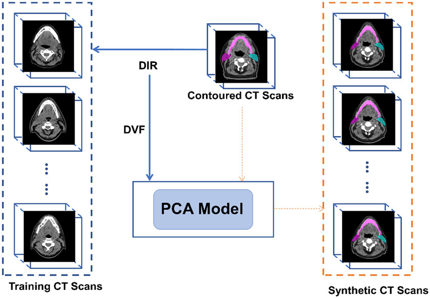
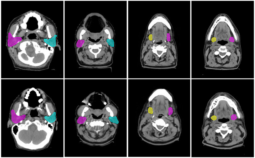
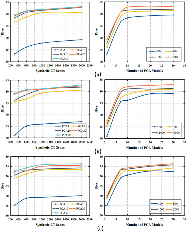
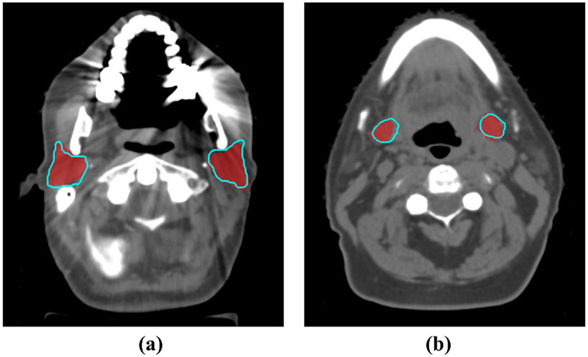
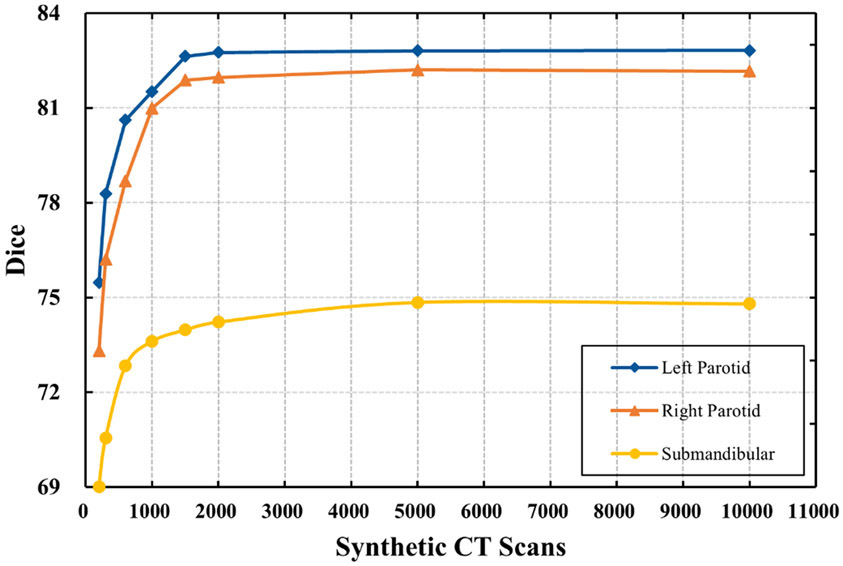
Methods: Thirty head and neck computed tomography (CT) scans with well-defined contours were deformably registered to 200 CT scans of the same anatomic site without contours. Acquired deformation vector fields were used to train a principal component analysis (PCA) model for each of the 30 contoured CT scans by capturing the mean deformation and most prominent variations. Each PCA model can produce an infinite number of synthetic CT scans and corresponding contours by applying random deformations. We used 300, 600, 1000, and 2000 synthetic CT scans and contours generated from one PCA model to train V-Net, a 3D convolutional neural network architecture, to segment parotid and submandibular glands. We repeated the training using same numbers of training cases generated from 7, 10, 20, and 30 PCA models, with the data distributed evenly between each PCA model. Performance of the segmentation models was evaluated with Dice similarity coefficients between auto-generated contours and physician-drawn contours on 162 test CT scans for parotid glands and another 21 test CT scans for submandibular glands.
Results: Dice values varied with the number of synthetic CT scans and the number of PCA models used to train the network. By using 2000 synthetic CT scans generated from 10 PCA models, we achieved Dice values of 82.8% ± 6.8% for right parotid, 82.0% ± 6.9% for left parotid, and 74.2% ± 6.8% for submandibular glands. These results are comparable with those obtained from state-of-the-art auto-contouring approaches, including a deep learning network trained from more than 1000 contoured patients and a multi-atlas algorithm from 12 well-contoured atlases. Improvement was marginal when >10 PCA models or >2000 synthetic CT scans were used.
Conclusions: We demonstrated an effective data augmentation approach to train high-quality deep learning segmentation models from a limited number of well-contoured patient cases.
Keywords: Auto-segmentation; convolutional neural networks; data augmentation; deep learning; principal component analysis.
© 2021 American Association of Physicists in Medicine.
Figures
References
-
- lin D, Vasilakos AV, Tang Y, Yao Y. Neural networks for computer-aided diagnosis in medicine: A review. Neurocomputing. 2016;216:700–708. doi:10.1016/j.neucom.2016.08.039 - DOI
-
- Cardenas CE, McCarroll RE, Court LE, et al. Deep Learning Algorithm for Auto-Delineation of High-Risk Oropharyngeal Clinical Target Volumes With Built-In Dice Similarity Coefficient Parameter Optimization Function. Int J Radiat Oncol. 2018;101(2):468–478. doi:10.1016/j.ijrobp.2018.01.114 - DOI - PMC - PubMed
-
- Nikolov S, Blackwell S, Mendes R, et al. Deep learning to achieve clinically applicable segmentation of head and neck anatomy for radiotherapy. ArXiv180904430 Phys Stat. Published online September 12, 2018. Accessed June 25, 2020. http://arxiv.org/abs/1809.04430
MeSH terms
Grants and funding
LinkOut - more resources
Full Text Sources
Other Literature Sources
Medical
