. 2021 Mar;216(3):585-598.
doi: 10.2214/AJR.20.22783. Epub 2021 Jan 21.

Best Practices: Hip Femoroacetabular Impingement

Affiliations

Best Practices: Hip Femoroacetabular Impingement

Florian Schmaranzer et al. AJR Am J Roentgenol. 2021 Mar.

Abstract

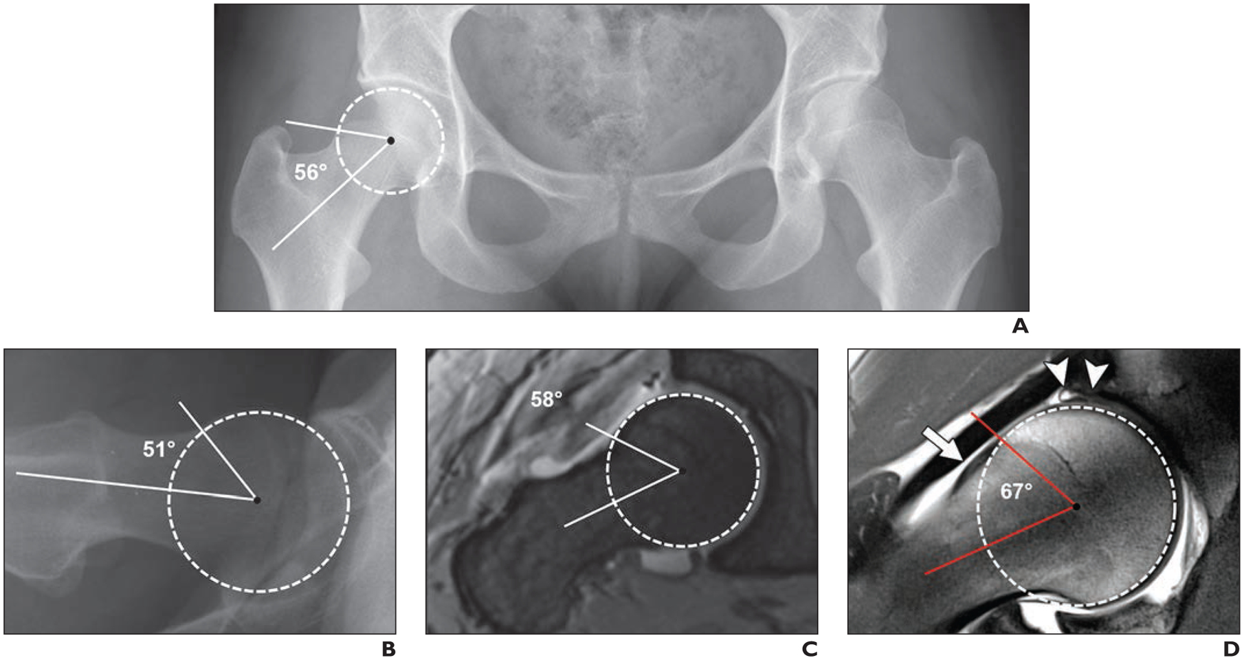
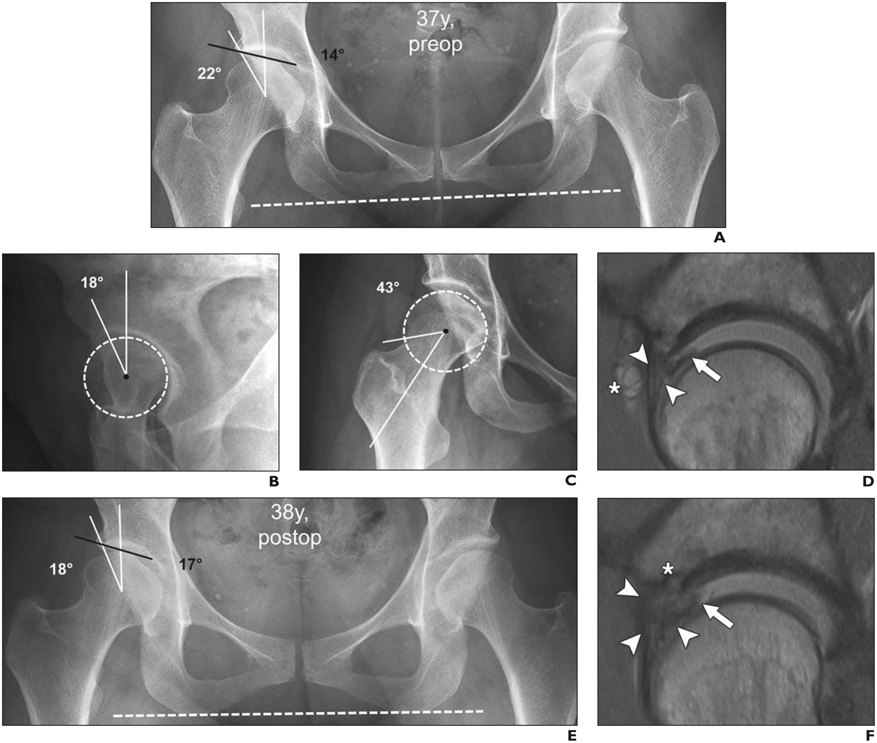
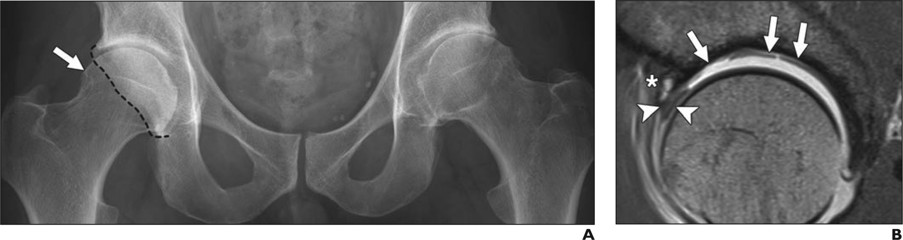
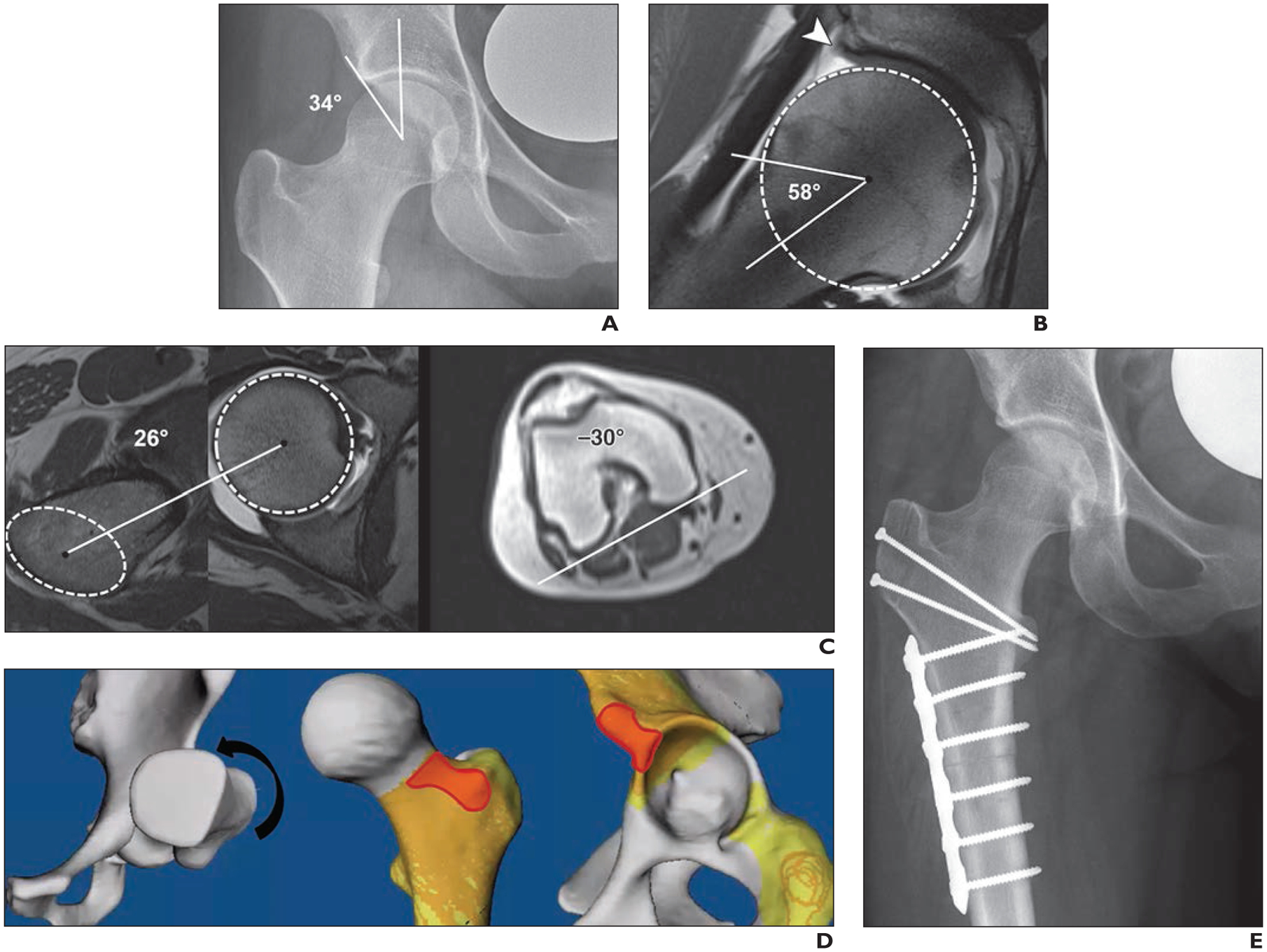
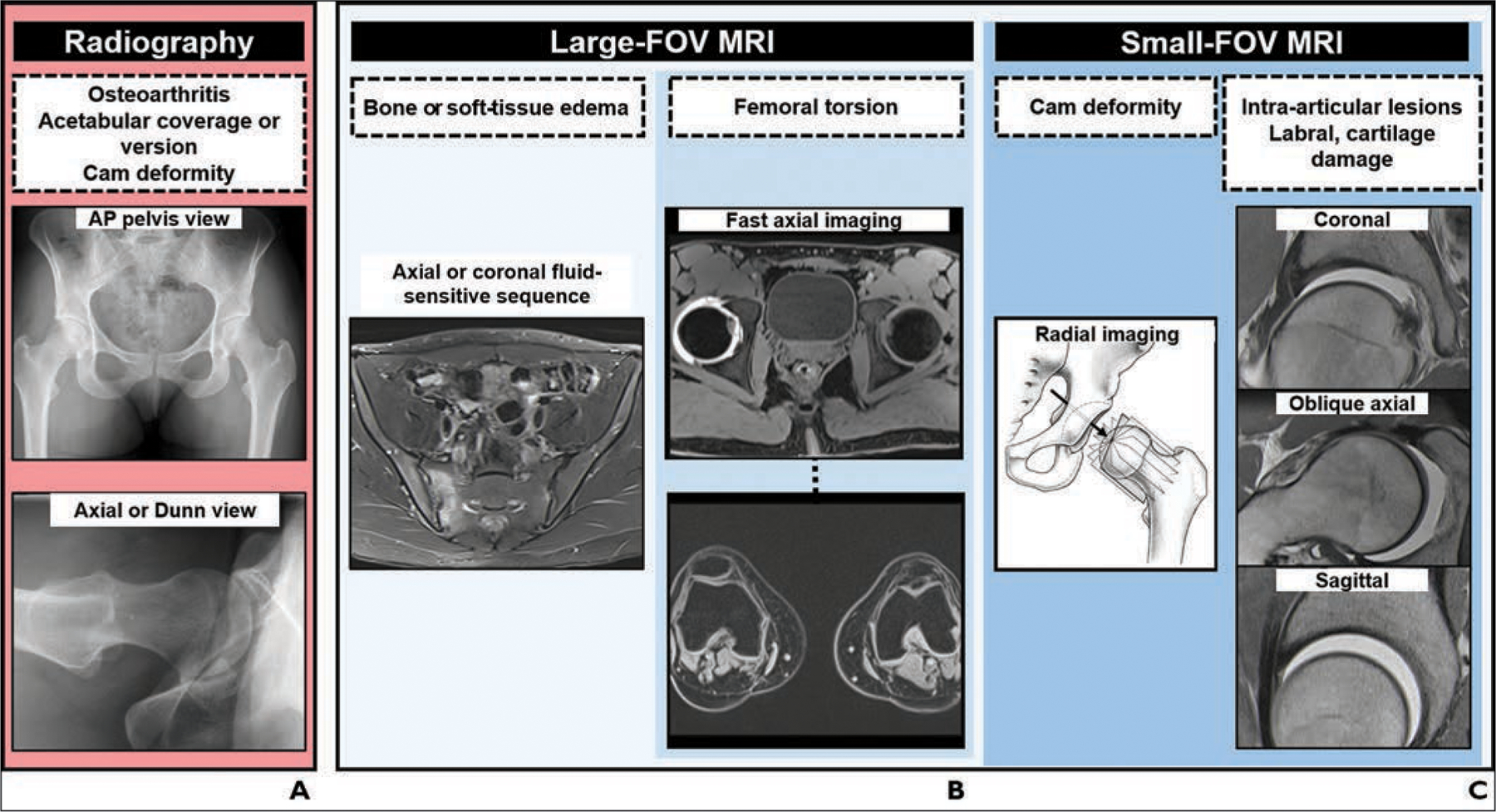
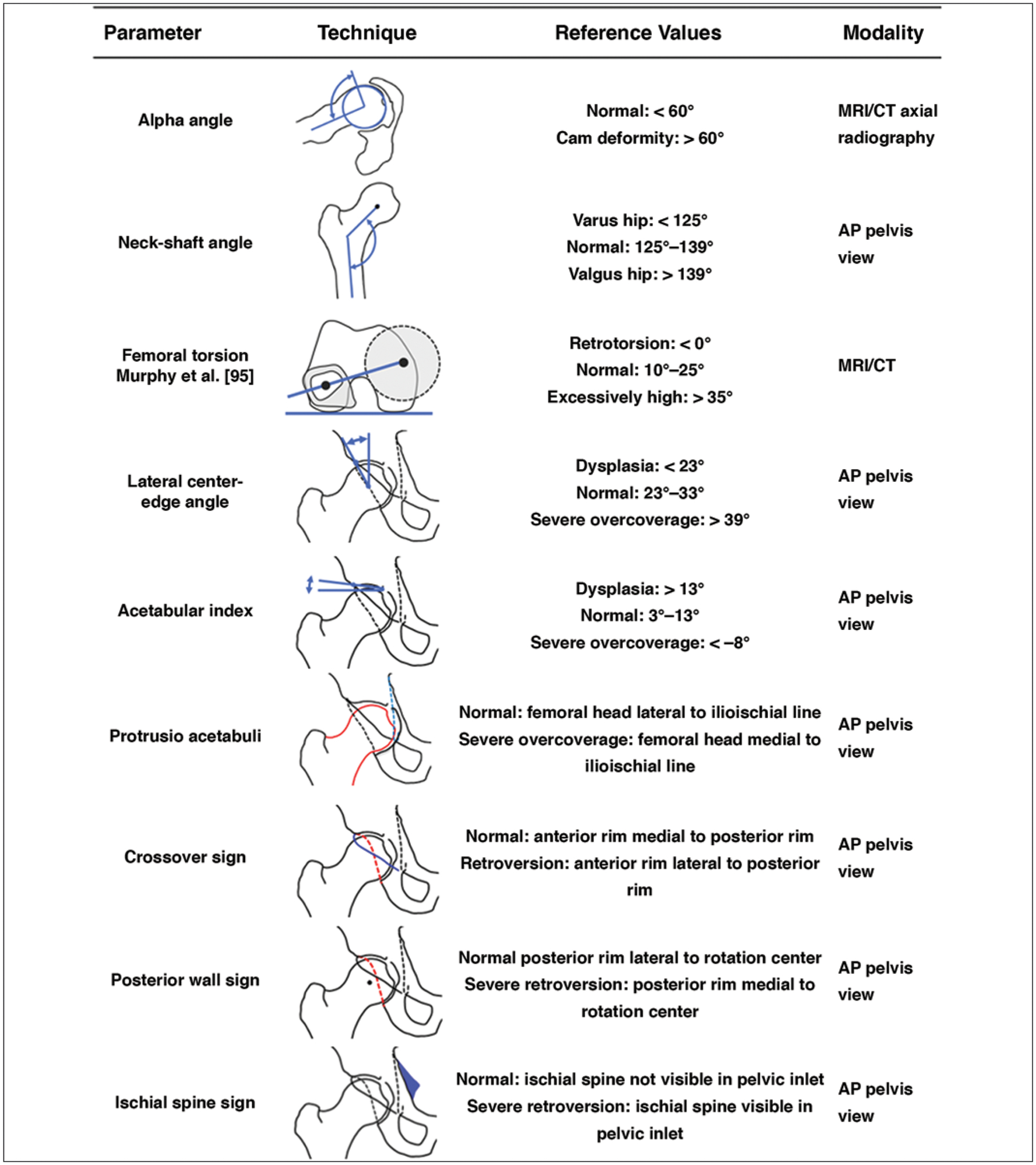
OBJECTIVE. Imaging plays a critical role in the assessment of patients with femoroacetabular impingement (FAI). With better understanding of the underlying pathomechanics and advances in joint-preserving surgery, there is an increasing need to define the most appropriate imaging workup. The purpose of this article is to provide guidance on best practices for imaging of patients with FAI in light of recent advances in corrective FAI surgery. CONCLUSION. Pelvic radiography with dedicated hip projections is the basis of the diagnostic workup of patients with suspected FAI to assess arthritic changes and acetabular coverage and to screen for cam deformities. Chondrolabral lesions should be evaluated with unenhanced MRI or MR arthrography. The protocol should include a large-FOV fluid-sensitive sequence to exclude conditions that can mimic or coexist with FAI, radial imaging to accurately determine the presence of a cam deformity, and imaging of the distal femoral condyles for measurement of femoral torsion. CT remains a valuable tool for planning of complex surgical corrections. Advanced imaging, such as 3D simulation, biochemical MRI, and MR arthrography with application of leg traction, has great potential to improve surgical decision-making. Further research is needed to assess the added clinical value of these techniques.

Keywords: MR arthrography; MRI; femoroacetabular impingement; imaging workup; radiography.


Figures

Fig. 1—
Fig. 1—
28-year-old woman with 2-year history of right-sided groin pain and positive anterior impingement test (pain with hip flexion, adduction, and internal rotation). Case emphasizes importance of appropriate imaging workup of patients with suspected femoroacetabular impingement to arrive at correct diagnosis. Alpha angles are measured between femoral neck axis (bottom line) and line that connects proximal part of asphericity (top line), determined by best-fitting circle (circle), with femoral rotation center. A, Anteroposterior radiograph of pelvis shows normal pelvic morphology, preserved hip joint space, and no arthritic change. B, Cross-table lateral radiograph of symptomatic right hip shows no obvious deformity. Patient was referred for MR arthrography to evaluate morphology of femoral head-neck junction. C, Oblique axial MR arthrogram shows normal femoral head-neck junction. D, Radial proton density–weighted MR image shows mild cam deformity (arrow) with alpha angle of 67°. Intrasubstance tear of superior labrum with adjacent cartilage damage (arrowheads) is evident. Patient was referred for hip arthroscopy for cam resection and labral repair. Cam deformities are frequently located in anterolateral aspect of femoral head-neck junction and therefore are not visible on standard anteroposterior and cross-table lateral radiographs.
Fig. 2—
Fig. 2—
37-year-old woman with right-sided groin pain with prolonged standing and provocative pain with flexion and internal rotation during clinical examination (positive anterior impingement test). This case of incorrect management of developmental dysplasia of hip (DDH) as femoroacetabular impingement (FAI) underlines importance of correct identification of DDH before FAI surgery. A, Anteroposterior radiograph of pelvis shows preserved joint space and DDH on right (lateral center-edge angle, 22° [white lines]; acetabular index, 14° [black line]). Dashed line indicates anatomic horizontal reference axis. B, False-profile-view radiograph shows deficient anterior coverage with anterior center-edge angle of 18°. Dashed circle indicates best-fitting circle of femoral head to define femoral rotation center. C, Forty-five-degree Dunn-view radiograph shows no cam deformity (alpha angle, 43°) at anterosuperior femoral head-neck junction. Dashed circle indicates best-fitting circle of femoral head to define femoral rotation center. D, Sagittal proton density–weighted image from direct MR arthrogram obtained with application of leg traction shows hypertrophic anterosuperior labrum (arrowheads) with mucoid degeneration and associated paralabral cyst (asterisk) and flap tear (arrow) at chondrolabral junction. Overall, these findings are indicative of hip instability rather than impingement conflict. Patient underwent arthroscopic labral repair with trimming of acetabular rim and reattachment of labrum. E, Anteroposterior radiograph of pelvis 1 year after arthroscopic labral repair with trimming of acetabular rim and reattachment of labrum shows iatrogenic progression of dysplasia of right hip after acetabuloplasty (lateral center-edge angle, 18° [white lines]; acetabular index, 17° [black line]). Dashed line indicates anatomic horizontal reference axis. F, Sagittal proton density–weighted MR arthrogram obtained with application of leg traction shows massive hypertrophy of labrum with diffuse contrast interposition, consistent with retear (arrowheads) and unstable chondrolabral flap (arrow). Anchor from labrum refixation (asterisk) is evident.
Fig. 3—
Fig. 3—
36-year-old man with gradually increasing bilateral groin pain over 2 years. Case shows value of MRI in surgical decision-making for patients with early signs of osteoarthritis on radiographs. A, Anteroposterior radiograph of pelvis shows advanced arthritis with moderate joint space narrowing on left. On right side, beginning joint space narrowing and mixed-type femoroacetabular impingement (FAI) with cam deformity (arrow) and prominent posterior wall (dashed line) indicating focal posterior overcoverage are evident. B, Sagittal proton density–weighted image from direct MR arthrogram obtained with application of leg traction of right hip to assess extent of intraarticular damage shows intrasubstance tearing of anterosuperior labrum (arrowheads). Excessive cartilage delamination and full-thickness cartilage loss (arrows) and subchondral cyst (asterisk) at acetabular rim are evident. These MRI findings are strong predictors of poor outcome after FAI surgery. Consequently, joint-preserving procedure on hip was not considered reasonable. Patient was scheduled for total hip arthroplasty of left hip and symptomatic treatment, including activity modification and physical therapy for right hip.
Fig. 4—
Fig. 4—
23-year-old male semiprofessional ice hockey player with right-sided hip pain and restricted range of motion. Case illustrates abnormal femoral torsion as cause of hip pain and emphasizes assessment of femoral torsion in workup of patients with femoroacetabular impingement. A, Anteroposterior radiograph of hip shows normal acetabular coverage (lateral center-edge angle of 34°) and normal femoral head-neck junction. B, Radial proton density–weighted MR arthrogram shows normal femoral head-neck junction (alpha angle of 58°) but degenerative fraying of labrum (arrowhead) and thinning at chondrolabral junction. Dashed circle indicates best-fitting circle of femoral head to define femoral rotation center. C, Measurement of femoral torsion according to Tomczak et al. [91] was performed. On axial MR images of hip (left, center), line connecting femoral head center, as determined with best-fitting circle (circle), with center of greater trochanter at base of femoral neck (ellipse) serves as proximal reference axis. On axial MR image of knee (right), line connecting posterior contours of distal femoral condyles serves as distal reference axis. Resulting calculation shows femoral retrotorsion of −4° (26° – 30° = −4°). D, Three-dimensional reformation from CT of osseous pelvis with virtual impingement simulation (flexion, 90°; adduction, 20°; internal rotation, 30° [arrow]) shows intraarticular and extraarticular anterior hip impingement (red). E, Patient underwent open surgical dislocation without trochanteric advancement; intraarticular abutment was confirmed intraoperatively, and 20° subtrochanteric derotational osteotomy was performed to increase femoral torsion and to restore normal range of motion. Anteroposterior radiograph of hip shows postsurgical change from subtrochanteric derotational osteotomy.
Fig. 5—
Fig. 5—
24-year-old man with groin and buttock pain for 1 year. Case emphasizes importance of screening for extraarticular hip abnormalities in young patients with suspected femoroacetabular impingement. A, Anteroposterior radiograph of pelvis shows cam deformity (arrow) and normal acetabular coverage. Subtle subchondral sclerosis (arrowheads) of right sacroiliac joint is evident. MR arthrography revealed intact labrum and cartilage (not shown). B, Axial STIR MR image of pelvis shows evidence of periarticular osteitis (asterisk), joint effusion, subchondral erosions of right sacroiliac joint, and subtle edema and sclerosis on left side (arrowheads). Patient was referred to rheumatologist, who confirmed diagnosis of seronegative spondyloarthritis.
Fig. 6—
Fig. 6—
26-year-old man with acute onset of groin pain. Case supports role of femoroacetabular impingement surgery in appropriate clinical setting and absence of joint degeneration and cartilage loss. A, Anteroposterior radiograph of pelvis shows preserved joint space and cam deformity (arrow). B, Midcoronal proton density–weighted MR image shows focal superior cartilage defect (arrowhead) and perilabral ossification (asterisk). Overall, no extensive joint degeneration was detected. C, Ten years after surgical hip dislocation, cam resection, and acetabular rim trimming with labral refixation, patient presented with tenderness over greater trochanter but no clinical signs of impingement. Radiograph shows no evidence of progression of osteoarthritis.
Fig. 7—
Fig. 7—
Charts show algorithm for imaging workup of patients with femoroacetabular impingement. A, Radiography. AP = anteroposterior. B, Large-FOV MRI. C, Small-FOV MRI. (Drawing adapted with permission of Wolters Kluwer Health, Inc. from Steppacher SD, Tannast M, Werlen S, Siebenrock KA, Femoral morphology differs between deficient and excessive acetabular coverage, Clinical Orthopaedics and Related Research, 466, 4, 782–790, journals.lww.com/clinorthop/, a publication of The Association of Bone and Joint Surgeons)
Fig. 8—
Fig. 8—
Chart shows imaging findings and reference values in femoroacetabular impingement. Alpha angle is measured between femoral neck axis (bottom line) and line that connects proximal part of asphericity (top line), determined by best-fitting circle, with femoral rotation center. Neck-shaft angle is measured between femoral neck axis (top line) and femoral shaft axis (bottom line). Femoral torsion is measured between top line connecting femoral head center, determined by perfectly fitting circle, to center of femoral neck base directly superior to lesser trochanter and bottom line connecting posterior contours of proximal femoral condyles. Lateral center-edge angle is measured between left line connecting femoral rotation center to lateral extension of sclerotic rim and vertical reference line. Acetabular wall lines shown are anterior rim (solid black lines) and posterior rim (dashed lines). Angle shown for acetabular index is formed between top line connecting medial and lateral sclerotic margins and horizontal reference line. For protrusio acetabuli, femoral head contour (red line) overlaps with ilioischial line (blue line). In crossover sign, anterior rim (blue line) projects laterally to posterior rim (red line). In posterior wall sign, posterior rim (red line) projects medially to rotation center. In ischial spine sign, ischial spine (blue shading) is visible in pelvic inlet. References values are recommendations and may differ by institutional approach [22, 23, 58]. AP = anteroposterior. (Translated by permission from Springer Nature Customer Service Centre GmbH: Springer Nature Radiologe [Impingement of the hip], Schmaranzer F, Hanke M, Lerch T, Steppacher S, Siebenrock K, Tannast M, Copyright 2016)

References

    1. Griffin DR, Dickenson EJ, O’Donnell J, et al. The Warwick agreement on femoroacetabular impingement syndrome (FAI syndrome): an international consensus statement. Br J Sports Med 2016; 50:1169–1176 - PubMed
    1. Ganz R, Parvizi J, Beck M, Leunig M, Nötzli H, Siebenrock KA. Femoroacetabular impingement: a cause for osteoarthritis of the hip. Clin Orthop Relat Res 2003; 417:112–120 - PubMed
    1. Steppacher SD, Anwander H, Zurmühle CA, Tannast M, Siebenrock KA. Eighty percent of patients with surgical hip dislocation for femoroacetabular impingement have a good clinical result without osteoarthritis progression at 10 years. Clin Orthop Relat Res 2015; 473:1333–1341 - PMC - PubMed
    1. Menge TJ, Briggs KK, Dornan GJ, McNamara SC, Philippon MJ. Survivorship and outcomes 10 years following hip arthroscopy for femoroacetabular impingement: labral debridement compared with labral repair. J Bone Joint Surg Am 2017; 99:997–1004 - PubMed
    1. Kaldau NC, Brorson S, Hölmich P, Lund B. Good midterm results of hip arthroscopy for femoroacetabular impingement. Dan Med J 2018; 65:A5483. - PubMed

Publication types

MeSH terms

LinkOut - more resources